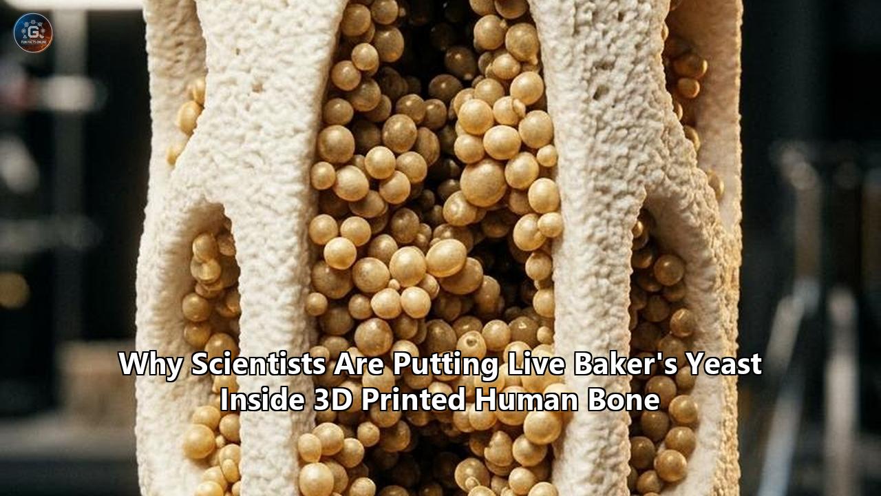
When orthopedic researchers at Clemson University loaded active, fermenting baker’s yeast into 3D-printed synthetic bone scaffolds, the setup appeared counterintuitive. For decades, the primary goal of orthopedic surgery has been maintaining sterile operating theaters and keeping microorganisms as far away from bone implants as possible. Introducing Saccharomyces cerevisiae—the ubiquitous single-celled fungus used to brew beer and bake sourdough—into an engineered skeletal matrix seemed to defy surgical logic.
Yet the study, spearheaded by biophysical chemist Dr. Jeffrey Anker alongside post-doctoral researcher Dr. Hannu Välimäki and their team, was designed to solve one of the most stubborn bottlenecks in regenerative medicine: the internal suffocation of artificial bone implants.
When surgeons attempt to reconstruct large skeletal defects resulting from high-impact trauma, blast injuries, cancer resections, or severe birth defects, standard synthetic bone grafts routinely fail from the inside out. While cells colonize the outer periphery of an implant with ease, the interior—any area deeper than a few hundred micrometers from the nearest blood vessel—rapidly runs out of oxygen. Deprived of blood flow, this central pocket turns into a necrotic dead zone where newly transplanted cells perish and tissue structural integrity collapses.
Testing physical solutions to this suffocation problem has historically taken months and cost hundreds of thousands of dollars. Culturing human stem cells and mature osteoblasts (bone-building cells) is slow, temperamental, and exorbitantly expensive. Human cells often fail silently deep within opaque ceramic materials, leaving bioengineers with little real-time data on how, when, and why the micro-environment turned lethal.
By using active baker’s yeast as a living metabolic surrogate, Anker’s research group found a way to compress those diagnostic timelines from months into minutes. Because live yeast cells metabolize oxygen at voracious, predictable rates, they act as high-speed stress-testers for the internal fluid dynamics and respiratory limits of artificial bone architectures. Paired with luminescent chemical sensor plates, the technique allows researchers to peer directly beneath opaque 3D-printed ceramic scaffolds and watch the suffocation process unfold across the graft’s micro-channels in real time.
The implications of this benchtop technique reach far beyond the novelty of microbial brewing organisms. By demonstrating that external perfusion pumps can reliably re-oxygenate the suffocating core of a 3D-printed bioactive glass implant, the team has established an operational blueprint for the next generation of active, vascularized human bone grafts.
+------------------------------------------------------------------------+
|                     THE CRITICAL-SIZED BONE CRISIS                     |
|                                                                        |
|  [ Traumatic Injury / Tumor Resection ]                                |
|                        │                                               |
|                        ▼                                               |
|  [ Implantation of 3D-Printed Synthetic Bone (>5mm) ]                  |
|                        │                                               |
|       ┌────────────────┴────────────────┐                              |
|       ▼                                 ▼                              |
|  Outer 200 µm Zone:               Core (>500 µm Deep):                 |
|  - Adequate passive diffusion     - Severe Ischemia & Hypoxia          |
|  - Capillary infiltration         - Rapid ATP depletion                |
|  - Cells survive & osteointegrate - Acidosis, Necrosis, Graft Failure   |
|                                         │                              |
|                                         ▼                              |
|           [ SOLUTION: Active Perfusion & Yeast Modeling ]               |
|           - Live yeast acts as high-speed metabolic probe              |
|           - Ratiometric luminescence maps real-time O2 flux            |
|           - Micro-pumps drive fluid through 3D print channels          |
+------------------------------------------------------------------------+
The Biological Wall: The 5-Millimeter Necrotic Core Problem
To understand why orthopedic bioengineers are turning to single-celled fungi, one must first look at the unforgiving physical laws governing human tissue survival.
Healthy mammalian bone is not an inert white stick of chalk. It is a highly dynamic, mineralized connective tissue permeated by an intricate vascular network. Inside natural human cortical bone, living osteocytes are housed within microscopic spaces called lacunae. These lacunae connect to one another through tiny fluid canals called canaliculi, which radiate outward toward central cylindrical structures known as osteons, or Haversian systems. Running through the center of every Haversian system is a dedicated blood vessel.
This microscopic architecture exists for a strict physiological reason: the Krogh diffusion limit.
Formulated in 1919 by Danish physiologist August Krogh, the principle dictates that oxygen, glucose, and essential water-soluble nutrients can only diffuse passively across a distance of roughly 100 to 200 micrometers (0.1 to 0.2 millimeters) through metabolically active tissue before being entirely consumed by surrounding cells. Beyond this 200-micrometer threshold, oxygen tension drops toward zero, cellular respiration grinds to a halt, adenosine triphosphate (ATP) production collapses, intracellular lactic acid builds up, and cells trigger programmed apoptosis or succumb to unprogrammed, inflammatory necrosis.
In minor hairline fractures or small drill-hole defects, the human body repairs bone autonomously. Platelets form a hematoma, inflammatory cells clear debris, mesenchymal stem cells migrate into the fracture gap, and capillary sprouting (angiogenesis) swiftly bridges the tiny divide.
However, when a patient suffers a "critical-sized bone defect"—clinically defined as a gap that will not heal spontaneously over the patient's lifetime without surgical intervention—the body's natural regenerative mechanics fail. In adult human long bones, such as the femur or tibia, a defect larger than 2 to 2.5 centimeters or a volumetric loss exceeding a few cubic centimeters crosses this fatal boundary.
                    PASSIVE DIFFUSION GRADIENT
Oxygen Source                                               Hypoxic Core
(Blood Vessel)                                              (Necrotic Zone)
      │                                                           │
   PO2: ~100 mmHg                                              PO2: 0 mmHg
      │                                                           │
      ▼                                                           ▼
      ═════════════════════════════════════════════════════════════
      0 µm            100 µm           200 µm           300+ µm
      [ Viable Cells ] [ Hypoxia Begins ] [ Apoptosis ] [ Total Necrosis ]
When surgeons confront critical defects, the current gold standard treatment is an autologous bone graft (autograft), typically harvested from the patient's own iliac crest (pelvis). The harvested bone contains the patient’s own mineral matrix, osteoinductive growth factors, and living osteoprogenitor cells.
Yet autografts are hobbled by steep physical and physiological limitations:
- Donor Site Morbidity: Harvest surgeries frequently result in secondary site infections, persistent neurovascular injuries, donor site herniation, hematomas, and severe chronic donor-site pain that can plague patients for years.
- Volume Scarcity: The human skeleton offers only limited expendable bone mass. In cases of extensive cancer resections or multiple severe fractures, there simply is not enough harvestable donor bone to plug the voids.
- Graft Resorption Without Ingrowth: Even harvested autologous blocks thicker than a few millimeters frequently suffer core ischemia before the recipient site's capillary beds can penetrate the dense cortical donor matrix, resulting in central structural collapse.
The alternative—allografts harvested from human cadaver donors—eliminates donor site pain but presents a different set of hurdles. Cadaver bone must be heavily freeze-dried, gamma-irradiated, or chemically treated to eliminate viral contamination risks and strip out human leukocyte antigens that would otherwise trigger life-threatening immune rejection. This aggressive processing completely kills off all living osteocytes and significantly degrades the bone’s structural and osteoinductive proteins. The resulting allograft acts as little more than a fragile, dead mineral scaffold that the host immune system slowly attempts to absorb, frequently failing to achieve mechanical union and succumbing to late-stage mechanical fatigue fractures.
These failures prompted the rise of 3D bone tissue engineering. Using patient-specific computed tomography (CT) scans, additive manufacturing systems can print porous ceramic, glass, or polymer scaffolds customized down to the micrometer to match a patient’s unique skeletal anatomy.
Yet the moment a bioengineered graft surpasses roughly 5 millimeters in thickness, it runs headfirst into the biological wall. When human stem cells are seeded into these scaffolds prior to implantation, the cells residing along the outer edge thrive on surrounding bodily fluids. But deep inside the porous interior, fluid remains stagnant. Passive diffusion cannot haul oxygen into the core or flush toxic metabolic byproducts out. Within 48 to 72 hours, the center of the graft becomes anoxic, forming a soft, rotting necrotic core.
Resolving this central suffocation problem is the single greatest hurdle standing between 3D-printed bone research and routine clinical adoption.
Why Bread Yeast? The Biological Proxy That Outperforms Human Cells in the Lab
To engineer a solution to this core suffocation, scientists must test how fluids, oxygen, and metabolic wastes interact within complex, three-dimensional micro-lattices.
For years, research labs have attempted to map these dynamics using human mesenchymal stem cells (hMSCs), human fetal osteoblasts (hFOBs), or rodent osteosarcoma cell lines. While these mammalian cell cultures offer high biological fidelity to human disease, using them to evaluate the raw fluid mechanics and oxygen physics of physical scaffolds introduces crippling experimental liabilities:
- Glacial Timelines: Human bone stem cells divide slowly, typically requiring an incubation period of 24 to 48 hours for a single doubling cycle. Differentiating these precursors into mature osteoblasts capable of depositing mineralized collagen matrices takes 14 to 28 continuous days in a cell incubator.
- Extreme Financial Cost: A single vial of character-certified human mesenchymal stem cells costs between $800 and $1,500. Maintaining them demands specialized basal media, fetal bovine serum (FBS)—which routinely costs over $600 per bottle—recombinant growth factors (such as BMP-2 or TGF-beta) running hundreds of dollars per microgram, and sterile cleanroom facilities.
- Biological Fragility: Mammalian cells are hypersensitive to slight fluctuations in temperature, pH, osmotic pressure, and fluid shear stresses. If a prototype perfusion pump experiences a minor flow surge or a bubble slips into the tubing, delicate human osteoblast membranes shear open, contaminating the experiment.
- Metabolic Invisibility: Mammalian cells consume oxygen at comparatively slow, variable rates depending on their differentiation stage and culture cycle. Mapping the exact boundaries of a hypoxic zone using human cells requires sacrificial analytical assays (such as destructive lactate dehydrogenase staining or gene expression lysis), preventing researchers from monitoring dynamic changes in the same graft over consecutive hours.
Faced with these investigative barriers, bioengineers turned to Saccharomyces cerevisiae.
From an evolutionary standpoint, baker's yeast is an exceptionally resilient single-celled eukaryote. Unlike bacteria, yeast cells contain a true nucleus, membrane-bound organelles, and complex mitochondria that carry out aerobic cellular respiration using enzymatic electron transport chains remarkably similar to those operating inside human cells.
When provided with oxygen and simple sugars (such as dextrose or glucose), live yeast undergoes vigorous aerobic respiration:
$$\text{C}_6\text{H}_{12}\text{O}_6 + 6\text{O}_2 \longrightarrow 6\text{CO}_2 + 6\text{H}_2\text{O} + 36\text{–}38\text{ ATP}$$
Because a single gram of baker's yeast contains approximately 20 to 30 billion living, metabolizing cells, its volumetric oxygen consumption rate is orders of magnitude higher than that of an equivalent mass of human bone tissue. When suspended in a simple, food-grade sugar solution, yeast cells double every 90 minutes and immediately begin consuming dissolved oxygen in accordance with classic Michaelis-Menten enzyme kinetics.
| Parameter | Primary Human Osteoblasts / Stem Cells | Baker’s Yeast (Saccharomyces cerevisiae) |
|---|---|---|
| Preparation Time | 2 to 4 weeks of sterile cell culture | 10 minutes of rehydration in sugar water |
| Volumetric Cost | ~$1,000–$2,500 per experimental batch | <$0.05 per experimental batch |
| Nutrient Media Needs | High-glucose DMEM, fetal bovine serum, antibiotics, growth factors | Water and 3% commercial table glucose |
| Shear Stress Sensitivity | High; cell detachment and membrane rupture occur easily | Extremely low; protected by thick chitin/glucan wall |
| Oxygen Consumption Rate | Low to moderate; takes days to register hypoxia | Extremely rapid; depletes local oxygen within minutes |
| Temperature Constraints | Strict 37.0°C; ambient cooling induces shock | Operates robustly across 18°C to 35°C |
| Biohazard Risk | Biosafety Level 2 (BSL-2); human pathogen risks | Biosafety Level 1 (BSL-1); generally recognized as safe |
Deploying live yeast in 3d printed bone scaffolds allowed the Clemson researchers to transform an opaque bioceramic structure into an active, breathing metabolic engine. Instead of waiting a month to learn whether a particular scaffold geometry starved its residents, the team could infuse billions of yeast cells into the internal channels, place the scaffold into a fluidic test chamber, and observe the immediate formation of a severe, localized hypoxic zone within minutes.
If an experimental pumping system could successfully push enough oxygen-saturated fluid through the scaffold to keep this voracious colony of baker’s yeast alive and breathing, that same physical design could effortlessly keep slower-breathing human osteoblasts alive in an operating room. The yeast served as a worst-case-scenario stress test: a biological stand-in operating at full metabolic throttle.
Robocasting and Bioceramics: 3D Printing the Skeleton's Architecture
The vehicles used in these experiments are not standard plastic toys produced on consumer filament printers. They are custom-engineered bioceramic structures manufactured using direct ink writing (DIW), an additive manufacturing method commonly known in materials science as robocasting.
Human bone possesses a complex Young's modulus (stiffness) ranging from 10 to 20 gigapascals (GPa) for dense cortical shafts down to 0.1 to 2 GPa for porous trabecular sponge. To mimic both the mechanical load-bearing capacity and the chemical composition of natural bone, scientists print these implants using bioactive glasses.
                ROBOCASTING DIRECT INK WRITING (DIW)

           [ Syringe Barrel: Bioactive Glass + Pluronic F127 ]
                                 │
                                 ▼ Pneumatic Pressure Controller
                           [ Fine Nozzle ]
                                 │ (~400 µm diameter)
                                 ▼
                     Extruded Shear-Thinning Filament
                                 │
          ┌──────────────────────┴──────────────────────┐
          ▼                                             ▼
    Layer N+1 ──► ░░░░░░░░░░░░░░░░░░░░░░░░░░░░░   (Pores: 300–800 µm)
    Layer N   ──► ░░░░      ░░░░      ░░░░
    Substrate ──► ═════════════════════════════
In the Clemson experiments, researchers utilized 1393B20 borosilicate glass, a specialized bioactive glass formulation. While traditional bioglass—such as Larry Hench's 45S5 Bioglass invented in 1969—is built entirely upon a silicate ($SiO_2$) backbone with calcium, sodium, and phosphorus oxides, 1393B20 substitutes a percentage of the silicon dioxide with boron trioxide ($B_2O_3$).
This specific elemental tweak yields two critical properties:
- Accelerated In Vivo Degradation: Borate glasses dissolve at a faster, more controllable rate than pure silicate glasses, gradually dissolving into benign non-toxic ions (borate, calcium, phosphate) as the host body replaces the artificial scaffold with natural hydroxyapatite crystals.
- Lower Sintering Temperature: The borate substitution lowers the glass transition temperature, allowing the printed scaffolds to fuse together solidly during thermal firing without crystallizing into brittle, unreactive ceramics.
Formulating the Extrudable "Ink"
To push brittle micro-scale glass particles through a 400-micrometer nozzle without clogging or collapsing, researchers suspend the glass powder in a specialized hydrogel vehicle: Pluronic F127.
Pluronic F127 is a synthetic triblock copolymer consisting of a central hydrophobic polypropylene oxide (PPO) block flanked by two hydrophilic polyethylene oxide (PEO) blocks:
$$\text{PEO}_{100}\text{–}\text{PPO}_{65}\text{–}\text{PEO}_{100}$$
In cold aqueous solutions (below 10°C), Pluronic molecules move freely as independent unimers. However, as the temperature rises to room temperature (20°C to 25°C), the hydrophobic PPO cores dehydrate and self-assemble into tightly packed spherical micelles. This self-assembly causes a physical transformation called reversible thermosensitive sol-gel transition.
At high solid loadings—such as the 30% Pluronic F127 to 70% bioactive glass powder ratio employed in the study—the ink displays pronounced shear-thinning (pseudoplastic) and thixotropic behavior. Inside the syringe barrel, the ink behaves like a firm gel. Under high shear stress when forced through the narrow printing tip by a pneumatic piston, the micellar networks slide over one another, dropping the fluid's viscosity dramatically and allowing it to flow like a smooth liquid.
The moment the ink exits the printing tip and the shear stress drops back to zero, it instantly solidifies, holding its exact cross-sectional shape and bearing the weight of successive layers without drooping or pooling.
Geometry, Sintering, and Trabecular Pores
The robocasting system deposits this paste in alternating, orthogonal cross-hatched paths layer by layer. The design parameters are selected to solve specific biological constraints:
- Macropore Dimension: The gap between printed struts is set between 300 and 800 micrometers. Studies have demonstrated that pore openings smaller than 100 micrometers inhibit vascular cell penetration, while pores larger than 1,000 micrometers fail to promote initial cellular attachment and weaken the scaffold's mechanical compressive strength.
- Total Porosity: The interior volume consists of approximately 50% to 60% open, fully interconnected voids, mirroring the structural architecture of natural human trabecular (cancellous) bone.
Once the green, wet scaffold is fully printed, it is dried and transferred to a high-temperature furnace. In this thermal stage, the polymer binder (Pluronic F127) completely burns off at temperatures between 400°C and 500°C, leaving no synthetic plastic residues behind. The furnace is then ramped up to roughly 600°C to 700°C, initiating viscous flow sintering. At this temperature, the bioactive glass particles soften, necking together into a monolithic, vitreous lattice possessing high compressive strength capable of withstanding the loads experienced by human limbs.
Yet, despite this open porosity, without active circulation, fluid inside these 3D-printed channels remains completely stationary. To solve this, researchers needed a way to visualize what happens inside these micro-tunnels when biological organisms move in.
Watching Cells Suffocate: The Ratiometric Luminescence Imaging Feat
One of the foundational challenges of bone tissue engineering is an optical one: bone is opaque.
Whether examining natural human cortical bone or a 3D-printed bioactive borosilicate matrix, physical light scattering prevents conventional light microscopy from penetrating deeper than a few hundred micrometers into the structure. Historically, researchers could not measure oxygen levels inside an intact, thick scaffold in real time. They had to rely on inserting invasive needle-type microelectrodes—which can alter local fluid flow, destroy delicate cell layers, and only provide a reading at a single pinpoint—or rely on theoretical mathematical simulations.
To shatter this blind spot, the Clemson team merged their yeast model with an advanced optical sensing methodology: ratiometric oxygen luminescence imaging.
                 RATIOMETRIC OXYGEN SENSING MECHANISM

      Excitation UV/Blue Light (~405 nm)
            │
            ├─────────────────────────┬─────────────────────────┐
            ▼                         ▼                         ▼
   [ Reference Dye ]         [ O2-Sensitive Dye ]               │
   (Inert Lumophore)         (Phosphorescent Dye)               │
            │                         │                         │
            ▼                         │                         │
   Emits Constant Red                 ▼                         │
   Signal (λ_ref)            Oxygen Quenching Collision         │
   Independent of O2                  │                         │
                                      ▼                         ▼
                             O2 Present (Normoxia):    O2 Absent (Hypoxia):
                             Energy transferred to O2  Full Phosphorescence
                             Luminescence QUENCHED     HIGH Signal (λ_O2)
The underlying physics relies on the phenomenon of dynamic luminescence quenching by molecular oxygen.
The researchers fabricated an optical sensor slide coated with a specialized polymer membrane containing two distinct fluorescent/phosphorescent dyes:
- An Oxygen-Sensitive Phosphor: Typically a heavy-metal transition complex, such as a platinum(II) or palladium(II) porphyrin (e.g., PtTFPP). When this molecule absorbs high-energy excitation light (usually in the blue or ultraviolet spectrum), its electrons jump to an excited triplet state. In the absence of oxygen, the molecule drops back to its ground state by emitting a bright, long-lived phosphorescent light.
- The Quenching Mechanism: If a molecular oxygen ($O_2$) molecule is present, it collides with the excited porphyrin molecule. Because ground-state molecular oxygen is naturally in a triplet electronic state ($^3\Sigma_g^-$), it undergoes an efficient, radiationless triplet-triplet energy transfer with the dye. The energy is harmlessly transferred to the oxygen molecule, promoting it to singlet oxygen, while the dye returns to its ground state without emitting a photon. The higher the oxygen concentration (partial pressure, $PO_2$), the more collisions take place, and the dimmer the phosphorescence becomes.
- An Oxygen-Insensitive Reference Dye: To ensure that variations in light intensity are caused strictly by oxygen changes—rather than uneven illumination, slight dye fading (photobleaching), or structural shadows cast by the printed glass struts—a second, non-reactive dye is incorporated into the film. This reference dye emits a constant fluorescent signal regardless of how much oxygen surrounds it.
The relationship between the measured luminescence intensity and the surrounding oxygen tension is calculated using the Stern-Volmer equation:
$$\frac{I_0}{I} = 1 + K_{SV} \cdot [O_2] = 1 + k_q \cdot \tau_0 \cdot [O_2]$$
Where:
- $I_0$ is the luminescence intensity in the total absence of oxygen (zero quenching).
- $I$ is the luminescence intensity measured at a specific oxygen concentration $[O_2]$.
- $K_{SV}$ is the overall Stern-Volmer quenching constant.
- $k_q$ is the bimolecular quenching rate constant.
- $\tau_0$ is the unquenched luminescence lifetime of the sensor molecule.
By capturing widefield microscopic images through separate emission filters, custom software divides the oxygen-sensitive signal by the reference signal pixel by pixel. This mathematical ratio yields an absolute, high-resolution 2D map of oxygen tension ($PO_2$) across every square micrometer directly underneath the bone scaffold.
The real-time visualization of yeast in 3d printed bone constructs revealed how fast a tissue environment can turn deadly. When the bioactive glass scaffold was soaked in a 3% glucose solution loaded with baker’s yeast and seated onto the sensor plate inside a micro-chamber, the initial oxygen reading sat near atmospheric equilibrium: a healthy, normoxic partial pressure of roughly $PO_2 \approx 21\text{ kPa}$ (~160 mmHg).
Within ten minutes of stopping external fluid flow, the cellular respiration of the yeast devoured the local oxygen reserve. The ratiometric cameras captured a dramatic color transition: the entire central footprint of the scaffold turned dark, with oxygen levels plunging to near absolute zero ($PO_2 \approx 0\text{ kPa}$).
The resulting spatial mapping demonstrated that while outer edge regions remained marginally oxygenated via passive atmospheric diffusion from the surrounding well, the center experienced total hypoxia. The metabolic consumption of the yeast had formed a sharp reaction-diffusion boundary, recreating the exact necrotic core conditions that have doomed thick synthetic bone implants in clinical trials.
The Fluid Dynamic Rescue: Testing Active Perfusion Inside Living Scaffolds
Having established a real-time visualization platform for core hypoxia, the Clemson team could directly evaluate the single physical mechanism capable of breaking the Krogh diffusion limit: active fluid perfusion.
In the human body, tissues avoid suffocation because the heart operates as a positive-displacement pump, maintaining high hydrostatic pressure gradients across arteriolar beds and driving blood through a network of 10-micrometer capillaries. In a large synthetic bone substitute, no such blood vessels exist immediately following surgery. Blood vessel ingrowth from surrounding host tissue is notoriously slow, proceeding at a rate of only roughly 0.1 to 0.5 millimeters per day. For a 10-millimeter or 20-millimeter bone graft, waiting weeks for natural capillary sprouting guarantees the core will be dead long before blood vessels ever reach the center.
To counter this, the researchers linked their yeast-loaded bone chambers to an external micro-perfusion pump system.
                    ACTIVE PERFUSION RESCUE CYCLE

  Oxygen Tension (PO2)
   21 kPa ┼──────────────────────────────┐                (Fresh Flush)
          │                              │\               Normoxia Restored
          │                              │ \
          │    Static Phase              │  \
          │    (Yeast consumes O2)       │   \            Static Phase Returns
          │                              │    \           (Rapid O2 Depletion)
    0 kPa ┼───\                          │     \─────────►
          └───┴──────────────────────────┴─────────────────────────► Time
            Flushing OFF (t=0)        Flushing ON (t=3 min)
The experiment evaluated cyclical, intermittent perfusion. With the scaffold resting in an initial state of complete, self-induced hypoxia, the external pump engaged for a brief three-minute flushing cycle, pushing fresh, oxygen-saturated medium horizontally through the 3D-printed channels at controlled flow rates.
The sensory readout was immediate and dramatic:
- The Influx Front: As the fluid moved through the porous grid, the ratiometric oxygen sensors documented a rapid upward surge in oxygen tension across all zones.
- Peripheral Saturation: At the outer boundaries, oxygen levels swiftly climbed back to the atmospheric ceiling of $PO_2 \approx 21\text{ kPa}$.
- Core Penetration: Most critically, the deep, suffocating center of the scaffold—which had been starved of oxygen—experienced a rapid reversal, climbing to a healthy $PO_2 \approx 9.5\text{ kPa}$ (~72 mmHg). In mammalian physiology, normal physiological tissue normoxia (physioxia) inside vascularized human bone marrow sits between 2% and 7% oxygen (roughly 15 to 50 mmHg). The three-minute wash had successfully transported life-sustaining levels of oxygen deep into the previously necrotic core.
The experiments also captured an intriguing physical nuance in fluid transport: the diffusion delay phenomenon.
When the pump was switched off, the outer edges of the scaffold began depleting their oxygen reserves immediately as yeast cells consumed the gas. In the central core, however, oxygen levels continued to rise slightly for roughly two minutes after fluid pumping had completely ceased.
This temporary counter-intuitive increase occurred because the fluid within the main channels had been saturated with fresh oxygen, which continued to diffuse laterally across roughly 700 micrometers into adjacent micro-porous voids according to Fick's Second Law of Diffusion:
$$\frac{\partial \phi}{\partial t} = D \frac{\partial^2 \phi}{\partial x^2}$$
Where:
- $\phi$ is the oxygen concentration.
- $t$ is time.
- $D$ is the diffusion coefficient of oxygen in aqueous media (approximately $2 \times 10^{-5}\text{ cm}^2/\text{s}$).
- $x$ is the physical diffusion distance.
Once that localized reservoir was consumed, the yeast's rapid metabolic engine dragged the entire scaffold back into deep hypoxia. Because the yeast's cellular response was so fast and resilient, the researchers could repeat this flush-and-starve cycle multiple times back-to-back, demonstrating near-perfect cycle-to-cycle reproducibility without having to discard or rebuild the experimental chamber.
The test established clear engineering proof: a temporary, low-volume external fluid flush can entirely eliminate the necrotic core inside a 3D-printed bone substitute without requiring a pre-existing capillary bed.
The Immune Cross-Talk: How Yeast Polymers Re-Educate the Human Immune System
While living yeast cells serve as diagnostic tools for fluid mechanics and oxygen consumption, Saccharomyces cerevisiae holds a second, direct therapeutic value in skeletal regeneration: its physical cell wall can chemically retrain the human immune system.
Historically, biomaterial researchers believed that an ideal bone implant should be entirely "bio-inert"—causing zero immune reaction whatsoever. Decades of clinical failures proved this concept misguided. When an artificial material is placed into the body, the immune system will inevitably recognize it. The true goal of contemporary regenerative medicine is not to suppress inflammation, but to steer it: an emerging discipline known as osteoimmunomodulation.
The study of yeast in 3d printed bone scaffolds has also spurred interest in the unique macromolecular polymers that make up the yeast cell wall: $\beta$-glucans and mannans.
                 MACROPHAGE POLARIZATION AXIS

  Uncommitted Host Macrophage (M0)
         │
         ├───► Triggered by Chronic Debris, Metal Wear, or Uncontrolled Damage:
         │     M1 PHENOTYPE (Pro-Inflammatory / Destructive)
         │     - Secretes: TNF-α, IL-1β, IL-6, reactive oxygen species (ROS)
         │     - Upregulates: RANKL signaling
         │     - Result: Osteoclast activation, severe bone loss, aseptic loosening
         │
         └───► Instructed by Yeast β-Glucan / Mannan Polymers (via Dectin-1):
               M2 PHENOTYPE (Pro-Regenerative / Anti-Inflammatory)
               - Secretes: IL-10, TGF-β, VEGF (Vascular Endothelial Growth Factor)
               - Downregulates: NF-κB and NFATc1
               - Result: Capillary sprouting (angiogenesis), osteoblast differentiation,
                 dense mineralized bone matrix deposition
The outer shell of Saccharomyces cerevisiae is composed largely of $\beta$-(1,3)/(1,6)-D-glucan, a complex branched polysaccharide. In natural biology, human immune cells identify $\beta$-glucans as pathogen-associated molecular patterns (PAMPs). When human monocytes and uncommitted macrophages (M0 phenotype) encounter carefully purified, micro-structured yeast $\beta$-glucan particles, the polymer binds specifically to a transmembrane receptor on the macrophage surface known as Dectin-1 (Dendritic cell-associated C-type lectin-1).
This binding event initiates a powerful biochemical cascade:
- Phenotype Switching (M1 to M2): In an ordinary bone break or biomaterial implantation, macrophages frequently stall in the M1 phenotype—a pro-inflammatory state characterized by high secretions of tumor necrosis factor-alpha (TNF-$\alpha$), interleukin-1 beta (IL-$1\beta$), and interleukin-6 (IL-6). M1 macrophages perpetuate chronic inflammation, degrade surrounding collagen, and promote fibrous scar formation. Controlled exposure to yeast $\beta$-glucan instructs macrophages to undergo a phenotype transition into the M2 phenotype—an anti-inflammatory, pro-healing state.
- Secretion of Angiogenic Factors: Once polarized to the M2 state, macrophages actively pump out vascular endothelial growth factor (VEGF) and transforming growth factor-beta (TGF-$\beta$). These signaling proteins act as homing beacons for endothelial cells, driving rapid blood vessel growth and capillary infiltration directly toward the scaffold.
- Suppression of Bone-Destroying Osteoclasts: One of the most common causes of bone graft failure is hyperactive osteoclastogenesis—where bone-eating cells (osteoclasts) dissolve the newly forming mineral faster than osteoblasts can synthesize it. Research has demonstrated that yeast $\beta$-glucan acts as a potent inhibitor of this destructive process.
When immature osteoclasts are stimulated by Receptor Activator of Nuclear Factor-$\kappa$B Ligand (RANKL), they normally trigger nuclear factor of activated T-cells 1 (NFATc1) via nuclear factor-$\kappa$B (NF-$\kappa$B) pathways, commanding the cells to mature and begin dissolving bone mineral.
Yeast $\beta$-glucans disrupt this pathway. By blocking NF-$\kappa$B translocation to the nucleus, the fungal polymer blunts NFATc1 expression, drastically reducing osteoclast differentiation while simultaneously encouraging mesenchymal stem cells to differentiate into mature bone-forming osteocytes.
Bioengineers are now isolating these purified yeast mannans and glucans, chemically cross-linking them with methacrylate groups (such as yeast mannan methacrylate, or YM-MA), and using them as bioactive hydrogel binders within 3D-printed bone constructs. The fungus, once viewed merely as a culinary ingredient, provides both the structural polysaccharide ink and the immunological signals necessary to bridge the gap between foreign synthetic glass and living human biology.
The Financial Anatomy of Bone Grafting: Market Pressures and Clinical Economics
The drive to harness non-traditional biological platforms like baker's yeast is rooted in the immense financial strains facing modern orthopedic healthcare.
Musculoskeletal conditions represent one of the largest economic burdens on global health systems. Worldwide, more than 2.2 million bone grafting procedures are carried out each year. The global market for bone graft substitutes—encompassing allografts, demineralized bone matrices (DBM), synthetic calcium phosphates, bioactive glasses, and recombinant morphogenetic proteins—is currently valued at more than $3.5 billion annually and is projected to surpass $5.2 billion before the end of the decade.
                GLOBAL BONE GRAFTING PROCEDURE MIX

  ┌────────────────────────────────────────────────────────┐
  │  Autografts (Iliac Crest / Local Harvest)  (~45%)     │
  ├────────────────────────────────────────────────────────┤
  │  Allografts (Cadaveric Donor Tissue)       (~35%)     │
  ├────────────────────────────────────────────────────────┤
  │  Synthetic Scaffolds & Bioceramics         (~20%)     │
  └────────────────────────────────────────────────────────┘

  Average Cost per Critical-Sized Defect Case:
  - Base Surgical Procedure:                $35,000 – $60,000
  - Autograft Complication (Infection/Pain): +$15,000 – $28,000
  - Revision Surgery for Graft Non-Union:    +$45,000 – $80,000
The underlying economic challenge is that traditional treatment modalities for critical-sized bone loss are inefficient, hazardous, and expensive:
- The Cost of Autograft Morbidity: Harvesting an iliac crest autograft adds an average of 45 to 90 minutes of operating room time, which in modern surgical suites costs between $60 and $100 per minute. Clinical health economic studies estimate that complications specifically associated with the harvest site—such as deep wound infection, persistent nerve paresthesia, or herniation—add between $15,000 and $28,000 to the total cost of care per patient over a five-year horizon.
- The Staggering Price of Recombinant Growth Factors: When synthetic calcium phosphate scaffolds are used without patient cells, orthopedic surgeons frequently soak them in recombinant human Bone Morphogenetic Protein-2 (rhBMP-2, commercialized as INFUSE). While rhBMP-2 is a powerful osteoinductive trigger, it is one of the most expensive liquid biologics in medicine, frequently billing at $3,500 to $6,000 per single-use kit. Furthermore, high doses of rhBMP-2 have been associated with dangerous clinical side effects, including uncontrolled swelling near cervical spines, ectopic bone formation in muscle tissue, and osteolytic bone resorption.
- The Catastrophic Cost of Revision Surgeries: When a thick, synthetic implant fails due to a central necrotic core, the graft experiences what orthopedists term "aseptic non-union" or fatigue failure. The dead scaffold shatters, requiring a complex revision surgery to cut away fibrous scar tissue, clean out fragments, and re-attempt stabilization. Hospital revision procedures routinely exceed $50,000 to $80,000 per intervention.
This is where the research into yeast in 3d printed bone systems shifts the economic equation.
In preclinical research and development, pharmaceutical and medical device companies spend tens of millions of dollars attempting to optimize scaffold channel diameters, porosity gradients, and perfusion protocols. Testing these designs in mammalian cell bioreactors or live large-animal trials (sheep, pigs, or dogs) costs hundreds of thousands of dollars per prototype run.
By utilizing rapid, food-grade yeast assays, research laboratories can run hundreds of microarchitectural and fluid-dynamic iterations in a standard lab setting at negligible cost. A single researcher can evaluate ten completely distinct 3D pore configurations in an afternoon for less than $10 worth of materials, pinpointing precisely how fluid flow rates alter central oxygenation profiles.
By eliminating the slow, high-cost mammalian cell barrier during the early prototyping phase, this approach drastically lowers the capital expenditure required to advance an orthopedic implant through preclinical testing.
From Bench to Operating Room: Overcoming Safety Hurdles and Technical Roadblocks
While the experimental results validate yeast as an exceptional diagnostic model, translating these discoveries into sterile human operating rooms introduces critical safety boundaries and regulatory realities.
No surgeon will ever inject live baker's yeast directly into an open human fracture. While Saccharomyces cerevisiae is classified as a Biosafety Level 1 (BSL-1) organism and is broadly non-pathogenic to healthy individuals, it remains an opportunistic fungal agent. In severely immunocompromised patients, individuals with central venous catheters, or patients suffering from severe burn trauma, systemic infections with Saccharomyces—known clinically as Saccharomyces fungemia or invasive yeast sepsis—can be fatal.
Therefore, the practical clinical path for this research operates along two completely distinct tracks:
               TWO-TRACK TRANSLATIONAL BLUEPRINT
                               │
       ┌───────────────────────┴───────────────────────┐
       ▼                                               ▼
  TRACK 1: THE DIAGNOSTIC SURROGATE          TRACK 2: THE CLINICAL IMPLANT
  - Live Yeast used in lab ONLY              - Pure, Synthetic Bioceramic Scaffold
  - Maps fluid mechanics & O2 limits         - Zero fungal cells implanted
  - Validates active perfusion protocols     - Incorporates isolated yeast polymers
  - Fast, ultra-cheap benchtop testing         (Purified β-glucans & mannans)
                                             - Driven by external micro-perfusion
Track 1: The Preclinical Engineering Standard
In the first track, live yeast remains strictly a laboratory test surrogate. It serves as a rapid calibration tool for medical device manufacturers. Before a company initiates a multi-million-dollar human clinical trial for a new 3D-printed orthopedic or craniomaxillofacial implant, the physical scaffold must be certified to provide adequate mass transfer and prevent internal hypoxia. Running the scaffold through standardized yeast respiration assays under varying flow rates allows bioengineers to mathematically certify that the scaffold's internal channels will sustain cellular life once implanted in humans.
Track 2: The Sterile, Perfusion-Driven Human Implant
In the second track, the physical insights gathered from the yeast model are translated directly into clinical implants. The scaffold that enters the patient’s body is a sterile, pure bioactive glass or bioceramic matrix completely free of live fungal organisms.
To replicate the fluid flow that kept the yeast alive, the implant is paired with a miniaturized, temporary external perfusion system:
- Implanted Micro-Tubing: During the reconstructive surgery, fine, flexible, medical-grade silicone tubing is threaded directly into the printed fluid distribution channels at the core of the bioceramic implant.
- Portable Perfusion Unit: The tubing exits the patient’s skin through a small incision and connects to a compact, wearable micro-perfusion pump, conceptually similar to the Negative Pressure Wound Therapy (NPWT) or "wound-vac" devices currently worn by millions of patients recovering from deep surgical wounds.
- Intermittent Flushing Protocol: Based on the timing profiles established by the yeast experiments, the unit periodically pumps tiny volumes of sterile, oxygen-enriched physiological saline supplemented with low-dose antibiotics and autologous serum into the center of the scaffold.
- Temporary Deployment: This active external perfusion maintains core normoxia during the critical first two to three weeks post-operation. Once native capillaries have fully infiltrated the scaffold's open pores and established a natural microvascular blood supply, the tubing is simply decoupled and withdrawn in an outpatient clinic, leaving behind an integrated, fully vascularized bone graft.
The Regulatory Path Forward
Bringing such a system through the United States Food and Drug Administration (FDA) requires navigating the framework for Combination Products.
Because the system unites a permanent structural implant (a 3D-printed bioceramic Class III device) with an active external infusion unit (a Class II or III delivery system) and bioactive immunological compounds (purified yeast $\beta$-glucan modulators), the premarket review must pass through both the Center for Devices and Radiological Health (CDRH) and the Center for Drug Evaluation and Research (CDER).
Manufacturers must demonstrate:
- Exhaustive Pyrogenicity Testing: Ensuring all isolated yeast cell wall polymers are entirely stripped of fungal endotoxins or residual fungal proteins that could trigger acute hyper-inflammatory systemic reactions or anaphylaxis.
- Mechanical Fatigue Testing: Verifying that the internal perfusion channels built into the 3D-printed bioceramic do not create stress concentration points that could cause the scaffold to crack under physiological cyclic loading (such as normal walking).
- Sterility Assurance Levels (SAL): Proving that the delivery lines maintain a sterility assurance level of $10^{-6}$, ensuring zero bacterial entry into the bone space throughout the multi-week perfusion treatment.
The Next Frontier: Smart Implants, X-Ray Chemical Sensing, and Biohybrid Medicine
The discovery that live baker's yeast can map internal scaffold suffocation marks the beginning of an era of smart, dynamic biomaterials.
Building on this foundational work, researchers are already preparing the next phase of this technology: X-ray excited luminescence chemical imaging (XELCI).
                 IN VIVO XELCI CHEMICAL MAPPING

  [ Focused X-Ray Beam Source ]
              │ (Penetrates deep soft tissue & bone without scatter)
              ▼
  ══════════════════════════════════════════════════════════════════
  Patient Skin & Muscle Tissue (Up to several centimeters thick)
  ══════════════════════════════════════════════════════════════════
              │
              ▼
   [ 3D-Printed Bone Implant with Scintillator & Sensing Layer ]
              │
              ├─► 1. X-rays hit internal radioluminescent phosphor particles
              │      (Scintillators emit localized UV/Visible light)
              ├─► 2. Optical sensors measure local O2 or pH in the deep core
              └─► 3. Modulated red/near-infrared photons pass back out
              │
              ▼
  [ External High-Sensitivity Optical Detector / Camera ]
  Displays Real-Time O2 & Infection Map on Surgeon's Monitor
While the original Clemson study tracked luminescence through a clear glass slide using visible light in a petri dish, visible light cannot travel through thick human muscle or skin. To overcome this in living patients, the Anker research group has developed special scintillator particles embedded directly inside the scaffold coatings.
When a focused, low-dose medical X-ray beam passes harmlessly through human flesh, it collides with these scintillating nanoparticles, causing them to emit localized visible light from the inside out. This localized light excites the surrounding oxygen- and pH-sensitive dyes. The resulting optical signals travel back to an external detector, allowing physicians to scan an orthopedic patient with a standard radiography machine and generate an immediate, non-invasive map of the oxygenation, metabolic health, and infection status deep inside an implanted bone graft without making a single surgical incision.
The diagnostic insights gathered from yeast in 3d printed bone are already influencing other areas of materials science and additive manufacturing:
- Engineered Living Architectural Materials: Across multiple university laboratories, civil engineers and architectural designers (such as Jenny Sabin’s team at Cornell University and materials scientists at Chalmers University of Technology) are applying similar logic to the built environment. They are 3D-printing porous architectural ceramic wall tiles that mimic human bone osteon geometry and infusing them with live baker's yeast. These living, breathing ceramic walls pull toxic volatile organic compounds (such as airborne formaldehyde) out of indoor air, using the fungus to digest environmental pollutants within porous, bio-inspired building systems.
- High-Efficiency Biocatalytic Scaffolds: In biotechnology, additive manufacturing teams are using 3D-printed polymer and ceramic lattices laden with ultra-high densities of live yeast to construct continuous, solid-state fermentation bioreactors. These living materials demonstrate vastly superior mass transfer and metabolic conversion of sugars into bio-ethanol and high-value pharmaceuticals compared to conventional bulk fluid fermentation tanks.
- Closed-Loop Smart Implants: Future iterations of clinical bone scaffolds will integrate bio-resorbable micro-sensors and piezoelectric harvesting units directly into the 3D-printed glass lattice. These implants will monitor internal oxygen and pH levels autonomously. If the center begins drifting toward hypoxia, the implant will trigger a tiny internal micro-reservoir to release oxygen-generating chemical compounds (such as calcium peroxide or sodium percarbonate complexes) or pulse an integrated micro-fluidic channel to restore oxygen homeostasis automatically.
By looking past traditional boundaries and viewing a common baking microorganism not as a foreign contaminant, but as a sophisticated metabolic modeling engine, scientists have cleared a major path forward for regenerative medicine. The simple yeast that allowed ancient humans to bake their first bread is now helping modern bioengineers solve the physics of human tissue survival, bringing the goal of permanent, suffocation-free 3D-printed skeletal replacement closer to surgical reality.
Reference:
- https://www.researchgate.net/publication/230585560_Overcoming_hypoxia_in_3D_culture_systems_for_tissue_engineering_of_bone_in_vitro_using_an_automated_oxygen-triggered_feedback_loop
- https://pubs.acs.org/doi/10.1021/acsbiomaterials.4c01845
- https://pubmed.ncbi.nlm.nih.gov/40464360/
- https://pmc.ncbi.nlm.nih.gov/articles/PMC12264768/
- https://www.academia.edu/35272045/Clinical_Applications_of_Biomaterials_State_of_the_Art_Progress_Trends_and_Novel_Approaches
- https://www.researchgate.net/publication/379116031_Pore_graded_borosilicate_bioactive_glass_scaffolds_in_vitro_dissolution_and_cytocompatibility
- https://ossiform.com/p3d-scaffolds/
- https://www.lidsen.com/journals/rpm/rpm-02-03-021/rpm.2003021.pdf
- https://www.icthealth.org/news/3d-printed-scaffold-could-accelerate-bone-regeneration
- https://pmc.ncbi.nlm.nih.gov/articles/PMC12264768/
- https://accscience.com/journal/IJB/12/2/10.36922/IJB026050033
- https://pubs.acs.org/abseba/article/9/9/5222/331386/Bioactive-Materials-for-Bone-Regeneration
- https://pmc.ncbi.nlm.nih.gov/articles/PMC9307568/
- https://pubs.acs.org/abseba/article/9/9/5222/331386/Bioactive-Materials-for-Bone-Regeneration
- https://www.livescience.com/chemistry/this-yeast-based-3d-printed-biomaterial-could-one-day-replace-your-wallpaper-and-drapes
- https://www.mdpi.com/2073-4360/17/23/3094
- https://www.researchgate.net/publication/360452686_Biocatalytic_living_materials_built_by_compartmentalized_microorganisms_in_annealable_granular_hydrogels
- https://pubs.acs.org/aamick/article/16/5/5513/133521/Designed-Microbial-Biosynthesis-of-Hierarchical
- https://www.researchgate.net/profile/Jeffrey-Anker-2
- https://www.pnas.org/doi/pdf/10.1073/pnas.2522200122
- https://www.voxelmatters.com/3d-printable-architectural-material-developed-from-yeast/
- https://www.frontiersin.org/journals/bioengineering-and-biotechnology/articles/10.3389/fbioe.2023.1250667/full