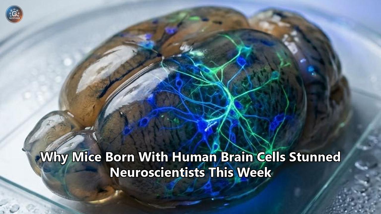
On September 16, 2026, a research team at Stanford Medicine published findings in Nature that dismantled a long-standing physical boundary in experimental biology. Led by neuroscientist Sergiu Pașca and postdoctoral fellow Konstantin Kaganovsky, the team revealed that they had successfully generated living mice whose cerebral cortices—the folded outer mantle responsible for cognition, sensory perception, and voluntary movement—are made up of more than 90 percent living human tissue.
By genetically knocking out the developmental program that forms a mouse’s own cortex and hippocampus before birth, the researchers engineered an anatomical vacuum inside the embryonic cranial vault. Into that biological void, they transplanted laboratory-grown human cortical organoids. Over the subsequent three months, the transplanted human cells underwent a fivefold volumetric expansion. They hooked into the host animal’s blood supply, organized into deep-layer networks, established electrical dialogue with the rodent thalamus, and projected nerve fibers along the entire neuroaxis into the cervical spinal cord.
The experiment produced animals carrying roughly 4 million human neurons in place of their missing 14 million mouse cortical cells. The graft colonized roughly half of the total volume of the rodent brain.
Within hours of the publication, the reaction across the neurobiology and clinical communities shifted from technical appreciation to intense debate. The team had resolved an intractable technical barrier: the inability to study complex, living human neural networks within a functioning, vascularized organism. Yet in doing so, they brought science directly against a formidable translational and bioethical frontier.
Studying human brain cells in mice is no longer a matter of tracking isolated clusters of cells scattered through rodent tissue. It now involves managing wholesale functional replacements of mammalian brain structures with human-derived circuits.
The Crisis Behind the Experiment: Why Modern Brain Science Hit a Wall
To understand why the Stanford team undertook such a radical engineering effort, one must examine the quiet crisis that has plagued translational neuroscience for decades.
Modern medicine has developed effective treatments for cardiac disease, unlocked targeted oncological immunotherapies, and engineered antiviral regimens that turn lethal infections into manageable chronic conditions. Yet central nervous system (CNS) drug discovery has stalled. Therapeutics designed to treat psychiatric conditions and neurodegenerative disorders fail in clinical trials at an estimated rate of 96 percent—the highest attrition rate in the pharmaceutical sector.
+-----------------------------------------------------------------------------+
|               THE TRANSLATIONAL BOTTLENECK IN CNS DRUG DISCOVERY           |
+-----------------------------------------------------------------------------+
|                                                                             |
|  2D CELL CULTURES             STANDARD RODENT MODELS       CLINICAL TRIALS  |
|  - Flat, non-physiological    - Fast development (weeks)   - 96% failure    |
|  - Lack cellular diversity    - Lack human-specific cells    rate in CNS    |
|  - Zero circuit architecture  - Distinct ion channel kinetics therapeutics  |
|         │                              │                          ▲         |
|         ▼                              ▼                          │         |
|  ────────────────────────────  ──────────────────────────         │         |
|  IN VITRO 3D ORGANOIDS         THE ANATOMICAL BOTTLENECK          │         |
|  - Arrest at mid-gestation     - Host mouse cells mature 20x      │         |
|  - Lack functional vasculature   faster than human tissue         │         |
|  - Internal necrosis sets in   - Physical & metabolic crowding    │         |
|         │                              │                          │         |
|         └───────────────┬──────────────┘                          │         |
|                         ▼                                         │         |
|             DEVELOPMENTAL XENOCORTICATION                         │         |
|             - Esco2 knockout clears cortex                        │         |
|             - Vascularized human organoid graft                   │         |
|             - True in vivo human circuit testing ─────────────────┘         |
|                                                                             |
+-----------------------------------------------------------------------------+
The underlying reason for this structural failure is evolutionary divergence. For more than seventy years, neuroscientists have used standard rodent models to uncover basic molecular and biological pathways. Mice and rats provide unmatched genetic manipulability and standardized rearing environments. But the rodent brain is lissencephalic—completely smooth, lacking the complex sulci and gyri that characterize primate neuroanatomy.
More crucially, the human neocortex possesses cellular sub-populations, gene-expression profiles, and prolonged developmental chronologies that simply do not exist in rodents:
- Outer Radial Glia (oRGs): In humans, neurogenesis is driven by a vast population of oRGs located in an expanded outer subventricular zone. These specialized progenitor cells generate the billions of excitatory neurons that form our six-layered cortex. Mice possess only a vanishingly small vestige of these cells.
- Prolonged Neoteny: A mouse neuron completes its migration, dendritic arborization, and synaptogenesis over a span of two to three weeks. Human corticogenesis, by contrast, takes months in utero and continues through years of postnatal life. This deliberate, slow maturation allows human synapses to incorporate complex developmental signaling cascades.
- Specialized Functional Subtypes: Primate brains contain uniquely adapted structural cells—such as spindle-shaped von Economo neurons and rosehip neurons—that play key roles in high-level integrative circuits. These cell types are entirely absent from standard laboratory rodents.
When pharmaceutical companies test compounds for schizophrenia, autism spectrum disorders, or frontotemporal dementia on standard mouse models, they are testing human disease mechanisms on neural machinery that lacks the target circuits. An experimental drug may successfully normalize synaptic transmission in a mouse hippocampus, only to fail completely in human clinical trials because the underlying human targets operate within a distinct metabolic, transcriptional, and architectural landscape.
The Limits of the Culture Dish
To bypass the rodent mismatch, scientists spent the last fifteen years refining human induced pluripotent stem cell (iPSC) technologies. By reprogramming adult human skin or blood cells back into a pluripotent state, researchers learned to steer these cells into becoming three-dimensional brain spheroids, or organoids. These self-assembling structures mimic several basic features of early human corticogenesis in a plastic Petri dish.
Yet in vitro organoids quickly hit an unyielding physical ceiling.
Deprived of a beating circulatory system, organoids grown in liquid media can only absorb oxygen and nutrients through passive diffusion across their outer layers. Once an organoid grows beyond roughly two millimeters in diameter, its interior core starves, suffocates, and undergoes necrosis.
Furthermore, a cluster of cells floating in an incubator receives no meaningful electrical or sensory inputs. Neurons require functional activity—sensory input, feedback loops, and physical axonal connections—to trim unnecessary synapses, form organized layers, and fully mature. Disconnected from an intact physiology, in vitro brain organoids inevitably arrest at a stage resembling mid-gestational fetal development. They cannot form the long-range axonal highways, like the corticospinal tract, that define functional neuroanatomy.
The core challenge became clear: to understand how human brain circuits develop, malfunction, and respond to pharmacology, scientists had to provide those cells with an intact, living physiology. Yet they had to do so without running into the biological roadblocks that had compromised every prior experiment.
The Crowding Problem: Why Previous Brain Chimeras Failed
The idea of transplanting human neural tissue into rodents is not entirely new. In 2022, Pașca’s Stanford laboratory published an experiment in Nature demonstrating that human cortical organoids could be microinjected into the somatosensory cortex of newborn rat pups. Once inside the rat brain, the human cells plugged into the local rodent vasculature, grew six times larger than their culture-dish twins, and extended functional connections that responded when researchers blew puffs of air across the rats’ whiskers.
Yet that experiment, along with related transplantation studies conducted across the globe, ran directly into an insurmountable biological roadblock: competitive displacement.
THE MATURATION MISMATCH

MOUSETIME:  [Birth] === Weeks 1-3 (Rapid Maturation & Synaptic Locking) ===> [Adult Brain]
HUMANTIME:  [Birth] ================= Month 3 (Immature Fetal Phase) ====> [Years to Mature]
                      ▲
                      │ Competition for space, nutrients, and synaptic territory
                      └─ Mouse tissue physically displaces slower-growing human xenograft.
When human brain organoids are introduced into an otherwise healthy, intact rodent brain, the human cells find themselves trapped in an uneven evolutionary race. Rat and mouse brain cells divide and mature at roughly twenty times the speed of human cells.
By the time the transplanted human neural stem cells began to form rudimentary connections, the surrounding host rodent tissue had already finished expanding, sealed its critical windows of plasticity, and locked down the structural boundaries of the skull.
As a result, the transplanted human cells were systematically crowded out. They were confined to a cramped pocket of the host animal’s somatosensory cortex, never claiming more than a fraction of one hemisphere. Because they were hemmed in by billions of native rodent cells that had already staked out their synaptic territory, the human neurons could not establish long-distance axonal tracts through the brainstem or spinal cord.
The host rodent’s own developmental program physically choked off the potential of the human graft. The resulting chimeric animals remained overwhelmingly rodent, with human cells acting as isolated islands within an alien biological landscape.
To give the human cells the physical room and metabolic resources needed to build true large-scale systems, the Stanford researchers realized they could not simply add human cells to an existing brain.
They had to eliminate the competition entirely.
The Biological Workaround: Engineering the Apallial Mouse
To create an anatomical niche capable of housing an entire human cortex, Pașca, Kaganovsky, and their team carried out an ambitious genetic intervention: they developed a line of mice that completely fails to grow its own higher brain.
The researchers zeroed in on a gene called Esco2. This gene encodes an acetyltransferase essential for the proper cohesion of sister chromatids during cellular mitosis. When Esco2 is functioning normally, it ensures that rapidly dividing progenitor cells can faithfully duplicate and segregate their chromosomes as the cerebral cortex forms.
THE GENETIC "VACUUM" STRATEGY

     Emx1-Cre Driver  x  Esco2-Floxed Mouse
                     │
                     ▼
       Selective deletion of Esco2 in dorsal telencephalon
                     │
                     ▼
       Mitotic catastrophe & targeted apoptosis
       of glutamatergic cortical progenitors
                     │
                     ▼
        THE "APALLIAL" RECIPIENT
        - Neocortex: Absent (98% depleted)
        - Hippocampus: Absent
        - Subcortex, Thalamus, Striatum, Brainstem: INTACT
        - Cranium and Meningeal Vasculature: INTACT
                     │
                     ▼
        Engraftment of Human Cortical Organoids (P0-P3)
                     │
                     ▼
        Human tissue expands to occupy 90-92% of the cortical mantle
Using Cre-loxP recombination driven by an Emx1 promoter, the team selectively excised the Esco2 gene strictly within the dorsal telencephalic progenitors that give rise to the excitatory glutamatergic neurons of the cerebral cortex and hippocampus.
The result was swift cellular clearance. As the embryonic mouse brain attempted to build its neocortex, the Esco2-deficient progenitor cells suffered mitotic catastrophe and triggered targeted programmed cell death. The entire neocortex and hippocampal formation—roughly 14 million neurons—simply vanished before the animals were born.
The resulting animals, which the researchers termed "apallial" mice (from the pallium, the evolutionary precursor to the cortex), were born missing virtually their entire higher brain mantle.
Remarkably, these apallial mice did not die at birth. Because the Esco2 deletion was restricted strictly to the dorsal cortex, the subcortical structures necessary to sustain basic biological life—the hypothalamus, brainstem, basal ganglia, and cerebellum—remained structurally and functionally intact. The mice could breathe, nurse, groom, and move.
They survived with a striking internal anatomy: an intact cranium, intact meninges, and an operational network of cerebral blood vessels, but an empty cranial cavity filled only with cerebrospinal fluid.
The anatomical stage was set. Within days of birth (between postnatal days 0 and 3), the team surgically microinjected three to four laboratory-grown human cortical organoids directly into this cranial void.
Without native rodent cells to compete with or crowd them out, the transplanted human brain cells in mice took root in 86 percent of the recipient animals.
What the Stanford Data Revealed: Inside the Xenocortical Brain
Over the next twelve weeks, the transplanted human tissues remodeled the internal anatomy of the rodent hosts.
Rather than arresting in development, the human organoids coalesced, expanded by 500 percent in volume, and filled 90 to 92 percent of the vacant cortical cavity. The animals had become what the team termed "xenocortical" mice: creatures that were physically and genetically mice below the thalamus, but whose higher cognitive mantle was composed predominantly of living, active human neural tissue.
+-----------------------------------------------------------------------------+
|                 ANATOMICAL COMPARISON: WILD-TYPE VS. XENOCORTICAL           |
+-----------------------------------------------------------------------------+
|                                                                             |
|  WILD-TYPE MOUSE BRAIN                DEVELOPMENTAL XENOCORTICAL BRAIN      |
|                                                                             |
|         .---''''''---.                             .---''''''---.           |
|       .'  Mouse       '.                         .'  HUMAN     '.           |
|      /    Neocortex     \                       /    CORTEX      \          |
|     |  (14M Rodent Neurons\                    |  (4M Human Neurons\        |
|     |   Lissencephalic)    |                   |   >90% Volume)     |       |
|     (                      )                   (                    )       |
|      \    [Striatum]      /                     \    [Striatum]    /        |
|       '.  [Thalamus]    .'                       '.  [Thalamus]  .'         |
|         '--..Brainstem.'                           '--..Brainstem'          |
|                 ||                                         ||               |
|                 || (Mouse Motor Tracts)                    || (HUMAN AXONS  |
|                 ||                                         ||  DESCENDING)  |
|                                                                             |
+-----------------------------------------------------------------------------+
When the researchers analyzed these brains using high-field magnetic resonance imaging, histology, single-cell RNA sequencing, and tract-tracing assays, the findings stunned the neurobiology community.
1. Functional Vascular Integration
The first challenge for any massive tissue transplant is vascularization. Without immediate blood flow, millions of human neurons cannot survive. In the xenocortical mice, the host animal’s endothelial cells migrated deep into the human grafts, weaving a dense, functional capillary bed throughout the human tissue.
The resulting vascular system was a cooperative biological chimera: mouse endothelial cells lined the vessels, but they responded directly to angiogenic signals dispatched by the human neurons and astrocytes, creating an operational blood-brain barrier.
2. Deep Projections to the Spinal Cord
In prior organoid experiments, human neurons formed local connections but failed to construct long-distance axonal pathways. In these xenocortical mice, the human neurons followed the structural architecture of the mouse brain.
The human cells extended thick, bundled axonal tracts downward through the internal capsule, navigated past the thalamus, traversed the cerebral peduncles in the midbrain, and sent projection fibers down into the cervical spinal cord.
The human neurons recognized and obeyed evolutionary guidance cues—molecular signals like netrins, slits, and semaphorins—laid down by the mouse subcortex, using them to map out descending motor pathways.
3. The Emergence of Von Economo Neurons
The most unexpected cellular finding of the study was the appearance of von Economo neurons (VENs).
VENs are large, spindle-shaped projection neurons characterized by a single apical dendrite and a single basal axon emerging from opposite poles of an elongated cell body. First described by Austrian neurologist Constantin von Economo in 1925, these rare cells are found in humans, great apes, cetaceans (whales and dolphins), and elephants—species that display complex social structures, extensive communicative vocabularies, and large encephalization quotients.
In humans, VENs are localized almost entirely to the anterior cingulate cortex and the fronto-insular cortex. They are among the earliest cells lost in behavioral-variant frontotemporal dementia (bvFTD), a devastating neurodegenerative condition marked by the rapid erosion of empathy, social judgment, and personality.
VON ECONOMO NEURON (VEN) MORPHOLOGY & LOCALIZATION

          Apical Dendrite
                 │
                 │
           ┌─────┴─────┐
           │           │  Elongated, Spindle-Shaped Soma
           │   Soma    │  (Distinct expression of FEZF2, CTIP2,
           │           │   and VMAT2 markers)
           └─────┬─────┘
                 │
                 │
                 ▼
          Single Basal Axon
    (Projects across long distances to subcortical targets)
For more than a decade, stem cell biologists have tried and failed to grow bona fide von Economo neurons in laboratory dishes. The necessary transcriptional programs simply refused to activate in vitro.
Yet inside the xenocortical mice, these rare cells emerged naturally within the transplanted human tissue. Single-cell sequencing confirmed their identity: they expressed the signature genetic markers of human VENs, including FEZF2 and CTIP2, and displayed their classical spindle-shaped architecture.
"VENs cannot be made in laboratory culture," Pașca stated following the study's release. "Yet here they were, sitting in the xenocortical mice’s human tissue. Now we can generate these rare cells from a healthy person and study them in a living, behaving animal to learn more about what they’re doing".
The discovery proved that human neural stem cells do not require a human skull to generate our species' rarest neurons. Instead, they need the physical, mechanical, and biochemical realities of a living, vascularized body: pulsatile blood pressure, circulating hormones, systemic metabolic cues, and reciprocal electrical loops between the brain and internal organs.
Solving the Translational Gap: How the Model Works in Practice
The creation of mice bearing extensive human cortical networks is not simply a triumph of surgical and genetic engineering. It directly addresses the primary failure point of modern neuroscience: the lack of a reliable, high-fidelity platform for modeling human brain disorders in vivo.
To prove that these animals could serve as useful tools for disease research rather than remaining mere biological curiosities, the Stanford team deployed the model against two complex conditions: perinatal hypoxic injury and rare neurodegenerative disease.
Modeling Perinatal Hypoxia and Cerebral Palsy
Every year, thousands of infants suffer hypoxic-ischemic events during difficult childbirths or early premature development, leading to conditions like cerebral palsy. For decades, testing protective drugs has been stymied by a simple biological reality: rodent brains are remarkably resistant to low oxygen.
A newborn mouse pup can tolerate hours of moderate-to-severe hypoxia without sustaining permanent damage to its motor cortex, thanks to distinct protective enzyme kinetics and metabolic pathways. Human infants possess no such defense.
EXPERIMENTAL PARADIGM: PERINATAL HYPOXIC CHALLENGE

[Wild-Type Mouse]  ─── 5 Hours of 10% O2 ───> Rapid Recovery; No Long-Term Deficits
                                             (Inherent rodent metabolic resistance)

[Xenocortical Mouse] ─ 5 Hours of 10% O2 ───> Severe, Permanent Gait and Motor Ataxia
(Human Cortical Mantle)                       (Matches Human Perinatal Injury Profiles)
                                                   │
                                                   ▼
                                      Host Mouse Glia Cross Species Boundary
                                      to Mount Phagocytic Response at Human Sites
The Stanford researchers exposed xenocortical mice, apallial control mice, and wild-type control mice to five hours of moderate hypoxia (10 percent oxygen).
The results exposed the vulnerability of human neural networks:
- Species-Specific Cellular Death: Wild-type mice shrugged off the oxygen deprivation, recovering normal motor coordination within days. The xenocortical mice, however, suffered widespread, lasting cellular damage throughout their human cortical tissue, mirroring the clinical patterns of periventricular leukomalacia seen in human babies.
- Clinical Motor Impairment: Following the hypoxic insult, the xenocortical mice developed severe, persistent motor deficits, displaying a shaky, wide-based, uncoordinated gait that closely resembles the clinical manifestations of cerebral palsy.
- Cross-Species Neuroimmune Dialogue: When the human neurons suffered ischemic injury, native mouse microglial cells and astrocytes migrated across the species boundary. The rodent immune cells engulfed and cleared the dead human cellular debris, attempting to wall off the injury site.
This dynamic demonstrated that the human and rodent cells had formed a unified, communicative pathological unit.
For the first time, researchers possess an animal model in which experimental neuroprotective compounds can be administered systematically to test whether they can prevent oxygen-deprivation damage specifically in living human cortical circuits.
Patient-Derived Xenocortical Disease Modeling
Beyond hypoxic injury, the xenocortical platform offers a direct path toward personalized neuropsychiatric medicine. Because organoids are grown from reprogrammed skin or blood cells, scientists can take a skin punch biopsy from an individual diagnosed with a complex neurodevelopmental condition, generate iPSCs, assemble cortical organoids, and engraft them into apallial mice.
PATIENT-SPECIFIC DRUG SCREENING PIPELINE

[Patient Skin Biopsy] (e.g., Severe Autism / Frontotemporal Dementia)
         │
         ▼
[iPSC Reprogramming] (Retains patient-specific genetic architecture)
         │
         ▼
[3D Cortical Organoid Differentiation]
         │
         ▼
[Engraftment into Newborn Apallial Mouse]
         │
         ▼
[In Vivo Maturation & Human Circuit Assembly] (3-6 Months)
         │
         ▼
[High-Throughput Behavioral, Electrophysiological, & Drug Testing Platforms]
In previous work focused on Timothy syndrome—a rare genetic condition caused by mutations in the CACNA1C calcium channel gene that leads to severe autism, epilepsy, and cardiac abnormalities—Pașca’s lab transplanted patient-derived organoids into rats to identify specific dendritic defects and test candidate antisense oligonucleotides.
With the xenocortical mouse model, this pipeline moves into an entirely new phase. Researchers can now observe how patient-specific human circuits wire across long distances, monitor their spontaneous electrophysiological activity in real time, and systematically screen therapeutic drugs against genuine human neural targets in a living mammal.
Behavioral Testing: Did the Transplants Change the Animals' Behavior?
The emergence of a mouse whose higher brain is overwhelmingly human immediately sparked a predictable question: Did the mice acquire human-like cognitive abilities?
The short answer from the researchers is an emphatic no.
The team put the xenocortical mice through a battery of standard behavioral assays, including the Y-maze (measuring working memory and spontaneous alternation), open-field locomotor tracking, and the rotarod test (measuring motor coordination).
+-----------------------------------------------------------------------------+
|                      BEHAVIORAL PERFORMANCE MATRIX                          |
+-----------------------------------------------------------------------------+
|                                                                             |
|  Assay                 Wild-Type Mouse   Apallial Mouse   Xenocortical      |
|                                          (No Cortex)      (Human Cortex)    |
|  ─────────────────────────────────────────────────────────────────────────  |
|  Spontaneous           ~68-72%           ~50%             ~61-63%           |
|  Alternation (Y-Maze)  (Normal)          (Pure Guesswork) (Partial Repair)  |
|                                                                             |
|  Locomotor Gait        Coordinated,      Hesitant,        Hesitant,         |
|  and Balance           Confident         Wide-Based       Cautious          |
|                                                                             |
|  Rotarod Motor         Normal Latency    Severe Early     Moderate Latency  |
|  Learning              to Fall           Fall Times       to Fall           |
|                                                                             |
|  Sentience /           Rodent            Impaired Rodent  Immature Rodent   |
|  Cognitive Profile     Baseline          Baseline         Baseline          |
|                                                                             |
+-----------------------------------------------------------------------------+
The behavioral testing yielded distinct insights into the function and limits of the chimeric brain:
- Apallial Mice Were Profoundly Impaired: Mice carrying the Esco2 deletion that received no human transplants operated on pure instinct driven by their subcortical structures. In the Y-maze, their alternation rate hovered near 50 percent—the statistical equivalent of random coin flipping. They moved cautiously, struggled with novel environments, and exhibited severe spatial memory deficits.
- Transplants Provided Partial Circuit Repair: Xenocortical mice that received human organoid grafts performed significantly better than their non-transplanted, cortex-deficient littermates. In the Y-maze, their alternation rates improved to roughly 62 percent—statistically above chance, indicating partial restoration of working memory circuits. Their motor coordination improved, and they navigated their cages with greater stability.
- No Cognitive Enhancement Beyond Normal Mice: The human tissue did not turn the mice into super-intelligent rodents. The xenocortical mice performed no better than standard, unmanipulated wild-type laboratory mice; in fact, on several demanding motor and cognitive metrics, they performed somewhat worse.
The biological reason for this performance ceiling lies in developmental timing.
Human neurons do not speed up their developmental clock simply because they are placed inside a mouse. Even within the warm, nutrient-rich environment of the rodent brain, human cortical neurons mature according to their intrinsic genetic timeline. At three to six months post-transplantation, the human tissue inside the xenocortical mice was structurally and electrophysiologically equivalent to the brain tissue of a human fetus entering the third trimester of pregnancy.
The human neurons were immature. They had not yet undergone the extensive dendritic arborization, competitive synaptic pruning, or dense myelination required for advanced cognitive operations. The mice remained fundamentally mice: their sensory inputs, basal drives, subcortical emotional processing, and peripheral motor effectors were entirely rodent. The human cortex acted primarily as an operational relay, integrating into host subcortical commands rather than imposing a human mind upon the animal.
Pașca warned against over-interpreting the behavioral improvements: "The most important point is that these are still mice. They have a mouse nervous system, mouse sensory organs, and mouse subcortical structures. What is unusual is that most of the cortical tissue present in these animals is human-derived and that the human neurons grow, integrate, and form functional connections with the rest of the mouse nervous system".
The Ethical Dilemma: Defining the Boundaries of Neural Chimerism
Despite the scientific team's clear findings, the xenocortical mouse study sent shockwaves through bioethics. It demonstrated that an engineered animal could be brought to term and sustained into adulthood with a brain whose entire executive mantle is composed of human cells.
THE MORAL STATUS CONTINUUM IN CHIMERIC NEUROSCIENCE

LOW CONCERN                                                 HIGH CONCERN
────────────────────────────────────────────────────────────────────────
Isolated Human       Human Glial        Xenocortical     Non-Human Primate
Organoids In Vitro   Chimeras In Vivo   Rodents (Current) Cortical Chimerism
(No sensory loops;   (Enhanced LTP;     (92% Human       (Risk of human-like
 metabolic arrest)   rodent neurons)    Cortex; Fetal)   affect/sentience)
The study pushed experimental biology directly into a gray area that legal and philosophical frameworks are ill-equipped to handle.
Duke University bioethicist Nita Farahany, who participated in an outside ethical review of the project, captured the conceptual shock: "There is now a mouse, the cortex of which is filled with human cells. We can’t quite call that a mouse, and we certainly can’t call it a human".
Farahany noted that while the research represents an extraordinary leap forward for pharmacological screening, it forces society to confront difficult questions: "This is probably one of the most promising advances for animal models that we’ve seen for a very long time, for having a better way to be able to both model diseases and test drugs. But it also blurs categories that our ethical and legal systems have historically treated as absolute".
John Evans, a bioethicist at the University of California, San Diego, pointed to the special significance of the cortex. "The cortex is significant, as it’s the part of the brain that results in our humanness," Evans noted, highlighting that it governs high-level thinking, semantic processing, and self-awareness.
When human cells dominate that specific anatomical region, standard ethical evaluations based purely on the animal’s external appearance become insufficient.
The primary ethical questions revolve around three concerns:
1. The Question of Altered Moral Status
Current biomedical ethics grants laboratory mice minimal moral standing. They are protected by humane handling guidelines, requirements for anesthesia during surgery, and mandates to minimize pain and distress.
Human beings, by contrast, possess full moral status and fundamental human rights.
That moral divide relies on cognitive and affective capacities: self-awareness, the capacity to plan for the future, the experience of subjective suffering (phenomenal consciousness), and complex social communication.
If human brain cells within an animal were ever to mature to a point where they supported human-like affective valence or self-reflective awareness, maintaining that animal in a standard laboratory cage would violate international ethical norms.
The Stanford data showed that these mice remain developmentally immature, but the experiment proved that the human tissue continues to mature over time. What happens if these animals are kept alive for eighteen months or two years? Could the human tissue, given sufficient time, begin to form the synaptic density associated with conscious awareness?
2. Species-Inappropriate Mental States
A chimeric animal cannot be treated simply as an upgraded mouse or a diminished human. It represents a novel biological entity.
A mouse brain driven by human cortical circuits might experience sensory inputs it cannot properly interpret, or form developmental drives that its rodent body cannot execute.
Such a mismatch could result in unprecedented forms of distress—neurological disorientation, chronic anxiety, or behavioral frustration—that standard animal welfare scoring metrics are blind to.
3. The Slippery Slope Toward Non-Human Primates
While mice have substantial physical and biological limits on how much human tissue they can support, those limits change if these techniques are transferred to larger animals.
If scientists applied this developmental xenocortication method to marmosets, rhesus macaques, or pigs, the donor human cells would have access to a much larger blood supply, a vastly expanded physical skull, and developmental timelines that span years rather than weeks.
The likelihood of generating human-like cognitive states in a non-human primate chimera is considerably higher than in a rodent, making the establishment of clear red lines an urgent priority.
What Experts and Regulators Are Doing: The Governance Framework
Faced with these profound questions, leaders across neuroscience, veterinary medicine, and bioethics are not waiting for an ethical breach to occur. The publication of the Stanford study has mobilized a coordinated effort to establish clear operational guardrails, diagnostic tripwires, and regulatory boundaries.
+-----------------------------------------------------------------------------+
|                THE MULTI-TIERED REGULATORY DEFENSE FRAMEWORK                |
+-----------------------------------------------------------------------------+
|                                                                             |
|  TIER 1: MACROSCOPIC RED LINES                                              |
|  - Absolute ban on non-human primate xenocortication                        |
|  - Strict ban on allowing chimeric animals to breed                         |
|  - Strict prohibition of human chimeric germline transmission               |
|                                                                             |
|  TIER 2: PHYSIOLOGICAL TRIPWIRES                                            |
|  - Mandatory, chronic wireless EEG monitoring                               |
|  - Automated screening for human-like sleep architecture (spindles, slow-wave)|
|  - Tracking for organized, complex gamma-band coherence across the graft    |
|                                                                             |
|  TIER 3: BEHAVIORAL SURVEILLANCE                                            |
|  - High-resolution, machine-vision tracking for species-atypical actions     |
|  - Monitoring for novel vocalizations, gestures, or self-directed tasks     |
|  - Mandatory humane endpoint protocols if distress signatures appear        |
|                                                                             |
+-----------------------------------------------------------------------------+
The ISSCR and National Academies Guidelines
The International Society for Stem Cell Research (ISSCR) and the National Academies of Sciences, Engineering, and Medicine (NASEM) had already begun developing oversight frameworks for organoid and chimera research. In the wake of the developmental xenocortication study, these groups are moving to turn broad guidelines into mandatory, enforceable laboratory protocols:
- Categorical Prohibition on Primate Xenocortication: Regulators are drawing an immediate line at non-human primates. While transplanting human neural organoids into mice and rats remains permissible under strict oversight, using developmental xenocortication—clearing an animal's native cortex to make way for a human graft—in primates is being designated as a prohibited experimental category.
- Reproductive Containment: To eliminate any possibility of human-derived cells contributing to reproduction, all animals undergoing neural chimerism must be strictly barred from breeding. Furthermore, single-cell lineage tracking must verify that the transplanted stem cells cannot migrate out of the central nervous system to contribute to the animal's germline.
- Humane Endpoints Based on Neurological Phenotypes: Institutional Animal Care and Use Committees (IACUCs) are updating animal welfare protocols. If an animal bearing human neural tissue displays signs of unmanageable chronic pain, intractable seizures, catatonia, or severe stereotyped distress behaviors, veterinary staff are required to immediately euthanize the animal.
Establishing Objective Neurophysiological Tripwires
To move beyond philosophical guesswork, neuroscientists are implementing real-time physiological tripwires to monitor the inner state of chimeric animals:
- Continuous Chronic EEG Telemetry: Xenocortical animals must be fitted with wireless electroencephalographic (EEG) transmitters to continuously monitor the electrical activity generated by the human graft.
- Surveillance for Human-Specific Oscillatory Patterns: Laboratory mice exhibit characteristic high-frequency, low-amplitude background electrical rhythms. The human cortex, by contrast, operates with distinct, lower-frequency oscillatory dynamics, including deep slow-wave sleep rhythms, sleep spindles, and complex theta-gamma phase-amplitude coupling associated with cognitive processing. Researchers are using automated algorithms to detect whether the human graft begins producing synchronized, high-order oscillatory activity that resembles an awake, conscious human brain.
- Machine-Vision Behavioral Tracking: Using high-speed cameras and artificial-intelligence pose-estimation software, laboratories are monitoring xenocortical mice around the clock for any signs of species-atypical behavior. If an animal begins executing complex, non-stereotyped behavioral routines, exhibits changes in social dynamics, or generates vocalizations outside the normal mouse repertoire, the experiment is flagged for immediate review.
By anchoring ethical oversight to real-time physical, electrophysiological, and behavioral readouts, regulatory bodies aim to allow disease research to proceed while maintaining an early-warning tripwire against unexpected cognitive emergence.
The Road Ahead: The Next Phase of Humanized Neurobiology
The development of developmental xenocortication marks the beginning of an entirely new chapter in neuroscience. The ability to integrate human brain cells in mice at this scale provides an unprecedented platform for studying biology, but it also demands significant technical refinement.
THE NEXT DEVELOPMENTAL HORIZONS

[Current Model: Pure Cortical Graft]
- 90% human excitatory glutamatergic neurons
- Host mouse blood vessels and microglia
- Mouse subcortical structures
               │
               ▼
[Horizon 1: Multi-Region Assembloid Transplantation]
- Co-transplantation of Cortical + Striatal + Thalamic organoids
- Generation of complete, multi-synaptic human neural loops
               │
               ▼
[Horizon 2: Fully Humanized Cellular Environments]
- Introducing human-derived microglia and astrocytes
- Engrafting human vascular organoids
- Recreating the complete human neurovascular and immune niche
               │
               ▼
[Horizon 3: Clinical Translation & Direct Drug Discovery]
- High-throughput testing of antisense oligonucleotides (ASOs)
- Real-time screening of compounds for autism, schizophrenia, and FTD
- Patient-derived avatars running parallel to Phase 1 clinical trials
Over the next three to five years, researchers are preparing to advance this research through three distinct scientific milestones:
1. Multi-Lineage Human "Assembloids" in Vivo
The current xenocortical mice carry grafts composed predominantly of excitatory cortical neurons. Yet the human brain relies on a fine balance between excitation and inhibition.
The next generation of experiments will utilize "assembloids"—structures formed by fusing cortical organoids with human medial ganglionic eminence (MGE) organoids. This approach will allow human inhibitory interneurons to migrate through the graft, recreating the complex microcircuits whose breakdown is implicated in epilepsy, schizophrenia, and autism.
2. Tri-Chimeric Cellular Systems
While the human neurons in the current study connected with host mouse blood vessels and microglia, mouse and human immune cells communicate using different cytokine and chemokine languages.
To build a more authentic human niche, researchers are working to introduce human microglia (derived from the same patient iPSC lines) into the empty cranial cavity alongside the cortical organoids. This will allow scientists to study neuroinflammatory cascades—such as those driving Alzheimer's disease and amyotrophic lateral sclerosis (ALS)—within a fully humanized neuroimmune environment.
3. Direct Clinical Trial Co-Testing
The ultimate test of the xenocortical mouse will unfold in pharmaceutical development pipelines. Several academic medical centers are preparing protocols to run xenocortical testing in parallel with early-stage human clinical trials.
By generating "xenocortical avatars" from patient populations enrolled in trials for rare, genetically defined neuropsychiatric disorders, researchers can observe how experimental drugs distribute across living human brain tissue, verify target engagement, and track adverse reactions at the cellular level before increasing doses in human patients.
A New Baseline for Neuroscience
The creation of mice bearing brains composed predominantly of living human cortical tissue has forever altered experimental neurobiology. By turning an anatomical hurdle into a clever genetic solution, the team at Stanford has bypassed the limits of both the plastic culture dish and the classical laboratory rodent.
They have established that human neurons possess the autonomy to build complex, long-range circuits, produce our species' rarest specialized cells, and direct motor pathways within an alien host physiology.
Yet this scientific leap comes with inescapable responsibilities. As researchers cultivate deeper networks of human brain cells in mice, the boundaries separating distinct biological systems will continue to soften.
The task facing the international scientific community is no longer just mastering the technical challenges of stem cell biology and neurosurgery. It is building, funding, and enforcing an ethical and regulatory architecture robust enough to guide these living models—ensuring that this powerful window into the human brain serves human health without compromising our ethical obligations to the living world.
Reference:
- https://www.futura-sciences.com/en/human-brain-tissue-implanted-in-mice-scientists-shocked-by-what-happened-next_900237/
- https://med.stanford.edu/news/all-news/2026/09/model-to-study-brain-development.html
- https://www.theguardian.com/science/2026/sep/16/mice-part-human-brains-research
- https://endpoints.news/researchers-implant-human-neurons-in-mice-brains-to-study-disease/
- https://www.psychologytoday.com/us/blog/the-asymmetric-brain/202609/scientists-create-mice-with-human-brain-cells
- https://www.sciencenews.org/article/mice-brain-human-organoid-cells
- https://www.sciencealert.com/scientists-grew-human-brain-tissue-inside-mice-heres-what-happened
- https://allsci.com/news/research/human-cortical-organoids-stanford-researchers/
- https://english.elpais.com/science-tech/2026-09-16/mice-with-millions-of-human-neurons-in-their-brains-bring-scientists-closer-to-understanding-mental-disorders.html
- https://futurism.com/health-medicine/scientists-unleash-franken-mice-with-brains-part-human
- https://www.smithsonianmag.com/smart-news/scientists-created-mice-with-half-human-brains-the-hybrid-animals-could-revolutionize-our-understanding-of-neurological-disorders-180989518/
- https://www.technologynetworks.com/neuroscience/news/human-cerebral-organoids-transplanted-into-newborn-rats-integrate-with-their-brain-366506
- https://www.livescience.com/health/neuroscience/scientists-shrank-mices-brains-and-replaced-the-missing-tissue-with-human-organoids
- https://www.facebook.com/stanfordmedicine/videos/transplanting-human-derived-organoids-allows-scientists-to-study-brain-disorders/1485173771948998/
- https://med.stanford.edu/news/all-news/2022/10/human-rat-brain-neuron.html