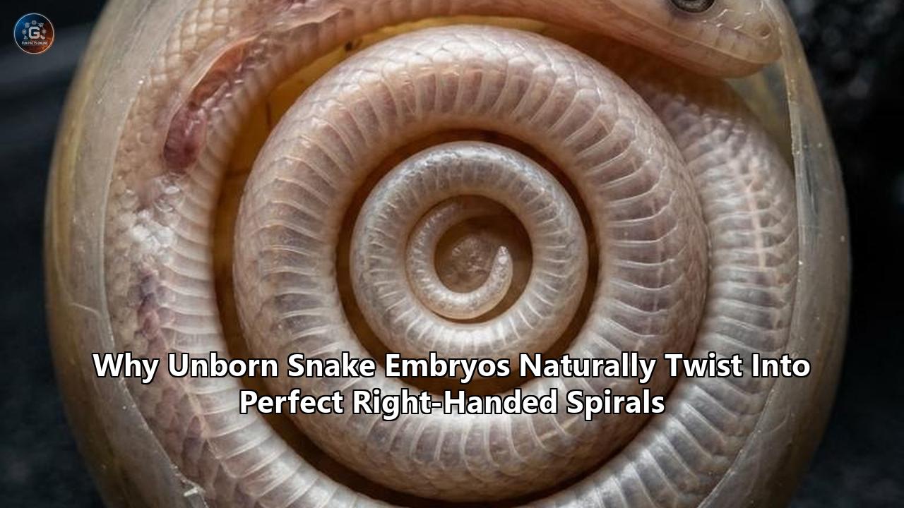
An international team of developmental biologists has solved a longstanding mystery in vertebrate morphology: why developing snake embryos invariably twist into right-handed, clockwise spirals during their earliest days within the egg. In a study published in Current Biology, researchers led by the Canadian Museum of Nature and the University of British Columbia revealed that this directional coiling is not driven by genetic chirality switches or early muscular twitching, but by a precise mechanical tug-of-war. The snake’s rapidly lengthening spinal column outpaces its digestive tract, forcing the embryo to buckle around a tethered visceral "pillar" against the asymmetrical placement of its yolk.
The findings explain how reptiles that hatch with up to 300 or more vertebrae manage to pack immense axial length inside a rigid, oval shell without tangling or compressing delicate organ primordia. By analyzing 913 embryos across 39 distinct species of snakes and limbless squamates, the team established that this dextral bias is an absolute rule of early snake embryogenesis before muscular movements emerge. The work directly connects tissue mechanics to macro-evolutionary innovation, providing an empirical case study of how physical forces interact with genetic instructions to shape the animal kingdom.
The Mechanics of the Visceral Pillar
The directional bias was uncovered after researchers combed through hundreds of museum specimens and laboratory archives. The team, directed by senior author Dr. Tetsuto Miyashita, an evolutionary biologist at the Canadian Museum of Nature, and lead author Alexandra Weber, a zoologist at the University of British Columbia, discovered that early embryos show complete uniformity. In a sample of 146 early-stage African house snake (Boaedon capensis) embryos, 100 percent coiled in a clockwise, right-handed direction. Across more than 200 early corn snake (Pantherophis guttatus) embryos, fewer than five percent deviated from this dextral orientation.
The uniformity presented an immediate biological puzzle: early squamate embryos lack functional skeletal muscle fibers and motor innervation. They are physically incapable of deliberately curling their bodies.
[Early Embryo Body Axis] ----(Accelerated Growth)----> Elongates rapidly
             |
             +--- Tethered by slower-growing "Visceral Pillar" (Gut Tube)
             |
             v
   [Mechanical Buckling] <--- Deflected rightward by Yolk on Left Side
             |
             v
[Uniform Dextral (Right-Handed) Spiral]
High-resolution micro-computed tomography (micro-CT) imaging performed by collaborator Dr. Raul Diaz at California State University, Los Angeles, uncovered the underlying anatomy. The scans exposed an unexpected internal arrangement: a detached, slow-growing column of gut tissue surrounded by vitelline blood vessels running directly through the center of the embryo's coil.
During early snake embryo development, somatic tissues along the dorsal axis proliferate at an accelerated rate to lay down the snake's repetitive vertebral precursors. The gut tube, derived from the endoderm, cannot maintain this pace. Rather than stretching to match the spine, the gut temporarily detaches from the posterior body wall and forms a taut central strut, termed the visceral pillar.
"It's like when you adjust the length of a strap and the longer, buckling side of the loop twists," explained Miyashita. As the dorsal axial column pushes forward against the mechanical resistance of the visceral tether, it experiences compressive stress. In physics, an elastic rod under axial compression buckles once it passes a critical load threshold—a dynamic described by classical Euler buckling formulations.
The direction of that buckle is determined by internal geometry. Across all amniote embryos, the early yolk stalk and digestive architecture establish a consistent left-right asymmetry, positioning the bulky yolk mass on the embryo’s left flank. As compressive forces build along the growing body axis, the yolk acts as an immovable physical barrier. The lengthening spine buckles toward the path of least mechanical resistance: away from the yolk, toward the right. This creates an inescapable right-handed helix.
The Impact on Developmental and Evolutionary Biology
This discovery recalibrates the balance between genetic determinism and physical morphogenesis in developmental biology. For decades, the dominant paradigm in squamate evolution focused almost exclusively on genomic mapping—specifically the shifts in Hox gene expression boundaries that erase limb buds and homogenize vertebrae throughout the trunk. While genetics explains how a snake generates hundreds of somites, it does not explain how those expanding tissues navigate the spatial constraints of the egg.
Evo-devo researchers now have concrete evidence that large-scale macroevolutionary transitions depend on secondary mechanical adaptations. Without an automated, reliable packing mechanism, the evolutionary elongation of the snake body axis would have created developmental dead ends: embryos colliding with their own egg membranes, suffering kinked spines, or cutting off vitelline circulation through disorganized knotting.
The identification of the visceral pillar challenges the assumption that organs must develop synchronously with the body frame. By temporarily decoupling gut development from spinal elongation, the snake embryo uses a localized growth mismatch as a functional tool. The gut is not lagging due to metabolic deficiency; its delay is an evolved biomechanical adaptation that exerts the exact mechanical tension required to twist the organism into a protective spiral.
Researchers investigating other squamates—such as glass lizards, slow worms, and amphisbaenians (worm lizards)—are revisiting their collections. Preliminary data from the study indicated that other limbless reptiles with elongated bodies display varying degrees of embryonic coiling. Determining whether these disparate lineages converged on the same visceral pillar mechanism or engineered alternative physical solutions will map the mechanical pathways of limbless evolution.
+-----------------------------------------------------------------------------------+
|                        THE DEXTRAL COILING SEQUENCE                               |
+-------------------+---------------------------------------------------------------+
| Phase 1: Somite   | Rapid proliferation of paraxial mesoderm;                     |
| Elongation        | body axis extends while endoderm growth lags behind.  |
+-------------------+---------------------------------------------------------------+
| Phase 2: Pillar   | Gut separates from body wall, forming an internal             |
| Formation         | visceral tether anchored by vitelline vessels. |
+-------------------+---------------------------------------------------------------+
| Phase 3: Euler    | Axial compression exceeds threshold; spine buckles            |
| Buckling          | away from left-side yolk into a right-handed coil.    |
+-------------------+---------------------------------------------------------------+
| Phase 4: Muscular | Yolk depletes, muscles innervate, embryo wriggles             |
| Realignment       | freely, shifting near-hatching chirality to 50/50.    |
+-------------------+---------------------------------------------------------------+
Practical Implications for Herpetology and Veterinary Science
Beyond theoretical biology, these mechanical findings yield actionable insights for captive breeding facilities, zoological institutions, and conservation biology programs.
Optimizing Incubation Protocols
Captive breeding of threatened boid and colubrid species frequently encounters embryonic mortality or spinal deformities characterized by fused vertebrae, axial kinks, and ventral lordosis. Historically, breeders attributed these defects to micro-fluctuations in incubation temperature or uneven substrate hydration.
The discovery that early axial alignment depends on passive biomechanical tension shifts attention to the physical orientation of the egg:
- Minimizing Early Disturbance: Snake eggs are non-cleidoic and adhere to one another in clusters shortly after oviposition. Because the embryo sits atop the yolk on its left side, rotating or jarring eggs during the first three weeks of incubation can shift the yolk's hydrostatic pressure. If the yolk slumps or presses against the right flank, it disrupts the Euler buckling angle, risking structural kinks or fatal tissue shearing across the visceral pillar.
- Managing Substrate Osmolarity: The mechanical tension of the visceral pillar relies on precise fluid pressure within the extraembryonic membranes. High-osmolarity environments that leach moisture from the egg reduce internal turgor pressure, weakening the counterforce necessary for clean buckling and increasing the frequency of axial misalignments.
Diagnostic Screening in Veterinary Herpetology
Veterinary pathologists studying "congenital kinking" in commercial python and corn snake hatcheries now have a baseline developmental window for structural anomalies. By pinpointing the timing of visceral pillar formation, clinicians can differentiate between:
- Mechanical Packing Failures: Spinal abnormalities characterized by sharp, right-angle rotations that trace back to aberrant tension during early buckling phases.
- Genetic Somitogenesis Errors: Irregularities caused by mutations in the Notch or Wnt signaling pathways, which create asymmetrical somites independent of egg mechanics.
This diagnostic clarity enables commercial breeders and conservationists managing ex-situ populations of critically endangered squamates, such as the Antiguan racer (Alsophis antiguae), to isolate whether breeding failures stem from inbreeding depression or faulty incubation mechanics.
What Changes in Morphogenetic Modeling and Physical Sciences
The study's conclusions establish a quantitative biological system that links classical continuum mechanics with embryology. Physicists and applied mathematicians modeling biological pattern formation are using these findings to refine algorithms of tissue packing, chirality, and elastic instabilities.
    EULER BUCKLING IN CONFINED MORPHOGENESIS:

            F_crit = (pi^2 * E * I) / (K * L)^2

    Where:
      E = Young's modulus of embryonic paraxial tissue
      I = Area moment of inertia of the dorsal axis
      L = Length of the accelerating body column
      K = Effective length factor (governed by the visceral tether)
As the axial length ($L$) increases rapidly while the effective length ($K$) is restricted by the visceral tether, the critical axial load ($F_{\text{crit}}$) is quickly exceeded. Because the boundary condition is asymmetrical—due to the dense yolk mass on the left—the deformation pathway is mechanically compelled into a single rotational vector.
This biological reality provides a living template for soft-matter physics:
- Self-Organizing Chirality: The research demonstrates that true biological chirality does not always require chiral molecules, asymmetric cilia, or nodal signaling cascades at the cellular level. Macroscopic handedness can emerge purely through boundary conditions acting upon an isotropic buckling rod.
- Predictive Computational Embryology: Biomechanical modelers are incorporating differential tissue growth parameters into finite element simulations. By inputting the growth rate of the paraxial mesoderm against the tensile modulus of the detached endoderm, computational systems can accurately predict the exact pitch, diameter, and turning frequency of the developing embryo inside various egg geometries.
Short-Term Consequences for Research Methodologies
The publication of this work has triggered an immediate shift in squamate laboratory handling and developmental staging systems.
Revision of Embryological Staging Tables
Standardized squamate staging tables, such as the widely referenced Zehr (1962) system for Thamnophis sirtalis or the newer tables for Boaedon capensis, have historically organized embryonic milestones around external structures: limb bud emergence (or lack thereof), eye pigmentation, scale formation, and hemipene development.
These staging systems are undergoing active updates:
- Visceral Pillar Detachment: Researchers must now record the precise timing of the gut tube’s detachment as a formal developmental marker.
- Coiling Onset and Handedness: Staging criteria are incorporating the transition from linear growth to dextral curling, establishing micro-stages (e.g., Stage 18a versus 18b) determined by the angle of axial deflection relative to the yolk stalk.
New Experimental Biomechanical Assays
Laboratory teams are deploying micro-dissection and laser ablation tools to interrogate the mechanical forces directly within the egg. By severing the visceral pillar using targeted laser micro-beams in living cultured embryos, scientists can test whether the embryo spontaneously uncoils or buckles into an irregular, non-chiral shape.
Similarly, micro-cantilevers and atomic force microscopy (AFM) are being employed to quantify the stiffness of the vitelline blood vessels and the tensile strength of the gut column. Gathering these physical constants is essential for validating mechanical simulations of animal form.
+-----------------------------------------------------------------------------------+
|                        RESEARCH REALIGNMENTS                                      |
+--------------------------+--------------------------------------------------------+
| Traditional Focus        | Revised Focus Following the Discovery                  |
+--------------------------+--------------------------------------------------------+
| Pure genomic profiling   | Integrated mechanobiology and differential tissue      |
| (Hox, Shh, Noggin)       | growth rates                           |
+--------------------------+--------------------------------------------------------+
| External morphological   | Internal vascular-visceral structural tracking via     |
| staging criteria         | micro-CT imaging                               |
+--------------------------+--------------------------------------------------------+
| Assumption of late,      | Recognition of early, passive biomechanical packing    |
| muscle-driven coiling    | via elastic buckling                    |
+--------------------------+--------------------------------------------------------+
| Fixed chirality models   | Dynamic chirality models (early dextral bias shifting  |
| across development       | to neutral 50/50 distribution)         |
+--------------------------+--------------------------------------------------------+
The Disappearance of Handedness: A Secondary Realignment
One of the study's most consequential insights is that this right-handed bias is entirely transient.
As snake embryo development progresses past the initial organogenesis stages, two major biological transitions take place:
- Metabolic Depletion of the Yolk: The embryo consumes the yolk mass, reducing its physical volume and releasing the spatial barrier on the left flank. This affords the growing animal increased volume within the eggshell.
- Neuromuscular Maturation: The musculoskeletal system completes innervation. Somites differentiate into skeletal muscle blocks capable of coordinated contraction, and the digestive system catches up in length, slackening the visceral pillar.
Once these shifts occur, the growing snake begins to move actively. It repositions its coils, sliding over its remaining yolk reserves to maximize space.
EARLY DEVELOPMENT:                       LATE DEVELOPMENT:
(Passive / Mechanically Constrained)      (Active / Muscular Control)

     +-------------------+                    +-------------------+
     | Yolk on Left Side |                    | Depleted Yolk     |
     | + Visceral Pillar |                    | + Functional Musc.|
     +---------+---------+                    +---------+---------+
               |                                        |
               v                                        v
     [ 100% Right-Handed ]                    [ 50% Right / 50% Left ]
        Dextral Spiral                           Neutral Distribution
By the time the snake approaches hatching, the directional bias disappears. The team found that near-term embryos exhibit a balanced 50/50 split between right-handed and left-handed coils.
This distinction confirms that adult snakes are not fundamentally "right-handed" organisms. The right-handed spiral is an ephemeral developmental state—a mechanical bridge that shepherds the fragile organism through the most dangerous packing phase of its lifecycle, only to be dismantled once active muscular locomotion takes over.
Long-Term Consequences: Engineering, Medicine, and Anatomy
The broader implications of this work extend beyond herpetology, reaching into industrial engineering, medical technology, and comparative vertebrate anatomy.
Bio-Inspired Deployable Structures and Soft Robotics
Engineers designing soft robotic actuators and deployable space structures face a persistent problem: how to store extreme lengths of flexible, semi-rigid materials in compact capsules and unpack them without motor-driven winches.
The snake embryo's visceral pillar demonstrates a functional paradigm:
- Tether-Controlled Self-Packing: By engineering an internal elastomer core with an intentionally slower expansion rate than an outer active layer, synthetic systems can automatically coil into tight, damage-free spirals without external manipulation or active motor control.
- Endoscopic Medical Probes: Long, flexible micro-endoscopes designed to navigate tortuous anatomical pathways, such as the human gastrointestinal tract or neurovascular networks, can incorporate differential-tension jackets. Inducing a controlled Euler buckle using an internal steering tether allows the instrument to coil smoothly into holding recesses without pinching delicate surrounding tissues.
Illuminating Asymmetries in Human and Mammalian Organogenesis
The physical mechanics discovered in squamates provide a comparative baseline for understanding developmental errors in other vertebrates, including humans.
During human embryonic development, several tubular organ systems rely on differential growth to achieve their functional geometry:
- Cardiac Looping: The primitive heart tube must twist and loop into an asymmetrical configuration to properly divide into four functional chambers. Disruptions in this looping phase cause severe congenital heart defects, such as dextrocardia and transposition of the great vessels.
- Gut Rotation and Mesenteric Attachment: The human digestive tract undergoes an automated 270-degree counterclockwise rotation around the superior mesenteric artery before settling into the abdominal cavity. Failure of this rotation leads to intestinal malrotation, volvulus, and gut strangulation.
Understanding how the snake body axis safely executes extreme mechanical buckling around a vascular tether illuminates the physical limits of tissue elasticity and shear stress. It helps medical researchers distinguish between organ defects caused by primary biochemical pathway failures and those caused by secondary physical obstructions in the embryonic environment.
+-----------------------------------------------------------------------------------+
|                        CROSS-DISCIPLINARY TRANSLATION                             |
+--------------------------+--------------------------------------------------------+
| Scientific Discipline    | Practical Application of the Discovery                 |
+--------------------------+--------------------------------------------------------+
| Soft Robotics            | Design of self-packing actuators using differential    |
|                          | material expansion and internal tethering              |
+--------------------------+--------------------------------------------------------+
| Surgical Instrumentation | Flexible, auto-coiling steerable micro-catheters       |
|                          | for minimally invasive procedures                      |
+--------------------------+--------------------------------------------------------+
| Pediatric Pathology      | Biomechanical comparative models for intestinal        |
|                          | malrotation and cardiac looping failures               |
+--------------------------+--------------------------------------------------------+
| Conservation Science     | Optimized incubation protocols and physical protection |
|                          | guidelines for rare squamate clutches                  |
+--------------------------+--------------------------------------------------------+
Emerging Frontiers and Unresolved Questions
While the discovery of the visceral pillar and yolk-directed Euler buckling answers how early squamate embryos coil, it uncovers several unresolved questions that will guide research over the coming decade.
The Puzzle of Viviparous Squamates
Roughly 20 percent of extant snake species are viviparous, giving birth to live young rather than laying eggs. These include diverse groups such as boas, anacondas, sea snakes, and vipers.
Viviparous species do not inhabit the rigid, calcified or leathery shells of oviparous taxa; instead, their embryos develop inside thin membrane sacs within the maternal oviduct. How does the absence of a rigid outer shell alter the boundary conditions of buckling?
- Do viviparous embryos develop the same tight dextral spiral, or does the compliance of maternal soft tissue allow for greater morphological variation?
- Does maternal locomotion—such as the burrowing of sand boas or the undulating swimming of marine hydrophiines—exert external physical stresses that disrupt the visceral pillar mechanism?
Initial observations suggest that the visceral pillar forms even in viviparous taxa, pointing to an evolutionary origin that predates the diversification of modern reproductive modes. However, comprehensive micro-CT analyses of in-utero viviparous clutches remain a critical next frontier.
Extreme Somite Counts and Multiple Visceral Pillars
While common colubrids and boas possess between 200 and 300 vertebrae, certain extreme species, such as large pythons (Malayopython reticulatus) and extinct giant snakes like Titanoboa cerrejonensis, develop skeletal frameworks that demand exceptional packing solutions.
In animals that exceed 400 vertebrae, does a single visceral pillar provide sufficient tensile anchoring, or does the embryo generate multiple functional attachment zones along the gut axis to initiate secondary and tertiary buckling folds? High-throughput comparative imaging across giant squamates will reveal whether the visceral pillar scales up linearly or switches to complex multi-nodal packing regimes.
Decoupling the Evolutionary Toolkit
The study confirms that the unique morphology of snakes is not simply the consequence of switching off limb genes, but the result of a coordinated mechanical choreography. The spine lengthens, the gut pauses, the yolk deflects, and the embryo turns.
The next milestone for evolutionary biology lies in determining the genomic switches that control this gut-growth delay. Identifying the precise microRNAs or transcription factors that downregulate cell proliferation in the endoderm while upregulating somite genesis in the mesoderm will unify the physical mechanics of the visceral pillar with the molecular circuitry of life.
The research reframes how scientists view the developing embryo: not as an inert blueprint passively awaiting genetic execution, but as a dynamic structural engine where physical forces, spatial constraints, and evolutionary lineages negotiate the emergence of biological form. As research teams turn toward real-time mechanobiological mapping, the simple right-handed twist of the unborn snake stands as a fundamental model for how nature solves its most demanding architectural challenges.
Reference:
- https://www.eurekalert.org/news-releases/1141750
- https://news.ubc.ca/2026/08/scientists-learn-why-snakes-in-an-egg-all-coil-in-the-same-direction/
- https://gulfnews.com/world/americas/why-do-baby-snakes-all-coil-the-same-way-inside-their-eggs-scientists-finally-have-an-answer-1.500659386
- https://www.zmescience.com/science/news-science/why-snake-embryos-coil-right/
- https://www.sciencedaily.com/releases/2026/09/260901070516.htm
- https://www.discovermagazine.com/the-mystery-of-why-snake-embryos-curl-to-the-right-could-lie-in-their-digestive-tract-49616
- https://www.popsci.com/environment/why-snake-embryos-coil-right/