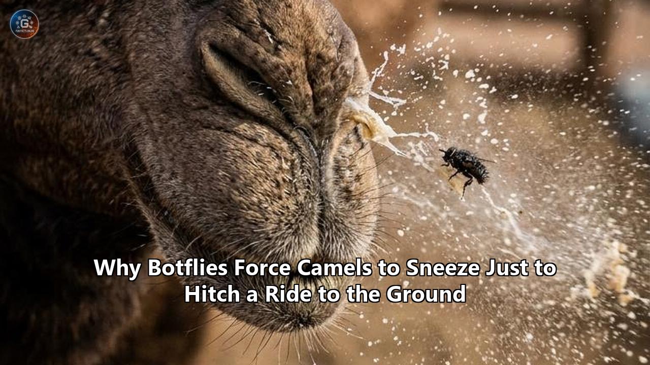
A massive epidemiological synthesis published by an international coalition of veterinary researchers has cast a spotlight on one of the most specialized host-parasite relationships in the arid world. The study, which compiled data from across Africa, the Middle East, and Central Asia, details the pervasive impact of Cephalopina titillator, commonly known as the camel nasal botfly. While pastoralists and desert veterinarians have long known of this parasite's existence, the new research maps out a biological spectacle: how this highly adapted fly forces camels into violent sneezing fits as a calculated, mechanical transport system to complete its lifecycle on the desert floor.
For a camel (Camelus dromedarius or Camelus bactrianus), a sneeze is rarely just a sneeze. Instead, it is the explosive climax of an internal residency that can last up to eleven months. During this time, dozens of fat, spiny larvae anchor themselves inside the camel’s nasopharynx, paranasal sinuses, and nasal cavities. When the larvae reach maturity, they face a perilous dilemma: they must exit the dark, moist safety of the camel’s head and reach the sandy soil below to undergo pupation.
To bridge this gap, the parasite does not simply crawl out; it orchestrates a physical eviction. By detaching their hooks and wriggling backward down the camel's narrow airway, the mature larvae trigger a severe, tickling irritation that forces the host to snort and sneeze with extreme force. The larvae are launched out of the nostrils, hitching a ride on a high-velocity stream of mucus straight to the ground, where they immediately burrow into the sand to escape the blistering desert sun.
Understanding this mechanism explains a bizarre natural phenomenon and highlights a significant, underreported veterinary crisis that threatens the economic security of millions of pastoralists.
The Subterranean Sanctuary: The Anatomy of a Camel’s Nose
To understand why the camel nasal botfly has evolved such a dramatic exit strategy, one must first look at the unique anatomy of the host. The respiratory system of the dromedary camel is an evolutionary masterpiece of water conservation and thermal regulation. In hyper-arid environments, camels must minimize respiratory water loss. They achieve this through a labyrinth of highly folded, scroll-like bones called nasal turbinates, covered in a highly vascularized mucosal lining.
This anatomical structure acts as a countercurrent heat and moisture exchanger. As the camel exhales, the cool nasal passages extract moisture from the warm breath, condensing it back into the body. As it inhales, the dry desert air is warmed and humidified before reaching the lungs.
For a parasitic insect, this warm, perpetually moist, and protected cavern is a prime piece of ecological real estate. The complex folds of the turbinates and the deep recesses of the paranasal sinuses provide an ideal microclimate, shielding the larvae from extreme external temperatures, which can exceed 50°C (122°F) in the summer, and from the freezing winds of desert nights.
However, this sanctuary is also a fortress. The nasal passages are narrow, winding, and lined with cilia designed to sweep foreign particles outward. To survive in this high-airflow, highly defensive environment, Cephalopina titillator has evolved highly specialized morphological tools:
- Cephalopharyngeal Skeleton (Mouth Hooks): The larvae possess a pair of curved, black, sclerotized mouth hooks. These hooks act as anchors, sinking deep into the mucous membranes of the nasopharynx to prevent the larva from being swept down into the trachea or blown out during normal breathing.
- Spinulose Bands: The outer cuticle of the larva is ringed with transverse rows of small, backward-pointing spines. These spines serve two purposes: they provide traction as the larva crawls through the mucous-covered passages, and they act as secondary anchors against the constant drag of inhaled and exhaled air.
- Spiracles: Located at the posterior end of the larvae are large respiratory plates called spiracles. This design allows the larvae to remain buried head-first in the mucosal tissue to feed while still drawing oxygen from the camel’s respiratory airflow.
Within this protected, nutrient-rich environment, the parasite undergoes a slow, highly coordinated development that is central to the survival of its species.
The Botfly Life Cycle: A Masterclass in Evolutionary Audacity
The survival of Cephalopina titillator is entirely dependent on its ability to navigate a tightly timed series of developmental stages, collectively referred to as the botfly life cycle. This cycle is a delicate balance of internal parasitism and external soil-dwelling phases, each timed to match the seasonal dynamics of the desert.
                     [ Adult Female Botfly ]
                                |
               (Larviposition: sprays L1 into nostrils)
                                v
                       [ L1 Larvae (Mucosa) ]
                                |
                   (Migrates to nasopharynx; molts)
                                v
                       [ L2 Larvae (Sinuses) ]
                                |
                 (Feeds & grows for 9-11 months; molts)
                                v
                       [ L3 Larvae (Mature) ]
                                |
            (Crawls forward, irritates mucosa, forced sneeze)
                                v
                       [ Larva on Ground ]
                                |
                     (Burrows into sand/soil)
                                v
                         [ Pupal Stage ]
                                |
                       (Eclosion / Emergence)
                                v
                      [ Adult Male/Female ]
Phase 1: The Aerial Ambush (Larviposition)
The botfly life cycle does not begin with an egg. Because eggs are highly vulnerable to drying out in the desert air, female nasal botflies are larviparous (or ovoviviparous). They hatch their eggs internally and carry active, microscopic first-instar (L1) larvae within their abdomens, ready for immediate deployment.
During the warm summer months—typically from June to September, depending on the region—fertilized female flies seek out camel herds. They do not land on the animal. Instead, they exhibit a characteristic hovering flight pattern, darting rapidly in front of the camel's muzzle.
With remarkable precision, the female fly sprays a fluid containing dozens of wriggling, 3.9-millimeter-long L1 larvae directly into or around the camel’s nostrils.
This aerial assault triggers immediate panic in the host. Camels can hear the high-pitched hum of the approaching fly and will shake their heads violently, stomp their feet, rub their noses against their forelegs, or bury their muzzles in the loose sand to prevent the flies from getting close. Despite these defensive maneuvers, the flies are often successful, depositing their microscopic cargo with a single, rapid pass.
Phase 2: The Deep Migration and Long Overwintering
Once deposited on the moist edge of the nostril, the tiny L1 larvae waste no time. Using their small spines and rapid, undulating body movements, they crawl upward through the nasal vestibule. They burrow through the thick nasal mucus, navigating toward the deeper, more protected cavities of the nasopharynx and paranasal sinuses.
Once they reach their destination, they use their sharp mouth hooks to secure themselves to the mucous membrane. Here, they molt for the first time, transitioning into second-instar (L2) larvae.
At this point, the botfly life cycle slows down significantly. The larvae enter a prolonged developmental phase that can last anywhere from 9 to 11 months. This extended residency is an evolutionary adaptation designed to carry the parasite through the harsh winter and dry seasons, when adult flies would not survive the open desert air.
During this overwintering period, the larvae feed continuously on the camel's epithelial cells, mucosal secretions, and inflammatory exudates. They undergo a second molt to become third-instar (L3) larvae.
As L3 larvae, they grow into robust, ivory-colored, cylindrical organisms measuring up to 3.5 centimeters in length. Their bodies are heavily segmented and armed with bands of thick, dark spines, making them look like armored, writhing caterpillars.
Phase 3: The Great U-Turn and the Forced Sneeze
By late spring or early summer of the following year, the L3 larvae have reached their maximum weight and are ready to transition to the pupal stage. However, they cannot complete this transition inside the camel. The host's body is too hot, too wet, and completely devoid of the soil needed to form a protective pupal casing.
To escape, the mature larvae must undertake a hazardous return journey. They detach their mouth hooks from the nasopharyngeal wall and begin crawling backward, making their way out of the sinuses and down the narrow nasal passages.
As these large, heavily spined, 3-centimeter-long organisms crawl over the highly sensitive sensory nerve endings of the camel’s nasal mucosa, they cause intense irritation. The crawling motion, combined with the scraping of their backwards-facing spines, triggers a massive physiological response in the host.
The camel experiences an unbearable, tickling itch within its nasal passages. It responds with intense snorting, head-shaking, and violent, explosive sneezing fits. This sneezing is exactly what the parasite needs.
As the camel forces a high-velocity blast of air through its nostrils, the detaching L3 larvae are swept up in the air current and ejected from the nose, landing on the ground several feet away.
Phase 4: Soil Burrowing and Pupation
Landing on the open desert sand is a moment of extreme danger for the ejected larva. Exposed to predators like birds and ants, and vulnerable to the dehydrating heat of the sun, the larva must act quickly.
The L3 larva exhibits strong negative phototaxis, meaning it instinctively moves away from light. Using its spiny bands for traction, it burrows rapidly into the loose sand or soil, typically sinking to a depth of several centimeters where the temperature is cooler and more stable.
Once safely underground, the outer cuticle of the larva begins to harden and darken, forming a protective, barrel-shaped casing known as a puparium. Inside this shell, the tissue of the maggot undergoes histolysis and histogenesis, reorganizing itself from a spiny, mucus-eating parasite into an adult fly.
Research has shown that this stage of the botfly life cycle is highly sensitive to environmental conditions. Under optimal conditions—specifically sand temperatures between 30°C and 36°C (86°F–97°F) and a relative humidity of 60% to 62%—the pupariation period takes only 1 to 9 days, and the pupation itself lasts between 5 and 13 days.
If the soil is too cold, too dry, or too compact, the pupa may fail to develop, or the emerging adult may be deformed.
| Developmental Stage | Location | Duration | Key Biological Activity |
|---|---|---|---|
| First Instar (L1) | Nose to Nasopharynx | Days to Weeks | Migrates inward, attaches to mucosa using mouth hooks |
| Second Instar (L2) | Nasopharynx & Sinuses | Several Months | Feeds on mucosal tissue, undergoes growth and development |
| Third Instar (L3) | Nasopharynx to Nostrils | Up to 11 Months (total larval time) | Reaches maximum size (3.5 cm), detaches, and irritates host to trigger expulsion |
| Pupa | Sandy Soil / Substrate | 5 to 13 Days (under optimal conditions) | Metamorphosis inside hard puparium; highly temperature-sensitive |
| Adult (Imago) | Free-living / Near Hosts | 8 to 12 Days (females live longer) | Non-feeding; mates and seeks host nostrils to deposit L1 larvae |
Phase 5: Emergence of the Adult Fly
At the end of the pupal stage, the adult fly uses a specialized, fluid-filled sac on its head called a ptilinum to push open the end of the puparium and dig its way up through the sand to the surface.
The emerged adult fly (or imago) is a robust, hairy insect resembling a honeybee, though it lacks functional mouthparts. Because they cannot feed, adult botflies have a very short lifespan. They must survive entirely on the energy reserves stored during their larval stage inside the camel.
Males live for an average of 8 days, while females live slightly longer, averaging around 12 days. Within this brief window, the flies must find a mate, and the females must locate a camel herd to deposit their larvae, restarting the complex botfly life cycle.
The Neurological and Biomechanical Triggers of the Sneeze
The mechanism by which Cephalopina titillator triggers its expulsion is a fascinating example of physiological hijacking. The parasite does not simply wait for a random sneeze; it actively forces the camel’s nervous system to cooperate.
Mechanical Stimulation of the Trigeminal Nerve
The nasal cavity of mammals is heavily innervated by the sensory branches of the trigeminal nerve (Cranial Nerve V), specifically the ophthalmic and maxillary branches. These nerves are highly sensitive to physical touch, chemical irritants, and temperature changes, serving as the body's primary defense against inhaled debris, dust, and pathogens.
When a mature L3 larva begins its journey out of the nasopharynx, its physical characteristics turn it into a walking irritant:
- Serrated Spines: The rows of backward-pointing spines on each of the larva’s 12 body segments act like tiny, coarse combs scraping against the delicate, highly sensitive epithelial cells of the nasal turbinates.
- Crawling Motion: The larval movement combines peristaltic body contractions with the extension and retraction of its sharp mouth hooks. This scraping and pulling action on the mucosal lining sends a rapid series of action potentials along the trigeminal nerve to the sneeze center in the brainstem (medulla oblongata).
Chemical Irritation and the Inflammatory Cascade
In addition to mechanical scraping, the larvae use chemical means to irritate their host. As they feed and move, they secrete excretory-secretory products (ESPs), which contain proteolytic enzymes designed to break down tissue and mucus.
These chemical secretions, combined with the physical damage caused by their spines, trigger a localized inflammatory response in the camel's nasal mucosa:
- Degranulation of Mast Cells: The physical trauma and foreign proteins from the larvae cause local mast cells to release inflammatory mediators, including histamines, prostaglandins, and leukotrienes.
- Vasodilation and Mucus Hypersecretion: These chemicals cause nearby blood vessels to dilate (leading to swelling and nasal congestion) and stimulate the mucosal glands to produce large quantities of thick, sticky, mucoid discharge.
- Sensitization of Nociceptors: The inflammatory soup lowers the activation threshold of the sensory nerve endings in the nose. What would normally be a minor tickle becomes an unbearable, painful itch.
The Physics of the Sneeze Blast
When the sensory input to the brain's sneeze center reaches a critical threshold, it triggers a coordinated, involuntary reflex. The camel takes a deep breath, its glottis closes, and its abdominal and respiratory muscles contract, building up immense pressure within the lungs.
Suddenly, the glottis opens, and a high-velocity blast of air is forced upward through the nasopharynx and out of the nostrils.
Because the mature L3 larvae have intentionally detached their mouth hooks to prepare for exit, they have no way to resist this sudden blast of air.
They are swept up in the high-pressure stream of air and mucus. The mucus, which has become thick and abundant due to the larval-induced inflammation, acts as a lubricant, reducing friction as the larvae are launched out of the narrow nasal passages.
Without this high-velocity ejection, the heavy, slow-moving larvae would likely become trapped in the dry outer nostrils of the camel, where they would quickly dehydrate and die under the desert sun. By forcing the camel to sneeze, the parasite ensures a rapid, lubricated transit from the inside of the host's head directly into the soil.
The Veterinary and Economic Toll of Camel Nasal Myiasis
While the survival strategy of the camel nasal botfly is biologically remarkable, its impact on camel health and pastoralist economies is severe. The infestation of live host tissue by fly larvae is known as myiasis, and in camels, Cephalopina titillator is the primary cause of nasopharyngeal myiasis.
                     [ Larval Infestation ]
                               |
             +-----------------+-----------------+
             |                                   |
             v                                   v
    [ Physical Blockage ]              [ Mucosal Damage & ESPs ]
             |                                   |
             +-----------------+-----------------+
                               |
                               v
                     [ Clinical Pathology ]
                               |
       +-----------------------+-----------------------+
       |                       |                       |
       v                       v                       v
[ Impaired Breathing ]   [ Chronic Pain & ]    [ Localized Tissue ]
(Snoring, Dyspnea)       [ Distressed Behavior ]   [ Inflammation ]
       |                       |                       |
       v                       v                       v
[ Reduced Grazing ]      [ Energy Depletion ]  [ Secondary Infections ]
       |                       |                       |
       +-----------------------+-----------------------+
                               |
                               v
                      [ Economic Impact ]
            (Weight loss, reduced milk/meat, death)
Prevalence and Distribution
Nasopharyngeal myiasis is endemic to almost every region where camels are raised, including North and East Africa, the Middle East, the Indian subcontinent, and the arid regions of Central Asia. Epidemiological studies reveal alarmingly high prevalence rates in domestic herds:
- Egypt: Studies of camels at municipal abattoirs have reported prevalence rates ranging from 41.6% to over 60%.
- Jordan and Saudi Arabia: Researchers have documented infestation rates as high as 46% to 82.6% in local camel herds, with the highest numbers of active larvae recovered during the cooler winter months.
- Ethiopia and Somalia: In these vital camel-rearing regions, the disease is known locally as Sengale or Sillill, and is recognized by herders as a major threat to animal health.
- China and Mongolia: Even the double-humped Bactrian camels of the cold deserts are highly susceptible, with recent studies showing significant larval burdens in herds across northwestern China.
Clinical Manifestations
A light infestation of a dozen larvae may cause only mild discomfort to a healthy camel, but heavy infestations—where a single camel’s head can host up to 250 squirming larvae—lead to severe pathology:
- Impaired Respiration (Dyspnea): The sheer volume of large L3 larvae, combined with the copious, sticky mucofibrinous secretions they trigger, can physically block the nasal passages. Infested camels often exhibit labored breathing, open-mouth breathing, and a characteristic, loud "snoring" sound, especially during exertion.
- Anorexia and Weight Loss: The constant irritation, pain, and difficulty breathing make camels highly restless. They spend less time grazing and resting, leading to rapid weight loss and muscle wasting.
- Nasal Discharge and Hemorrhage: The scraping of the larval spines and the attachment of their mouth hooks cause extensive mechanical damage to the nasal mucosa. This leads to chronic, purulent (pus-filled) nasal discharge, which is frequently stained with blood.
- Secondary Bacterial Infections: The damaged, raw mucosal tissue provides an ideal entry point for opportunistic pathogens. Infested camels often develop secondary bacterial sinusitis, tracheitis, or bronchopneumonia, which can be fatal if left untreated.
- Neurological Disorders ("False Gid"): In severe cases, the larvae can migrate deeply through the nasal passages, occasionally eroding the delicate bone of the cribriform plate. This allows the larvae or secondary bacterial infections to enter the cranial cavity, leading to meningitis, brain abscesses, and neurological symptoms such as circling, blindness, head pressing, and death.
Economic Consequences
For pastoralist communities in arid lands, the camel is not just livestock; it is a walking bank account, a source of nutrition, and a means of survival. Camels provide high-quality milk, meat, hide, and invaluable draft power for transport.
The systemic stress caused by Cephalopina titillator directly undermines these vital resources:
- Milk Production: Heavy infestations can cause milk yields to drop by up to 30%, directly impacting the food security of nomadic families who rely on camel milk as a daily staple.
- Meat Quality and Weight: Affected animals suffer from poor feed conversion and weight loss, reducing their market value at sale.
- Working Capacity: Sick, labored-breathing camels cannot be used for transporting goods or riding, disrupting local trade routes and daily pastoralist activities.
Treating and Preventing Nasal Myiasis: Traditional vs. Modern Approaches
Because of the high prevalence and economic impact of the camel nasal botfly, herders and veterinarians have developed a range of strategies to disrupt the botfly life cycle and relieve the suffering of infested animals.
Traditional Remedies
Nomadic herders have long relied on botanical and manual interventions to treat nasal myiasis:
- Manual Extraction: Experienced herders will sometimes manually remove visible L3 larvae from the outer nostrils of camels using curved wires or their fingers, though this does not address the larvae deep within the sinuses.
- Nasal Irrigations: Traditional treatments often involve blowing irritating powders or liquids into the camel's nostrils to force violent sneezing and expel the larvae prematurely. Common ingredients include tobacco dust, hot wood ash, or diluted salt solutions.
- Essential Oils: Pastoralists across North Africa and the Middle East have used locally available plant oils to treat infected animals. Modern research has begun to validate these traditional practices.
Modern Veterinary Interventions
Modern veterinary medicine has introduced highly effective chemical treatments that have transformed the management of camel nasal myiasis:
Systemic Parasiticides (Macrocyclic Lactones)
The introduction of systemic macrocyclic lactones has provided veterinarians with a powerful tool to eliminate botfly larvae from deep within the camel's respiratory system.
- Ivermectin: Administered via subcutaneous injection, Ivermectin is highly effective against all three larval stages (L1, L2, and L3) of Cephalopina titillator. The drug works by binding to glutamate-gated chloride channels in the parasite's nervous system, causing flaccid paralysis. The paralyzed larvae detach their mouth hooks and are easily swept out by the camel's normal breathing or mild sneezes.
- Doramectin: Similar to Ivermectin, Doramectin has shown exceptional efficacy. In vitro and in vivo studies have demonstrated that even low concentrations of Doramectin can achieve 100% mortality of L2 and L3 larvae within 24 to 30 hours of treatment.
Scientific Evaluation of Essential Oils
In search of more sustainable and environmentally friendly treatments, researchers have evaluated the larvicide potential of various essential oils. A notable study published in Parasitology Research compared the efficacy of several essential oils against Cephalopina titillator larvae:
- Lavender Oil (Lavandula angustifolia): Showed exceptional efficacy, matching the performance of synthetic parasiticides. In laboratory tests, 100% of L2 larvae died within 18 hours of treatment, and 100% of L3 larvae died within 24 hours.
- Camphor Oil (Cinnamomum camphora): Demonstrated strong larvicidal activity, proving twice as effective as other tested traditional remedies like onion oil.
- Onion Oil (Allium cepa): While traditionally used, onion oil required significantly longer exposure times to achieve larval mortality compared to lavender and camphor.
These natural essential oils represent a promising alternative for organic camel farming and for pastoralists who may lack access to or cannot afford synthetic veterinary drugs.
Shifting Sands: Climate Change and the Future of the Parasite
As global climates shift, the dynamics of host-parasite relationships are undergoing profound changes. The future of Cephalopina titillator and the camels they infest will be shaped by several emerging environmental and socioeconomic factors:
1. The Expansion of Camel Husbandry
As climate-change-induced desertification makes cattle and sheep rearing increasingly difficult in arid regions of Africa and the Middle East, many livestock owners are transitioning to camel husbandry. Camels are far more resilient to extreme heat, prolonged droughts, and poor-quality forage than other domestic herbivores.
This shift is driving a global expansion of the camel population. However, as camel densities increase and herds are managed in closer proximity, the transmission rates of parasites like Cephalopina titillator are expected to rise. Higher host density provides adult female botflies with more targets, potentially leading to more severe and frequent infestations.
2. Temperature Anomalies and Pupation Success
The pupal stage of the botfly life cycle is highly sensitive to soil temperature and humidity. As global temperatures rise, some desert soils may exceed the optimal 36°C (97°F) limit for pupation during peak summer months.
If sand temperatures rise too high, it could lead to increased pupal mortality and a decrease in the adult fly population.
Conversely, warmer winters in temperate and semi-arid zones could extend the active season of adult flies, allowing them to complete multiple generations per year and expanding their geographic range further north into Southern Europe and Central Asia.
3. Advancements in Molecular Diagnostics
Historically, diagnosing nasal myiasis in live camels has been difficult, often relying on the observation of clinical signs (nasal discharge, sneezing) or post-mortem examinations at abattoirs.
However, veterinary researchers are developing more advanced diagnostic tools:
- Polymerase Chain Reaction (PCR): Researchers are utilizing PCR assays targeting the mitochondrial cytochrome c oxidase subunit I (COX1) gene and 28S rRNA gene sequences to identify and classify Cephalopina titillator larvae with high precision.
- Serodiagnosis: Efforts are underway to develop enzyme-linked immunosorbent assays (ELISA) that can detect specific antibodies against larval proteins in the camel's blood. This would allow for early, non-invasive detection of infestations, enabling herders to treat animals before severe tissue damage and respiratory distress occur.
Conclusion: The Precision of Parasitic Adaptation
The bizarre relationship between the camel and its nasal botfly is a testament to the uncompromising precision of evolutionary adaptation. Every step of the botfly life cycle—from the female’s rapid, mid-air delivery of live larvae to the spiny, armor-clad residency of the mature maggots deep within the sinuses—is designed to exploit the host's anatomy while shielding the parasite from the harsh realities of the desert environment.
The forced sneeze is not a biological accident; it is a vital ecological bridge. It is the mechanism by which a slow, fragile larva successfully navigates the transition from a warm, wet sanctuary inside a camel's head to the hot, dry sands of its pupal home.
As research continues to untangle the molecular, physiological, and ecological threads of this relationship, veterinarians and pastoralists are gaining the tools they need to better manage this parasite, protecting both the health of these resilient desert beasts and the livelihoods of the communities that depend on them.
Reference:
- https://www.frontiersin.org/journals/veterinary-science/articles/10.3389/fvets.2026.1863158/full
- https://grokipedia.com/page/cephalopsis_titillator
- https://pmc.ncbi.nlm.nih.gov/articles/PMC9520952/
- https://scispace.com/pdf/seasonal-prevalence-of-the-larvae-of-the-nasal-fly-4cyy3lzb77.pdf
- https://www.researchgate.net/publication/364656340_Prevalence_and_Morphological_Characterization_of_the_Camel_Nasal_Botfly_Cephalopina_titillator_Diptera_Oestridae_Collected_from_Abattoirs_in_Egypt
- https://www.youtube.com/shorts/xMuGpSmo9nk
- https://pmc.ncbi.nlm.nih.gov/articles/PMC11606288/
- https://realbrownbeauties.com/camel-nasal-myiasis/
- https://bu.edu.eg/portal/uploads/Science/Entomology/1378/publications/Abla%20Dsowk%20Ibrahim_cephalopina%202013.pdf
- https://pmc.ncbi.nlm.nih.gov/articles/PMC11442609/