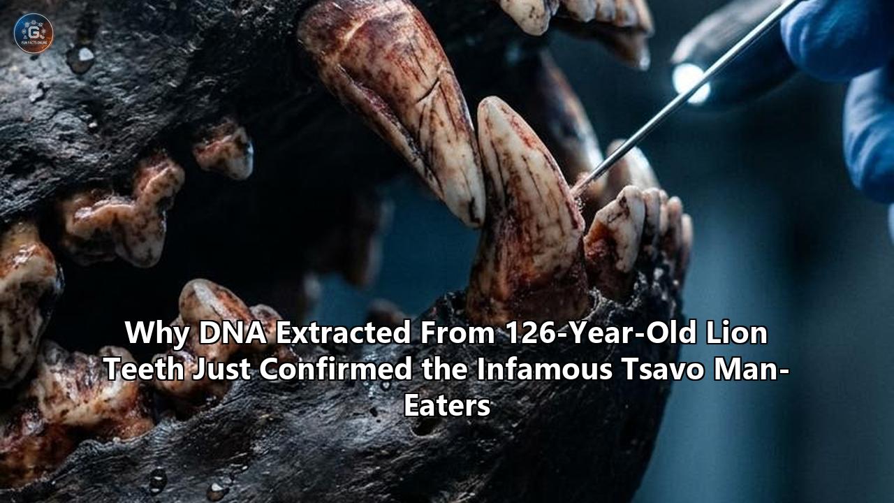
Inside the exposed, necrotic pulp cavity of a canine tooth belonging to a lion shot in December 1898, scientists have isolated the genetic remnants of human flesh.
The finding, published in Current Biology by an interdisciplinary team from the University of Illinois Urbana-Champaign and Chicago’s Field Museum of Natural History, converts one of the most sensationalized chapters of colonial-era folklore into verifiable molecular fact. By developing novel protocols to extract and sequence ancient mitochondrial DNA preserved inside compacted hair shafts wedged deep within the carnivores’ shattered teeth, researchers verified that the pair of maneless male lions known as the Tsavo man-eaters consumed humans alongside at least five other species of wild mammals.
The genomic data definitively settles questions surrounding the carnivores' diet, which had lingered through decades of disputed railway ledgers, imperial memoirs, and Hollywood myth-making.
Yet the analysis yielded surprises that extend beyond human predation. Wedged alongside human hair in the tooth cavities of these century-old predators were the mitochondrial signatures of Masai giraffe, plains zebra, waterbuck, fringe-eared oryx, and wildebeest. The wildebeest sequence presented an immediate ecological puzzle: in the late 1890s, the nearest known grazing range for wildebeests was more than 50 miles away from the Tsavo River. Its presence validates historic diary entries describing months-long lulls in human attacks, indicating the predators maintained hunting territories far broader than previously recorded.
Equally revelatory was the utter absence of African buffalo DNA—a species that constitutes over half of the modern Tsavo lion diet. That void provides genomic confirmation of the ecological collapse triggered by the continental rinderpest pandemic of the 1890s, an outbreak that starved apex predators and drove injured carnivores straight toward human encampments.
Deciphering the Tsavo man-eaters DNA represents a watershed achievement in museomics—the extraction of degraded genomic material from historical museum artifacts. More than a century after Lieutenant-Colonel John Henry Patterson pumped bullet after bullet into the predators along the banks of the Tsavo River, modern molecular tools are finally reconstructing the complex ecological crisis that transformed two injured predators into historical legends.
1898: Siege at the Tsavo River and the Mechanics of Panic
The historical episode began with the expansion of the British Empire's East Africa Protectorate. In March 1898, John Henry Patterson, a civil engineer and lieutenant-colonel in the British Army, arrived at the Tsavo River in present-day Kenya. His assignment was formidable: supervise the construction of a permanent railway bridge across the river, linking the Indian Ocean port of Mombasa with the interior of Uganda.
The colonial project—dubbed the "Lunatic Line" by British parliamentarians due to its astronomical expense and perilous logistics—depended on thousands of indentured Indian railway laborers, known as coolies, alongside local African porters and European engineers.
The landscape was punishing. Tsavo, an indigenous Kamba word loosely translated as "place of slaughter," was a dry scrubland dominated by dense Commiphora and Acacia thorn thickets, scorching heat, and malarial swamps.
Within days of Patterson’s arrival, two unusually large, maneless male lions began stalking the camps.
The attacks escalated according to a chilling, predictable cadence. The lions operated as a coordinated hunting pair. Striking under the cover of night, they bypassed watch fires, pushed through heavy thorn defenses (bomas) erected around the tents, seized sleeping men by the neck or head, and dragged them out into the darkness to be consumed.
Initial attacks targeted single individuals on the periphery of the sprawling camps. Within months, the predators grew remarkably bold. Workers stationed guards, built ten-foot-high fences woven from interlocking whistling thorn branches, and beat metal pots to create clamor throughout the night. The lions responded by leaping over the barriers or forcing their shoulders through the barbed branches, dragging their prey back out across the thorn walls.
In his 1907 memoir, The Man-Eaters of Tsavo, Patterson detailed the psychological paralysis that seized the encampment:
"The condition of affairs at Tsavo had now become such that it was impossible to proceed with the bridge. A veritable reign of terror prevailed, and the coolies absolutely refused to work."
In December 1898, the labor force struck, boarding an outbound construction train and declaring they would not lay another foot of track until the "devils"—believed by many workers to be shape-shifting spirits or ancestral guardians resisting colonial desecration—were slain. Construction was halted for nearly three weeks.
Patterson abandoned his engineering duties to become a full-time hunter. Sitting night after night in elevated shooting platforms (machans) constructed from timber or tethered to baobab limbs, he baited traps with cattle, goats, and occasionally used himself or his staff as human decoys. His hunts were marked by terrifying close encounters; on multiple occasions, the lions bypassed tethered cattle to stalk Patterson’s blind from behind.
On December 9, 1898, Patterson finally shot the first lion, designated decades later in the Field Museum’s catalog as FMNH 23987. It required multiple heavy-caliber rifle rounds to bring down the animal, which measured nine feet, eight inches from nose to tail.
Twenty days later, on December 29, Patterson tracked and killed the second lion, FMNH 23988, after a protracted confrontation in which the wounded predator absorbed multiple rifle shots before collapsing mid-charge.
With the predators dead, the bridge was completed, the railway continued its trajectory toward Lake Victoria, and Patterson’s accounts of the Tsavo man-eaters passed into global mythology.
1899–1924: From Trophies to Taxidermy in the Field Museum Vaults
For a quarter of a century following their deaths, the physical remains of the Tsavo lions served as private domestic decor.
Patterson kept their skins as floor rugs in his home, while their skulls sat on display as hunting trophies. The pelts sustained heavy wear, stepped on by visitors and treated with contemporary, crude chemical preservatives that damaged the fragile keratinous fibers of the hair.
In 1924, Patterson delivered a lecture at the Field Museum of Natural History in Chicago, recounting his ordeal in the East African bush. Sitting in the audience was the museum’s president, Stanley Field. Impressed by Patterson’s narrative and recognizing the commercial draw of the animals, Field arranged to purchase both lion skins and their accompanying crania for $5,000—a sizable sum at the time, equivalent to more than $90,000 today.
When the Field Museum received the specimens, staff taxidermist Julius Friesser faced a daunting task.
Because the skins had been trimmed, cured, and trampled as rugs for twenty-five years, they had lost considerable surface area and structural elasticity. Friesser constructed wood and plaster manikins based on anatomical measurements of modern African lions.
He discovered that the Tsavo lions were completely devoid of the prominent cranial and thoracic manes typical of adult male lions depicted in Western natural history museums. To compensate for the distorted, flattened hide shapes, Friesser mounted the cats in low, stalking postures.
Once placed on public display in the museum’s lower hall, the lions remained static curiosities for the next seven decades.
Colonial literature accepted Patterson’s sensational casualty figures without question: his book claimed the pair had devoured 135 people, comprising 28 Indian railway workers and dozens of African porters and local residents.
Zoologists debated why the pair lacked manes, with some asserting they represented an aberrant subspecies, while others dismissed them as subadults whose mane growth had been arrested by malnutrition.
For the better part of the 20th century, the skulls and mounts remained silent, classified as imperial relics rather than valuable repositories of biological data.
The 1990s Turning Point: Pathology in the Drawers
The scientific rehabilitation of the Tsavo specimens began in the early 1990s through the curiosity of Thomas Gnoske, an ecologist and collections manager at the Field Museum.
Gnoske sought to resolve the perpetual question of lion manes. While reviewing historical specimens in the museum's mammal range, he pulled the skulls of FMNH 23987 and FMNH 23988 from their cabinet drawers.
First, Gnoske examined the dental wear and cranial suture fusion of the specimens. He confirmed that both animals were not subadults, as earlier theorists suggested; they were fully grown, mature adults.
Working with field observations from Tsavo East National Park, Gnoske established that the absence of manes was a localized physiological and climatic adaptation. Male lions in the Tsavo ecosystem frequently lack manes to facilitate heat dissipation in the arid lowlands and prevent entanglement in the vicious hooks of thorn scrub.
+------------------------------------------------------------------------------------------------------+
|                                   MUSEUM SPECIMEN PROFILE: THE TSAVO LIONS                            |
+----------------------+-----------------------------------------------+-------------------------------+
| Catalog Number       | FMNH 23987 (First Shot: Dec 9, 1898)         | FMNH 23988 (Second Shot: Dec 29, 1898)        |
+----------------------+-----------------------------------------------+-------------------------------+
| Apparent Morphology  | Maneless adult male                           | Maneless adult male                           |
| Physical Trauma      | Severe canine fracture; alveolar abscess;     | Skull fracture; misshapen tooth alignment;    |
|                      | lower jaw bone infection                      | fractured upper canine                        |
| Dietary Role         | Primary man-eater (~24.2 humans)              | Secondary consumer (~10.5 humans)             |
| Tooth Hair Findings  | Dense, compacted mats wedged in exposed pulp  | Compacted hair deposits in broken margins     |
| Relatedness          | Maternally identical brother to 23988         | Maternally identical brother to 23987         |
+----------------------+-----------------------------------------------+-------------------------------+
The pivotal revelation, however, occurred when Gnoske brought the skulls into direct light to inspect their dentition.
The jaw of the first lion, FMNH 23987, displayed catastrophic physical trauma. A canine tooth had snapped off entirely during life, leaving the root canal exposed. Deep within the mandible, a severe alveolar abscess had hollowed out a necrotic cavern in the surrounding bone, creating an excruciating condition that would have rendered the animal incapable of applying the thousands of pounds of per-square-inch crushing force required to clamp the windpipe of a thrashing 1,500-pound African buffalo or adult zebra.
The second lion, FMNH 23988, showed less severe pathology, but possessed fractured upper canines and cranial asymmetry.
Peering into the jagged root cavities and broken fissures of the damaged teeth, Gnoske observed dark, fibrous material packed tightly into the crevices.
During life, the lions’ normal grooming behaviors, combined with the process of tearing into prey, had pushed loose hair shafts into the exposed pulp cavities. The hairs did not rinse away.
Instead, over weeks and months of feeding, layer upon layer of hair was driven into the cavities, forming compressed, felt-like plugs.
In 2001, Gnoske and Julian Kerbis Peterhans, an adjunct curator at the Field Museum and professor at Roosevelt University, published a study documenting this cranial pathology.
They proposed the broken tooth hypothesis: rather than selecting humans out of innate malice or acquired taste, the lions turned to bipedal, soft-skinned mammals because their physical infirmities precluded hunting large, dangerous, thick-skinned African ungulates.
The dental defects, which caused constant pain, essentially forced an apex predator to find prey that offered minimal resistance.
Crucially, Gnoske and Kerbis Peterhans noted that the compacted masses of hair wedged within the broken jaws were an archaeological cache—a dietary time capsule preserved across the decades.
2001–2009: The Isotopic Reckoning and the Body Count Debate
Before genetic sequencing could be applied to century-old degraded hair, researchers employed another analytical tool: stable isotope biogeochemistry.
For a century, Patterson’s written claim of 135 dead remained unchallenged in popular culture.
However, official records from the Uganda Railway Company compiled in 1899 by the railway's chief engineer, Sir George Whitehouse, documented 28 Indian coolie deaths attributed to lion predation. The number of unrecorded African casualties—casual day laborers, indigenous scouts, and enslaved people migrating through the region—remained an agonizing historical unknown, with estimates floating between zero and more than a hundred.
In 2009, a team led by Nathaniel Dominy of the University of California, Santa Cruz, Bruce Patterson (MacArthur Curator of Mammals at the Field Museum, no relation to John Henry Patterson), and Justin Yeakel turned to stable isotope analysis of bone collagen and hair keratin to audit the historical record.
Their method relied on a foundational rule of ecological chemistry: "you are what you eat."
Stable isotopes of carbon ($\delta^{13}\text{C}$) and nitrogen ($\delta^{15}\text{N}$) are permanently incorporated into the tissues of an animal based on the isotopic signatures of the foods it metabolizes.
       ISOTOPIC SIGNATURE DISCRIMINATION

       Low δ13C / Low δ15N          -----> Wild Browsers (Giraffe, Dik-dik)
       Intermediate δ13C / Low δ15N -----> Wild Grazers (Zebra, Waterbuck, Buffalo)
       High δ13C / High δ15N        -----> Humans (Diet rich in C4 plants: maize, millet,
                                           and livestock fed on tropical grasses)
In East Africa, wild herbivores feed primarily on savanna grasses (which use the $C_4$ photosynthetic pathway) or shrubs and trees ($C_3$ pathway).
Humans living in the railway camps consumed a diet fundamentally distinct from wild African wildlife. The Indian laborers and local Africans ate significant quantities of $C_4$ domestic crops (such as maize and sorghum), along with domestic animals fed on these cultivated grains, combined with a high trophic position that elevated their $\delta^{15}\text{N}$ ratios.
Because bone collagen integrates dietary signals over several years of an animal's life, whereas hair keratin captures the diet of the final few months before death, comparing bone to hair allowed the researchers to isolate changes in the lions’ feeding habits over time.
By using an isotopic mixing model, the 2009 study systematically separated the lions' historic consumption into baseline wild prey and human victims.
The conclusions overturned Patterson’s numbers:
- The first lion, FMNH 23987—the individual with the severe tooth abscess and broken canine—derived roughly 30% to 35% of its caloric intake during its final months from human tissue, translating to an estimated 24.2 humans consumed.
- The second lion, FMNH 23988, which possessed intact canines and less debilitating wear, derived just 10% to 13% of its diet from humans, amounting to an estimated 10.5 humans.
Together, the lions accounted for approximately 35 human victims, closely matching the 28 documented Indian railway fatalities alongside a modest number of unrecorded African laborers.
The isotope data revealed that Patterson had exaggerated his figures by nearly 400% to burnish his reputation as a great white hunter and boost sales of his book.
Moreover, the data confirmed that predation was unequal: the broken-toothed lion did the majority of the human-killing, while its partner acted as an opportunistic scavenger or accomplice, feeding primarily on traditional wild ungulates.
Still, stable isotope analysis had an insurmountable limitation: it could only provide broad dietary categories. It distinguished a human from a generic browser, but it could not differentiate an oryx from a zebra, nor could it identify the precise animal species trapped in the lion's teeth.
The 2010s: Microscopic Morphometry and Its Limits
With the body count recalibrated, attention returned to the physical material wedged within the dental lesions.
In the 2010s, a joint effort between the Field Museum and East African researchers commenced an anatomical examination of the preserved hairs.
The team included Ogeto Mwebi, a senior research scientist at the National Museums of Kenya in Nairobi, and Nduhiu Gitahi, an animal scientist at the University of Nairobi.
Working with Thomas Gnoske and Julian Kerbis Peterhans, they carefully extracted sub-samples of the dense hair plugs using fine micro-forceps, transferring the century-old fibers to laboratory environments for structural evaluation.
The initial approach relied on hair microscopy—an established forensic discipline based on the morphological patterns of mammalian hair shafts.
Mammalian hair consists of three distinct anatomical zones:
- The Cuticle: The outer layer of overlapping keratin scales, whose shape, margin patterns, and spacing vary widely among taxonomic families.
- The Cortex: The structural middle layer that houses melanin pigment granules.
- The Medulla: The central core canal, which can be continuous, interrupted, laddered, or amorphous, providing diagnostic cross-sectional indices for species identification.
       MAMMALIAN HAIR MORPHOLOGY

       +---------------------------------------------+
       | [Cuticle]  Overlapping scale patterns       |
       | +-----------------------------------------+ |
       | | [Cortex] Structural keratin & pigment   | |
       | | +-------------------------------------+ | |
       | | | [Medulla] Central canal structure   | | |
       | | +-------------------------------------+ | |
       | +-----------------------------------------+ |
       +---------------------------------------------+
Mwebi and Gitahi mounted the extracted Tsavo hairs onto microscope slides and photographed the cuticular scale margins using cast impressions and scanning electron microscopy (SEM).
The morphological examination proved exceedingly difficult. The specimens had resided inside the mouth of living carnivores, subjected to saliva, physical chewing, digestive enzymes, bacterial decay, and the subsequent chemical preservatives applied to the skulls after Patterson brought them to England.
Many of the hair shafts had sheared off, lost their cuticular scales, or sustained structural crushing.
Despite these hurdles, preliminary microscopic assessments revealed cuticle patterns indicative of several wild ungulates. The researchers identified traits suggestive of:
- Eland (Taurotragus oryx)
- Impala (Aepyceros melampus)
- Warthog (Phacochoerus africanus)
- Bushpig (Potamochoerus larvatus)
- Zebra (Equus quagga)
Yet microscopy alone hit an analytical ceiling. Mammalian hairs within the same family—such as different species of antelopes or bovids—frequently possess cuticular patterns that look virtually identical when degraded.
Critically, distinguishing between degraded human hair and non-human primate hair, or separating specific antelopes from one another, carried an unacceptable margin of subjective error.
The physical structure of the hair yielded hints, but it could not provide unambiguous forensic proof.
To turn the hair into definitive evidence, researchers needed to extract the chemical instructions preserved inside the keratin itself.
2020–2024: The Genomic Escalation at Urbana-Champaign
The investigation reached its climax through a collaboration with the University of Illinois Urbana-Champaign.
The laboratory of Ripan S. Malhi, an anthropological geneticist and professor affiliated with the Carl R. Woese Institute for Genomic Biology, had spent decades refining techniques for isolating degraded ancient DNA from historical specimens.
Alongside Alida de Flamingh, a postdoctoral conservation geneticist and molecular biologist at Illinois, the team initiated a comprehensive genomic analysis of the Tsavo tooth hairs.
Extracting authentic genetic material from hair shafts over a century old is notoriously difficult.
In forensic and archaeological contexts, DNA extraction has traditionally centered on the hair root or follicle, which contains nucleated cells packed with both nuclear and mitochondrial DNA.
The hair shaft itself—the portion found lodged in the teeth—is primarily dead, cornified tissue composed of densely cross-linked keratin proteins.
While the hair follicle yields abundant nuclear DNA, the shaft holds only tiny fragments of fragmented, degraded DNA trapped inside the keratin matrix.
+------------------------------------------------------------------------------------------------------+
|                                   METHODOLOGICAL LEAP: HAIR ROOT VS. SHAFT                           |
+--------------------------+-------------------------------------+-------------------------------------+
| Metric                   | Traditional Forensics               | Ancient DNA Museomics               |
+--------------------------+-------------------------------------+-------------------------------------+
| Biological Target        | Hair Follicle (Root)                | Hair Shaft (Acellular Keratin)      |
| Sample Age Requirement   | Fresh to moderately aged            | Historical / Ancient (126+ years)   |
| DNA Type Utilized        | Nuclear DNA dominant                | Mitochondrial DNA (mtDNA) dominant  |
| Sample Size Needed       | Intact single hairs (>1-2 cm)       | Fragments shorter than a fingernail |
| Primary Challenge        | Sample availability                 | Deamination, fragmentation, lysis   |
+--------------------------+-------------------------------------+-------------------------------------+
"Traditionally, when people want to get DNA from hairs, they'll focus on the follicle, which is going to have a lot of nuclear DNA in it," Malhi explained upon the study's release. "But these were fragments of hair shafts that were more than 100 years old".
De Flamingh and Malhi recognized that where nuclear DNA fails, mitochondrial DNA (mtDNA) offers an invaluable alternative.
Mitochondria are the energy-producing organelles present in hundreds to thousands of copies per cell, whereas the nuclear genome exists in only two copies per cell.
Because mtDNA exists in such massive copy numbers, its survival probability across historical time scales is orders of magnitude higher.
Furthermore, the dense keratin matrix of the hair shaft, while difficult to chemically dissolve, acts as an airtight capsule that protects mtDNA from exogenous bacterial contamination and environmental oxidation.
The team isolated four individual hair shafts and three compacted clumps from the tooth cavities of both lions, working in dedicated, ultra-clean paleogenomics facilities to prevent contamination by modern mammalian DNA.
The technical hurdles were formidable:
- Chemical Decontamination: Every hair fragment had to be washed with dilute sodium hypochlorite (bleach) and sterile water to strip away exterior environmental contaminants, skin oils from museum handlers, and airborne mold spores, without dissolving the degraded hair shaft.
- Keratin Digestion: The researchers employed optimized digestion buffers containing proteinase K and dithiothreitol (DTT) to break down the resilient disulfide bonds of the keratin without destroying the minute, fragmented strands of DNA preserved inside.
- DNA Authentication: When historical DNA decays, it accumulates predictable biochemical damage, notably cytosine-to-thymine (C-to-T) transitions through hydrolytic deamination near the ends of molecules. De Flamingh evaluated the extracted DNA libraries for these specific signatures. The presence of authentic post-mortem deamination proved that the extracted sequences were not modern contaminants from the laboratory, but ancient sequences over a century old.
- Targeted Mitogenome Mapping: Because the DNA was too fragmented for broad-spectrum whole-genome sequencing, the team aligned high-throughput sequencing reads against a customized mitochondrial reference database containing complete mitochondrial genomes of candidate East African mammals.
Even hair fragments smaller than a pinky fingernail yielded usable sequence data.
The forensic readouts reconstructed complete and near-complete mitochondrial profiles, finally lifting the veil on the Tsavo man-eaters' true dietary record.
The Species Roster: What the DNA Confirmed
The paleogenomic findings, published in late 2024, provided a taxonomic inventory of what the two Tsavo lions consumed during their 1898 reign of terror.
The results validated aspects of the historical folklore while overturning others.
       SPECIES IDENTIFIED FROM TSAVO MAN-EATERS DENTAL HAIR CLUMPS

       [HUMAN]               Mitochondrial DNA confirmed (Homo sapiens)
       [GIRAFFE]             Masai giraffe (Giraffa tippelskirchi) - min. 2 individuals
       [ZEBRA]               Plains zebra (Equus quagga) - local Tsavo haplogroup
       [WILDEBEEST]          Blue wildebeest (Connochaetes taurinus) - closest herd >50 mi
       [ORYX]                Fringe-eared oryx (Oryx beisa callotis)
       [WATERBUCK]           Defassa / Common waterbuck (Kobus ellipsiprymnus)
       [LION]                Panthera leo (reciprocal grooming / brother lineages)
       -----------------------------------------------------------------------------
       [BUFFALO]             ABSENT (Syncerus caffer) - decimation via 1890s Rinderpest
1. Human (Homo sapiens)
The extraction produced definitive, unambiguous human mitochondrial DNA from hairs lodged within the broken teeth.
For the first time in history, the status of the Tsavo cats as man-eaters moved from an empirical assumption based on historical journals to an absolute genetic confirmation.
Human hair had been physically ingested, crushed, and compacted into the tooth canal during life.
2. Masai Giraffe (Giraffa tippelskirchi tippelskirchi)
Giraffe hair was detected across multiple samples.
The mitochondrial resolution was so precise that the Illinois team mapped the sequences directly to the Masai giraffe subspecies endemic to southern Kenya and northern Tanzania.
Furthermore, divergence in the mitochondrial sequences demonstrated that the lions had fed on at least two distinct giraffe individuals.
Giraffes represent massive, high-yield prey items, but their hunting carries exceptional risk: a single defensive kick from an adult giraffe can crack a lion’s skull or snap its spine.
The presence of giraffe DNA indicates that the lions either actively ambushed giraffes in dense riverine thickets—where the ungulates' mobility was restricted—or systematically scavenged dead animals.
3. Plains Zebra (Equus quagga)
Sequencing recovered complete zebra mitochondrial genomes.
Phylogeographic analysis placed these zebras within the expected East African geographic clade typical of the Tsavo ecosystem.
Zebra skin is tough and fibrous, and their hair forms stiff, bristly coats that lodge easily into tooth margins, matching the dense plugs discovered by Gnoske.
4. Fringe-Eared Oryx (Oryx beisa callotis) and Waterbuck (Kobus ellipsiprymnus)
Both antelopes were standard residents of the Tsavo drylands in the late 19th century.
Waterbuck congregate along the banks of permanent watercourses, such as the Tsavo and Athi rivers, which were the primary staging grounds for Patterson’s bridge-building crews.
Oryx inhabit the surrounding arid bushlands. These sequences confirmed that even while attacking humans, the cats maintained predation on native wild bovids.
5. Lion (Panthera leo)
The genomic sweep identified lion hair mixed inside the teeth of both lions.
The sequences were identical to the endogenous mitochondrial genomes of the Tsavo lions themselves.
Critically, both male lions shared the exact same maternally inherited mitochondrial haplotype, providing strong evidence that the two man-eaters were biological brothers born of the same mother.
The presence of their own and each other's hair inside their dental fissures confirmed extensive reciprocal allogrooming.
Male lion coalitions—often composed of brothers who disperse together from their natal pride—frequently groom one another around the face, neck, and mane areas to reinforce social bonding and clean parasites.
Over time, this mutual grooming pushed hairs from their coats into their damaged teeth alongside the remains of their prey.
The Wildebeest Mystery: Solving a Colonial Discrepancy
Among all the prey identified by the genomic investigation, none caught the research team more off guard than the blue wildebeest (Connochaetes taurinus).
During the late 19th century, the ecological distribution of wildebeest was thoroughly mapped by colonial naturalists, game wardens, and expeditionary hunters.
Wildebeests are migratory bulk grazers dependent on open, short-grass savannas.
The immediate environment of Tsavo, characterized by dense, thorny Commiphora-Acacia bush and rugged volcanic rock fields, is poor wildebeest habitat.
In the 1890s, the nearest known resident wildebeest populations grazed on the plains surrounding the Athi-Kapiti plateau and the Machakos district—a distance of more than 50 miles (80 kilometers) northwest of the Tsavo-Athi river confluence where Patterson was building his bridge.
                     GEOGRAPHIC DISCREPANCY RESOLVED

   [Athi-Kapiti Plains]                               [Tsavo River Bridge]
   Wildebeest grazing territory                       Railway Encampment
   (1890s Range Limit)                                (Patterson's Site)
            |                                                 |
            +----------------- ~50+ MILES --------------------+
                              (80 Kilometers)
                                     |
    * Historical logs: Lions vanished for up to 6 months
    * DNA shows: Lions hunted wildebeest on open plains
    * Conclusion: Carnivores operated over massive geographic range
"The closest grazing area for wildebeest was over 50 miles from where the lions were killed in 1898," Alida de Flamingh highlighted in the wake of the discovery. "This suggests that the Tsavo lions may have either traveled farther than previously believed, or that wildebeest were present in the Tsavo region during that time".
The recovery of wildebeest DNA illuminates a perplexing gap in the historical record.
In his handwritten field journals and subsequent book, Patterson recorded that the lion attacks were not continuous throughout the nine months.
At one point during the summer of 1898, the killings at the Tsavo bridge camp abruptly ceased for several months.
Patterson noted that rumors trickled in from other parts of the district that lions were striking outposts miles down the line, but the Tsavo camp enjoyed a temporary lull.
When the lions reappeared in late autumn, their attacks reached a fever pitch: instead of stalking separately, the pair began bursting into the bomas together, coordinating their strikes with brazen disregard for gunfire and human crowds.
The genetic presence of wildebeest confirms that during these unrecorded lulls, the lions ranged dozens of miles up-country into open savanna country, actively hunting standard migratory herds before returning to the concentrated labor camp at Tsavo.
Rather than being stationary pests confined to a small bend in the river, the Tsavo lions operated across an expansive home range that encompassed both the arid lowlands and distant game plains.
The Missing Buffalo and the 1890s Rinderpest Catastrophe
While the presence of wildebeest solved a behavioral question, an absence in the genetic data highlighted a broader historical event: the analysis detected not a single strand of African buffalo (Syncerus caffer) DNA in the sequenced tooth samples, and only one single hair tentatively assigned to buffalo through optical microscopy.
In modern African ecosystems—and in Tsavo East and West National Parks today—the African buffalo is the preferred prey of adult male lions.
Studies tracking modern lion prides in Tsavo indicate that buffalo can constitute anywhere from 40% to 65% of the total biomass consumed by male coalitions.
Buffalo are abundant, nutrient-dense, and inhabit the riverine forests where lions seek daytime refuge.
Why had these two apex predators ignored the most ubiquitous food source in their environment?
The answer lies in one of the most catastrophic ecological disasters in African history: the Great Rinderpest Panzootic of 1888–1897.
                THE 1890s ECOLOGICAL COLLAPSE CASCADE

[1887-1889] Italian invasion of Eritrea introduces Indian cattle carrying Rinderpest
                              |
[1890-1895] Viral panzootic sweeps across sub-Saharan Africa
                              |
[1896-1897] 90% to 95% of native African bovids (Buffalo, Bushbuck, Cattle) perish
                              |
[1898]       Apex predators face catastrophic prey collapse
             Tsavo lions suffer dental trauma + starve
                              |
[1898]       British build Uganda Railway, importing thousands of laborers
                              |
[1898]       PREDATOR SHIFT: Compromised, starving lions turn to human camps
Rinderpest, a paramyxovirus related to measles that infects cloven-hoofed animals, was accidentally introduced into the Horn of Africa in the late 1880s by Italian forces importing domestic cattle from India to provision troops fighting in Eritrea.
The virgin-soil epidemic spread through native and domestic ungulates like fire through dry brush.
Between 1890 and 1897, the virus moved down the Rift Valley, wiping out an estimated 90% to 95% of all cattle across eastern and southern Africa, alongside nearly the entire wild buffalo population.
Julian Kerbis Peterhans reviewed Patterson's handwritten field journals for corroboration.
"Patterson kept a handwritten field journal during his time at Tsavo," Kerbis Peterhans observed. "But he never recorded seeing buffalo or indigenous cattle in his journal".
The genomic void in the teeth confirmed this ecological vacuum.
The primary natural prey base that sustained adult male lions in Tsavo had been eradicated just months before the railway company arrived.
The lions were operating in a desolate landscape stripped of wild bovids.
When the British government imported thousands of poorly sheltered laborers into the heart of this depleted ecosystem, they essentially created an artificial prey concentration directly in front of two starving predators—one of which was physically crippled by broken teeth and severe facial abscesses.
The Tsavo man-eaters DNA provides molecular evidence of how an introduced pathogen triggered a behavioral shift in an apex predator, driving it toward human predation.
Ethical Frontiers: Decoupling Ancestry from Exploitation
The recovery of human mitochondrial DNA from inside the lions' jaws brought researchers to an immediate ethical crossroad.
In modern genomic science, the extraction of human DNA from historical and archaeological contexts is governed by increasingly strict ethical frameworks, particularly when working with marginalized, colonized, or vulnerable populations.
The victims at Tsavo were not anonymous ancient hominins from the Pleistocene; they were individuals who lived just 126 years ago—a span encompassing only four or five human generations.
The victims were indentured Indian laborers from Punjab and Gujarat, alongside African porters from regional Kenyan communities.
Their direct grandchildren and great-grandchildren are alive today in Kenya, India, and across the global South Asian diaspora.
When the genetic analysis identified human mitochondrial DNA, the team possessed the technical capability to sequence the human molecules at deeper resolution.
They could have characterized specific mitochondrial haplogroups, mapped potential regional origins within India or East Africa, or attempted nuclear sequencing to identify phenotypic traits or even trace specific descendant families.
Malhi and de Flamingh made a deliberate decision to halt further analysis of the human DNA.
+------------------------------------------------------------------------------------------------------+
|                                   BIOETHICAL DECISION MATRIX IN THE TSAVO STUDY                      |
+--------------------------+-------------------------------------+-------------------------------------+
| Action                   | Scientific Feasibility              | Ethical Outcome Taken               |
+--------------------------+-------------------------------------+-------------------------------------+
| Species Identification   | Successfully sequenced mtDNA        | Published: Confirmed Homo sapiens   |
| Human Haplogroup Mapping | Technically feasible via deep read  | Halted: Requires community consent  |
| Individual Profiling     | Conceivable via nuclear recovery    | Halted: Protects family privacy     |
| Community Engagement     | Ongoing cross-institutional dialog  | Active: National Museums of Kenya   |
+--------------------------+-------------------------------------+-------------------------------------+
"There are potentially descendants, or a descendant community that may or may not want this type of analysis done, or maybe they do—we just don't know yet," Ripan Malhi noted to scientific reporters.
Rather than prioritizing academic curiosity, the research team implemented a strict consultative boundary.
The human genomic data was decoupled from identity profiling.
The researchers published the confirmation that Homo sapiens DNA was present—affirming the historical trauma of the victims—while refusing to map specific lineages until formal consultations can occur with regional Kenyan historical institutions, descendant organizations, and legal stakeholders.
This stance represents a growing movement within biological anthropology and museum curation: acknowledging that the descendants of individuals killed during colonial industrial expansion retain rights over their ancestors' biological remains, even when those remains reside as microscopic fragments inside the tooth cavities of taxidermy specimens.
Dental Museomics and the Future of Paleobiology
Beyond settling historical debates about Patterson’s casualty records, the extraction of the Tsavo man-eaters DNA proves the efficacy of a new paleobiological methodology: the recovery of prey DNA from the dental cavities and broken tooth margins of historical museum specimens.
Museum collections across the globe house millions of carnivore skulls spanning hundreds of years of ecological collection, alongside fossilized carnivores preserved in permafrost, dry caves, and asphalt deposits.
Historically, researchers studying the diets of extinct or historic carnivores were limited to:
- Macro-wear and micro-wear analysis of tooth enamel, which indicates whether an animal chewed bones or soft meat, but cannot identify individual species.
- Stable isotope biogeochemistry, which categorizes broad photosynthetic pathways, but cannot separate closely related ungulates.
- Morphological analysis of gut contents or coprolites, which are rarely preserved.
The techniques established during the Tsavo investigation demonstrate that broken, diseased, or maloccluded teeth act as long-term genetic repositories.
Any apex predator that lived with fractured enamel, periodontal abscesses, or irregular dental spacing accumulated hairs during grooming and feeding.
The keratin surrounding those hairs protects the fragmented mitochondrial genomes of consumed prey from environmental degradation for centuries.
"This methodology can potentially be used on hairs from broken teeth of more ancient carnivores from hundreds to thousands of years ago," Malhi observed. "[It] opens up a new avenue of inquiry into the past".
               POTENTIAL TARGETS FOR DENTAL MUSEOMICS

[Pleistocene Asphalt]     Saber-toothed cats (Smilodon fatalis) from La Brea Tar Pits
                                      |
[Permafrost Carnivores]   Late Pleistocene Cave Lions (Panthera spelaea) from Siberia
                                      |
[Historic Museum Vaults]  Extinct Tasmanian Tigers (Thylacinus cynocephalus)
                                      |
[Contemporary Ecology]    Modern lions and leopards involved in human-carnivore conflict
Paleontologists are already preparing to apply this protocol to specimens far older than the Tsavo cats.
Targets include late-Pleistocene saber-toothed cats (Smilodon fatalis) and dire wolves (Aenocyon dirus) from the Rancho La Brea tar pits, whose skulls frequently display broken canines and impacted dental margins packed with sediment and micro-debris.
If preserved hair or keratin fragments can be isolated from these fossilized teeth, the methodology could reveal the precise dietary preferences of ice age predators with single-species genomic clarity.
What Comes Next for the Tsavo Remains
The molecular analysis of the Tsavo man-eaters remains an active investigation.
While the 2024 study extracted and sequenced four hairs and three clumps to confirm the prey species roster, thousands of hair fragments remain tightly packed inside the dental cavities of FMNH 23987 and FMNH 23988.
The next scientific phase involves micro-stratigraphic sequencing.
Just as geological strata record eras of sedimentation, the hair masses in the lions' teeth accumulated chronologically.
The hairs driven deepest into the base of the tooth canal were ingested earliest in the lion's adult life, whereas the hairs resting at the outer margins of the pulp cavity were packed during its final days of feeding in December 1898.
       CHRONOLOGICAL ACCRETION OF DENTAL HAIR MASSES

       Outer Margin: Final Meals (December 1898)
       ├── Hairs from human victims killed during final encampment strikes
       ├── Lion hair from final bouts of social grooming
       |
       Middle Layers: Mid-Life Predation (Summer 1898)
       ├── Blue wildebeest hair from distant Athi-Kapiti hunts
       ├── Plains zebra and waterbuck
       |
       Deepest Cavity: Earliest Trauma (Months / Years Prior)
       └── Earliest meals following the initial canine tooth fracture
By peeling back the layers of hair using micro-dissection tools, the Illinois and Field Museum teams hope to build a chronological timeline of the lions’ diet.
Such a reconstruction could reveal precisely when the broken-toothed cat stopped hunting large ungulates, when the first human was consumed, and whether the shift to human predation was a gradual behavioral adaptation or an immediate response to the acute pain of its fractured canine.
Simultaneously, the geopolitical trajectory of the specimens remains unresolved.
For decades, Kenyan cultural authorities, wildlife conservationists, and civic organizations have lobbied for the return of the Tsavo lion mounts and skulls to their country of origin.
Advocates assert that the remains represent an integral component of Kenya’s colonial history and natural heritage, arguing they should be permanently housed in the National Museums of Kenya in Nairobi or in a dedicated interpretive center in Tsavo East National Park.
The Field Museum has historically maintained that the specimens were legally purchased from Patterson in 1924 and that Chicago’s conservation infrastructure provides the environmental controls necessary to preserve the century-old hides and fragile skeletal structures.
The recent genomic revelations will fuel these repatriations discussions.
By demonstrating that the specimens hold not only the remnants of African wildlife, but the tangible physical and genetic remains of Kenyan and Indian workers, the extraction of the Tsavo man-eaters DNA transforms the Chicago exhibit from a relic of British hunting lore into a shared site of forensic memory.
The hairs inside the predators' teeth are no longer museum curiosities; they are ancestral records, demanding a reevaluation of how science, history, and museums treat the physical remnants of colonial survival.
Reference:
- https://www.eurekalert.org/news-releases/1060053
- https://connectsci.au/news/news-parent/1350/Kenya-s-man-eater-lions-of-the-19th-century
- https://www.jpost.com/science/science-around-the-world/article-833489
- https://www.iflscience.com/ancient-dna-from-the-infamous-tsavo-man-eater-lions-teeth-reveals-human-hair-76321
- https://pubmed.ncbi.nlm.nih.gov/39395415/
- https://www.sciencealert.com/human-dna-found-in-lions-teeth-confirms-a-tragic-legend-of-history
- https://allthatsinteresting.com/tsavo-lions-diet
- https://las.illinois.edu/news/2024-10-16/genomic-study-identifies-human-animal-hair-man-eater-lions-teeth
- https://www.smithsonianmag.com/smart-news/two-lions-went-on-a-man-eating-spree-in-1898-now-dna-evidence-reveals-their-diets-180985269/
- https://www.facebook.com/groups/ScienceTechnologyAndSocietyDiscussionCorner/posts/10169627617135008/
- https://www.earth.com/animals/diets-of-man-eating-lions-revealed/
- https://www.iflscience.com/ancient-dna-from-the-infamous-tsavo-man-eater-lions-teeth-reveals-human-hair-76321
- https://www.livescience.com/animals/lions/new-dna-findings-shed-light-on-tsavos-infamous-man-eating-lions
- https://las.illinois.edu/news/2024-10-16/genomic-study-identifies-human-animal-hair-man-eater-lions-teeth
- https://allthatsinteresting.com/tsavo-lions-diet