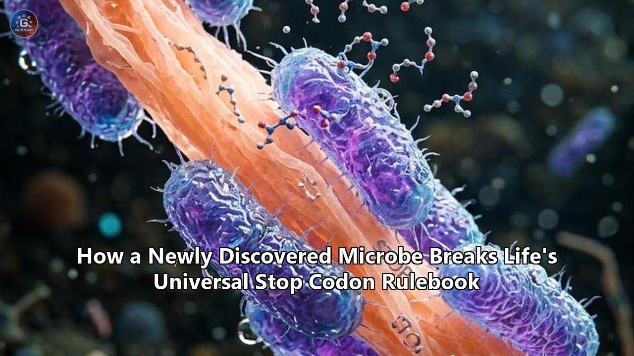
A microscopic organism pulled from a freshwater pond in Oxford University Parks in the United Kingdom has overturned a foundational principle of modern genetics. When molecular evolutionary biologist Dr. Jamie McGowan and his colleagues at the Earlham Institute and the University of Oxford sequenced the genome of an uncultivated ciliate protist, designated Oligohymenophorea sp. PL0344, they expected a standard validation run for a low-input single-cell DNA sequencing pipeline. Instead, they uncovered an unprecedented biological anomaly: the organism splits the function of two genetic signals that researchers long believed were mechanistically inseparable, using them to insert two completely different amino acids into its growing proteins.
For more than six decades, the central dogma of molecular biology has treated translation termination as an unambiguous full stop. In virtually every organism on Earth, three specific triplet sequences of messenger RNA—UAA, UAG, and UGA—serve as punctuation marks. They instruct the cellular ribosome to halt protein synthesis, release the finished polypeptide chain, and allow the molecular machine to fold its product into a functioning enzyme, receptor, or structural filament. Even when extreme evolutionary pressures have driven certain species to reassign these signals to encode amino acids, nature has consistently adhered to a strict constraint: UAA and UAG always changed in tandem, decoding into the exact same amino acid.
Oligohymenophorea sp. PL0344 rejects that constraint entirely. In this pond-dwelling protozoan, UAA instructs the ribosome to insert lysine, while UAG directs the addition of glutamic acid. Translation termination is left exclusively to the third signal, UGA."This is extremely unusual," Dr. McGowan stated following the verification of the organism’s transcriptome and nuclear genome. "We're not aware of any other case where these stop codons are linked to two different amino acids. It breaks some of the rules we thought we knew about gene translation—these two codons were thought to be coupled."
The implications of this discovery reach far beyond protist taxonomy. By demonstrating that the canonical code is neither immutable nor rigidly constrained by historical biochemical assumptions, this single-celled organism reveals serious blind spots in current computational biology, exposes vulnerabilities in global metagenomic databases, and provides synthetic biologists with a natural template for re-engineering cellular protein factories.
The Challenge: A 60-Year Dogma Meets Structural Anomaly
To understand the problem exposed by Oligohymenophorea sp. PL0344, one must examine the molecular mechanics that govern how genetic code is deciphered. When Francis Crick proposed the "wobble hypothesis" in 1966, he outlined the physical rules that dictate how transfer RNA (tRNA) molecules match their anticodons to messenger RNA (mRNA) codons inside the ribosome. Because the third base position of a codon experiences less spatial restriction on the ribosomal surface, non-standard hydrogen bonding occurs. Under these rules, a tRNA molecule bearing a uracil (U) at its first anticodon position (position 34) naturally pairs with both adenine (A) and guanine (G) at the third position of an mRNA codon.
Because UAA and UAG share an identical 5'-UA-3' core and end in purines (adenine and guanine), they share a physical symmetry. Over billions of years of biological evolution, this symmetry kept them chemically tethered. Whenever a suppressor tRNA mutated to read one of these codons, its wobble interaction inevitably swept up the other. In ciliates such as Tetrahymena thermophila or Paramecium tetraurelia, both UAA and UAG were reassigned to glutamine. In other non-canonical single-celled species, both were co-opted for tyrosine or leucine. No organism had ever been observed splitting the pair into distinct, competing sense assignments.
Standard Genetic Code:
Codon UAA  ───>  STOP (Ochre)
Codon UAG  ───>  STOP (Amber)
Codon UGA  ───>  STOP (Opal)

Canonical Ciliate Variant (e.g., Tetrahymena):
Codon UAA  ───>  Glutamine (Q)
Codon UAG  ───>  Glutamine (Q)
Codon UGA  ───>  STOP

Oligohymenophorea sp. PL0344:
Codon UAA  ───>  Lysine (K)        [Positively charged]
Codon UAG  ───>  Glutamic Acid (E) [Negatively charged]
Codon UGA  ───>  STOP              [Sole functional terminator]
The physical reality of this division poses a massive biochemical problem. Lysine is a basic, positively charged amino acid with a flexible, nitrogen-tipped side chain; glutamic acid is an acidic, negatively charged residue that forms electrostatic salt bridges and catalytic centers. Inadvertently swapping one for the other inside a folded protein typically destabilizes hydrophobic cores, disrupts enzymatic active sites, and triggers toxic protein aggregation.
For an organism to survive while using both codons simultaneously, its translational machinery must maintain absolute fidelity. If the cell's suppressor tRNAs wobbled carelessly between UAA and UAG, every protein produced within its cytoplasm would become a scrambled mixture of conflicting polarities, resulting in immediate cellular death. Yet Oligohymenophorea sp. PL0344 does not just survive; it thrives in wild aquatic ecosystems alongside standard freshwater fauna.
The uncultivated pond organism shattered the long-held assumption that a microbe stop codon operates within an immutable chemical pair, proving that cellular translation machinery can evolve surgical discernment where biophysicists once assumed wobble ambiguity was unavoidable.
What Went Wrong in Genomic Pipelines
The discovery of Oligohymenophorea sp. PL0344 has exposed a compounding systematic error across computational biology. For the past three decades, high-throughput sequencing projects have relied on automated gene-prediction software—such as Prodigal, GeneMarkS, and AUGUSTUS—to parse through terabytes of raw DNA and assemble open reading frames (ORFs).
These algorithms rely on rigid pre-programmed translation tables. In standard bioinformatics protocols, whenever an automated parser encounters an in-frame UAA or UAG codon, it assumes the ribosome must disengage. The algorithm inserts an asterisk, chops the putative gene into fragments, and marks the downstream sequence as non-coding space.
Because of this automated assumption, millions of genes across planetary microbiome databases are actively misannotated. When researchers sequence environmental DNA from deep-sea sediments, soil horizons, or animal digestive tracts, uncultivated microbes with variant genetic codes are processed through standard computational filters.
The structural fallout manifests in three critical ways:
- Truncated Proteomes: When a protein-coding sequence contains internal UAA or UAG codons that actually code for lysine or glutamate, gene-callers categorize the real gene as a broken pseudogene or a sequence artifact. The algorithm predicts truncated, inactive fragments instead of the full-length, biologically active enzyme.
- Invisible Metabolic Pathways: Key metabolic enzymes responsible for carbon cycling, nitrogen fixation, or secondary metabolite production go undetected because their open reading frames are peppered with what algorithms classify as lethal nonsense mutations.
- Skewed Evolutionary Models: Phylogenetic trees constructed from ribosomal proteins and single-copy core genes suffer from systematic errors when codons are mapped incorrectly, leading evolutionary biologists to miscalculate evolutionary distances and divergence timelines across the eukaryotic tree of life.
When Dr. McGowan's team first pulled the raw sequencing data into their computers, the automated output looked nonsensical. Crucial housekeeping genes—conserved proteins responsible for cytoskeletal integrity, cellular replication, and energy production—appeared to be riddled with stop signals right in the middle of their catalytic domains.
"When he uploaded the organism's DNA sequence to his computer, McGowan noticed that right in the middle of the nucleotide sequence, lots of the genes contained what would normally be a stop codon," noted reports tracking the Earlham Institute analysis. "It looked strange."
Under conventional analysis pipelines, Oligohymenophorea sp. PL0344 would have been discarded as degraded DNA or filtered out as sequencing artifact. The failure was not in the ciliate’s biology; the failure was baked directly into the analytical software used by geneticists worldwide.
How Automated Algorithms Fail Variant Microbes:

Target Gene: [ATG] ... [AAA] ... [UAA] ... [GAG] ... [UAG] ... [UGA]
Real Output:  Met  ...  Lys  ...  Lys  ...  Glu  ...  Glu  ...  STOP (Functional Enzyme)

Standard Pipeline Execution:
Step 1: Reader encounters [ATG] ──> Begins gene prediction.
Step 2: Reader encounters [UAA] ──> "Premature Stop Codon Detected."
Step 3: Prediction truncated at residue 28; classified as non-functional fragment / pseudogene.
Result: Critical enzyme discarded; remaining downstream codons flagged as non-coding junk.
The problem highlights how automated gene-calling algorithms misinterpret a microbe stop codon as a fatal punctuation mark, blinding global databases to functional enzymes produced by unconventional microbial lineages.
The Biological Friction: Surviving Without Runaway Translation
The reassignment of stop codons to amino acids introduces severe physiological hazards within the cell. If a microorganism converts UAA and UAG into sense codons, it strips itself of two-thirds of its termination vocabulary. This raises an obvious mechanistic question: what keeps the cell from destroying its own proteome through runaway translation?
When a ribosome reaches the authentic end of a messenger RNA transcript, it must disengage. If it fails to terminate, it translates straight through into the 3' untranslated region (3' UTR), appending hydrophobic or dysfunctional peptide tails to the protein. These tail-extended proteins frequently misfold, expose aberrant degradation signals, and precipitate out of solution, exhausting the cell's chaperone machinery and triggering apoptosis or metabolic arrest.
Deep bioinformatic analysis of Oligohymenophorea sp. PL0344 revealed the specialized adaptations that allow the ciliate to maintain order in the absence of canonical termination signals:
1. Dedicated Suppressor tRNAs with Altered Loop Geometry
To decode UAA as lysine and UAG as glutamic acid without cross-reactivity, the ciliate developed specialized suppressor tRNAs. McGowan and his team identified multiple suppressor tRNA genes bearing specific anticodon loops designed to eliminate wobble ambiguity.
Rather than relying on generic tRNAs that loosely recognize both purine-ending triplets, the organism evolved structurally tailored transfer RNAs that restrict their base-pairing interactions exclusively to their designated targets. Post-transcriptional base modifications—chemical alterations introduced to nucleotide bases at positions 34 and 37 of the tRNA anticodon loop—play an essential role in stabilizing these interactions, preventing the dangerous wobble cross-talk that would otherwise substitute lysine for glutamate.
2. Eukaryotic Release Factor Specialization (eRF1)
In typical eukaryotes, translation termination is governed by eukaryotic release factor 1 (eRF1), a protein shaped remarkably like a tRNA. eRF1 is an omnipotent factor; its protein structure features specific peptide motifs that recognize all three stop codons—UAA, UAG, and UGA—with high affinity.
For Oligohymenophorea sp. PL0344 to function, its eRF1 had to undergo drastic structural remodeling. Mutations concentrated in the N-terminal decoding domain of its eRF1 dismantled the pocket responsible for recognizing UAA and UAG, leaving only the binding pocket for UGA intact. By paralyzing the release factor’s capacity to interact with UAA and UAG, the cell cleared the way for suppressor tRNAs to access the ribosomal A-site unimpeded, eliminating competitive pauses that would stall protein production.
3. The 3' UTR "Catch-Basin" Architecture
The ciliate’s most pronounced structural failsafe is located directly past the finish line of its genes. Because the cell operates with only one functional termination signal—UGA—any ribosomal readthrough past a legitimate stop signal could prove fatal.
To protect against this vulnerability, Oligohymenophorea sp. PL0344 employs a genomic failsafe: an overwhelming enrichment of tandem, consecutive UGA codons strategically positioned immediately downstream of coding sequences in the 3' UTR. If a translating ribosome occasionally slips past an authentic termination signal, it encounters a dense cluster of backup UGA stops within a few nucleotides. This architectural arrangement ensures that translational termination occurs before extended, toxic protein tails can be assembled.
Understanding the biological mechanisms that allow a microbe stop codon to be reassigned without collapsing cellular translation has provided evolutionary biologists with a blueprint of how complex molecular machines adapt when basic genetic grammar is radically modified.
Genomic Architecture in Oligohymenophorea sp. PL0344:

 mRNA Sequence:
 ───[Open Reading Frame]───||───[Authentic Stop]───||───[Safety Net UTR]───>
 ... GAU (UAA) GAA (UAG) GCU      (UGA)             (UGA)  (UGA)  Poly-A
     Asp [Lys] Glu [Glu] Ala       STOP              STOP   STOP
                                 Primary          Backup Terminals
                              Termination        Preventing Runaway
The ciliate is not entirely alone in its linguistic rebellion. Related research on other atypical microbes shows that life has repeatedly tested the boundaries of the stop codon rulebook. In the flagellated parasite Blastocrithidia nonstop, discovered by an international research consortium, all three canonical stop codons were reassigned to sense codons (UAA and UAG to glutamate; UGA to tryptophan), with UAA paradoxically serving a dual role as both an amino acid and a context-dependent terminator.
Meanwhile, at the University of California, Berkeley, molecular microbiologist Dr. Dipti Nayak demonstrated that the methane-producing archaeon Methanosarcina acetivorans treats the UAG stop codon as an environmental coin flip. Depending on the concentration of methylamines in its environment, it either stops protein synthesis or inserts pyrrolysine, the rare 22nd genetically encoded amino acid.
"Objectively, ambiguity in the genetic code should be deleterious; you end up generating a random pool of proteins," Dr. Nayak noted in a study detailing ambiguous amber codon usage. "But biological systems are more ambiguous than we give them credit to be and that ambiguity is actually a feature—it's not a bug."
The Solution: Rewriting Computational and Synthetic Biology
Faced with proof that microscopic organisms do not follow uniform genetic rules, the scientific community has begun overhauling both computational infrastructure and synthetic biology platforms. Resolving the problems created by non-canonical translation systems requires a multi-pronged strategy led by bioinformaticians, structural biologists, and genetic engineers.
1. Algorithmic Overhauls and Dynamic Gene-Callers
The first operational step has targeted computational annotation pipelines. Traditional gene finders rely on static lookup tables assigned at the beginning of an analysis. To fix this, computational biologists are deploying dynamic, context-aware gene-prediction tools like Codetta and customized machine-learning frameworks.
Instead of assuming a static code, these advanced algorithms evaluate the functional conservation of protein alignments across homologous species before deciding whether a sequence represents an in-frame stop or an amino acid. If a suspected stop codon appears consistently in evolutionarily preserved regions where related species feature a charged lysine or glutamate residue, the algorithm automatically flags a prospective codon reassignment and dynamically builds a custom translation table for the target organism.
The National Center for Biotechnology Information (NCBI) and the European Bioinformatics Institute (EMBL-EBI) are actively updating their translation tables. Historically, the NCBI maintained roughly 30 alternative genetic codes, largely covering variations in mitochondrial genomes, mycoplasmas, and canonical ciliates. The discovery of Oligohymenophorea sp. PL0344 has driven the formalization of new eukaryotic nuclear translation tables, ensuring that automated databases stop discarding valid protist genomes as defective sequences.
Computational Pipeline Evolution:

Legacy Static Model:
Raw Reads ──> Fixed Table Lookup (Code 1 or 6) ──> Rigid ORF Prediction ──> Truncated Data

Modern Dynamic Framework:
Raw Reads ──> Multiple Sequence Alignment ──> Conservation Profiling ──> Dynamic Codon Reassignment ──> Validated Proteome
2. Deep Metagenomic Re-annotation Campaigns
Armed with adaptive algorithmic pipelines, researchers are returning to massive public datasets to mine for overlooked biology. Teams connected to the global TARA Oceans Project and the Darwin Tree of Life project are systematically reprocessing uncultivated microbial datasets.
The results are already reshaping microbial phylogeny. In follow-up research published in PLOS Genetics, Dr. McGowan and his colleagues re-analyzed marine metagenomes from the Phyllopharyngea class of ciliates. They identified at least three independent, uncoupled reassignments of the UAG codon to leucine within uncultivated ocean-dwelling species, demonstrating that uncoupled stop codon evolution is not an isolated fluke confined to a single Oxford pond, but a recurring theme across global aquatic ecosystems.
| Organism / Clade | Reassigned Codon(s) | Decoded Meaning | Functional Stop Codon(s) | Primary Adaptive Mechanism |
|---|---|---|---|---|
| Oligohymenophorea sp. PL0344 | UAA / UAG | Lysine (K) / Glutamic Acid (E) | UGA | Diverged tRNAs, altered eRF1, tandem 3' UTR stops |
| Tetrahymena thermophila | UAA / UAG | Glutamine (Q) | UGA | Wobble-capable single suppressor tRNA (UUA) |
| Blastocrithidia nonstop | UAA, UAG, UGA | Glutamate (UAR) / Tryptophan (UGA) | UAA (Dual-role) | Mutated eRF1/eRF3, context-dependent readthrough |
| Phyllopharyngea spp. (Marine) | UAG | Leucine (L) | UAA, UGA | Distinct suppressor tRNALeu (CUA), independent of UAA |
| Methanosarcina acetivorans | UAG (Conditional) | Pyrrolysine (Pyl) | UAG, UAA, UGA | Specialized PylRS / tRNAPyl system tied to methylamine diet |
3. Synthetic Biology and Orthogonal Protein Engineering
While computational scientists fix existing databases, synthetic biologists are actively translating these microbial survival strategies into new biotechnology. For decades, synthetic biology labs—pioneered by researchers like Dr. Peter Schultz at Scripps and Dr. Jason Chin at the MRC Laboratory of Molecular Biology—have worked to re-engineer bacterial genomes to incorporate non-canonical amino acids (ncAAs).
The primary obstacle has always been efficiency. In standard genetic code expansion, researchers delete a stop codon—most commonly the amber codon, UAG—and introduce an engineered orthogonal tRNA/aminoacyl-tRNA synthetase pair to insert a custom synthetic building block. However, endogenous release factors frequently compete with the synthetic tRNA at the ribosome, causing truncated peptides, low protein yields, and cellular toxicity.
Microbial solutions derived from Oligohymenophorea sp. PL0344 and Methanosarcina acetivorans offer natural workarounds. Because these single-celled organisms have already solved the structural problem of completely stripping release factors of their ability to bind UAA and UAG without killing the host, their modified eRF1 architectures serve as templates for creating hyper-efficient synthetic cell lines.
Using these structural blueprints, synthetic biologists are creating fully orthogonal genetic translation systems. In these re-engineered cells, a microbe stop codon reassignment allows industrial fermenters to incorporate multiple non-standard amino acids simultaneously—such as fluorescent probes, click-chemistry handles, and post-translationally modified residues—into a single therapeutic protein with zero cross-talk and high yields.
Applications in Synthetic Biology:

[Engineered Cell Line]
   │
   ├──> Stripped Release Factor (eRF1-ΔUAG/UAA): Ignores designated codons.
   │
   ├──> Orthogonal tRNA-1 (CUA anticodon): Inserts Synthetic Amino Acid A at UAG.
   │
   ├──> Orthogonal tRNA-2 (UUA anticodon): Inserts Synthetic Amino Acid B at UAA.
   │
   └──> Retained UGA Factor: Terminates translation precisely at gene end.

Outcome: Dual non-canonical amino acid incorporation for advanced biotherapeutics.
4. Therapeutic Blueprints for Human Genetic Disease
Beyond industrial biotechnology, this natural evolutionary mechanism provides new strategies for human medical therapy. In clinical medicine, roughly 11% of all inherited human genetic disorders—including cystic fibrosis, Duchenne muscular dystrophy, spinal muscular atrophy, and numerous familial cancers—are classified as nonsense-mutation diseases. They are caused by single-base point mutations that convert an ordinary sense codon into a premature termination codon (PTC), causing the ribosome to abort synthesis before completing a critical protein.
Pharmaceutical developers have spent decades searching for "nonsense suppression" therapies: small-molecule drugs or modified tRNAs that induce ribosomal readthrough at premature stop codons to restore production of full-length proteins. The central barrier has always been toxicity. If a drug causes the ribosome to ignore premature stop codons, it often causes the ribosome to ignore natural, authentic stop codons as well, generating dangerous proteomic chaos across every organ system in the patient’s body.
The architectural layout observed in Oligohymenophorea sp. PL0344 models a resolution to this therapeutic impasse. The organism clearly distinguishes between internal, reassigned codons and true terminal stops using a combination of tailored tRNAs, modified termination factors, and tandem safety-stop architectures.
Biomedical researchers studying engineered suppressor tRNAs are leveraging these natural structural configurations to design next-generation therapeutics. By mimicking the subtle base modifications and loop dynamics found in ciliate suppressor tRNAs, genetic medicine platforms are engineering anti-PTC tRNAs that exhibit high specificity for premature nonsense mutations inside diseased genes while allowing authentic cellular stop complexes to terminate translation normally at genuine 3' UTR boundaries.
The Uncharted Microbial Frontier
The extraction of Oligohymenophorea sp. PL0344 from an ordinary campus pond demonstrates that our understanding of biology’s core operating system remains incomplete. For over a half-century, basic biology education has presented the genetic code as an immutable granite tablet, handed down virtually unchanged since the Last Universal Common Ancestor (LUCA).
The biological reality is far more dynamic. The genetic code functions less like a fixed stone monument and more like a malleable operating system—one capable of being modified, patched, and reprogrammed by evolutionary pressures.
Significant questions remain unresolved. Because Oligohymenophorea sp. PL0344 has so far resisted long-term laboratory culture, researchers have not yet been able to grow enough biomass to perform exhaustive peptide sequencing via mass spectrometry. Confirming every predicted tRNA charging dynamic in living, dividing cultures will require either microfluidic culture breakthroughs or the heterologous expression of the ciliate's translation machinery in model organisms like Tetrahymena or Escherichia coli.
Further environmental sampling is already underway. Consortia running high-throughput single-cell sequencing across under-sampled microbial ecosystems are identifying additional variants that test our models of translation.
As these tools advance, the scientific community is shifting away from rigid standard templates toward an adaptive computational framework. What began as a routine test of a single-cell sequencing pipeline in an Oxford pond has broken open a decades-old consensus, proving that when it comes to life’s basic rulebook, the microscopic world is still writing new chapters.
Reference:
- https://www.technologynetworks.com/genomics/news/organism-found-in-university-pond-rewrites-the-rules-of-dna-379612
- http://www.sciencedaily.com/releases/2026/10/261007233136.htm
- https://journals.plos.org/plosgenetics/article?id=10.1371/journal.pgen.1010913
- https://en.clickpetroleoegas.com.br/scientists-discovered-purely-by-chance-that-a-microscopic-organism-taken-from-a-pond-in-oxford-does-not-follow-one-of-the-universal-rules-of-btl96/
- https://www.biorxiv.org/content/10.1101/2022.12.16.520718v2
- https://www.sciencedaily.com/releases/2026/02/260227071920.htm
- https://www.pnas.org/post/journal-club/translucent-microbe-deviates-universal-genetic-code
- https://www.icr.org/article/dna-stop-signs-change-message
- https://www.earlham.ac.uk/news/end-genes-routine-test-reveals-unique-divergence-genetic-code
- https://journals.plos.org/plosgenetics/article?id=10.1371/journal.pgen.1011512
- https://elifesciences.org/articles/91405
- https://academic.oup.com/mbe/article/40/4/msad064/7084900
- https://elifesciences.org/reviewed-preprints/93502
- https://journals.asm.org/doi/10.1128/mbio.00885-25
- https://www.sciencedaily.com/releases/2026/10/261007233136.htm
- https://pubmed.ncbi.nlm.nih.gov/39689125/
- https://web.osu.cz/~Yurchenko/pdf/NAR%202024.pdf
- https://www.technologynetworks.com/proteomics/news/this-microbe-breaks-a-fundamental-rule-of-the-genetic-code-407578
- https://pmc.ncbi.nlm.nih.gov/articles/PMC12152300/
- https://www.ornl.gov/news/scientists-uncover-novel-genetic-code-microbes-opening-new-biotech-frontier
- https://innovativegenomics.org/news/new-genetic-code-microbes/