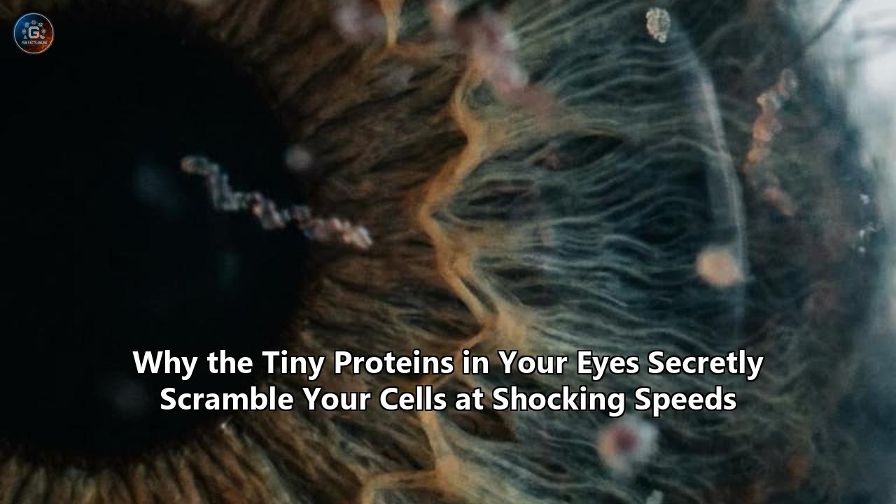
In the quiet, microscopic architecture of the human retina, a frantic metabolic salvage operation is constantly taking place. Every second, millions of phototransduction receptors are bombarded by photons, triggering the biochemical cascade that translates light into sight. Yet, behind this textbook marvel of sensory biology lies a hidden physical crisis: the light-sensitive membranes of our eyes are subjected to intense mechanical and chemical stress that, if left uncorrected, would collapse the delicate structures of our cells and lead to irreversible blindness.
For decades, the mechanics of how the eye stabilizes these membranes and clears toxic byproducts remained one of the most enduring mysteries in biophysics. However, a study published in Nature Structural & Molecular Biology has blown this mystery wide open, revealing a startling double life led by the most abundant proteins in human eyes.
A collaborative research team led by investigators at Weill Cornell Medicine and Ruhr University Bochum in Germany has developed a single-vesicle fluorescence imaging technique. For the first time, researchers can isolate and measure the activity of individual membrane proteins in real time.
The findings are as astonishing as they are counterintuitive. Opsin—the primary light-detecting G protein-coupled receptor (GPCR) apoprotein in rod photoreceptor cells—acts as a hyperactive, ATP-independent lipid scramblase. While typical active transport proteins drag molecules across cell membranes at a rate of 1 to 100 molecules per second, a single monomer of opsin "scrambles" lipids across the membrane bilayer at speeds exceeding 10,000 lipids per second. It performs this molecular gymnastics without consuming a single unit of cellular energy.
This discovery shatters the traditional, signaling-centric view of GPCRs. It reveals a biological master switch where these proteins in human eyes transition from highly tuned light detectors to physical membrane stabilizers. It also exposes a critical, previously invisible layer of regulation governed entirely by the surrounding lipid environment—specifically, cholesterol.
To understand why our eyes depend on these proteins to scramble our cells at such shocking speeds, we must look beyond the textbook diagrams and enter the complex, crowded, and highly asymmetric world of the cellular membrane.
The Architecture of Asymmetry: Why Membrane order is a Matter of Life and Death
Every cell in the human body is defined by its membrane: a thin, self-assembling bilayer of phospholipids that acts as both a barrier and a gatekeeper. This bilayer is not a homogenous, static wall. Instead, it is a dynamic fluid, with lipids diffusing laterally at rapid speeds.
Crucially, the two leaflets of the bilayer—the inner, cytosolic monolayer and the outer, extracellular monolayer—are highly asymmetric in their chemical composition. Cells expend a massive amount of metabolic energy to maintain this asymmetry, placing specific lipid species in one leaflet while sequestering others in the opposing leaflet.
  EXTRACELLULAR LEAFLET (Lumen of Disk)
  [PC] [SM] [Glycolipids] [NRPE (trapped)]
  ~~~~~~~~~~~~~~~~~~~~~~~~~~~~~~~~~~~~~~~~~~~~~~~ (Lipid Bilayer Core)
  [PE] [PS] [PI] [Phosphatidic Acid]
  CYTOSOLIC LEAFLET (Cytoplasm)
In a typical mammalian cell, zwitterionic phospholipids like phosphatidylcholine (PC) and sphingomyelin (SM) are strictly corralled in the outer, extracellular leaflet. Conversely, aminophospholipids like phosphatidylethanolamine (PE) and the negatively charged phosphatidylserine (PS) are sequestered in the inner, cytosolic leaflet. This precise spatial organization is critical for cellular signaling, membrane potential, and organelle function.
However, maintaining this asymmetry is a monumental thermodynamic challenge. While lateral diffusion in the plane of the membrane is effortless, transverse diffusion—commonly referred to as "lipid flip-flop," where a lipid moves from one leaflet to the other—is energetically unfavorable.
A phospholipid molecule consists of a highly polar, hydrophilic headgroup and two long, hydrophobic fatty-acid acyl chains. For a lipid to flip spontaneously across the membrane, its polar, water-loving headgroup must leave its protective hydration shell, force its way through the oily, hydrophobic core of the bilayer, and re-emerge on the other side.
This thermodynamic barrier is massive, with an activation energy ($\Delta G^\ddagger$) of approximately 80 to 100 kJ/mol for standard zwitterionic lipids. Uncatalyzed, a single lipid molecule takes hours, days, or even weeks to spontaneously cross the bilayer.
This slow, natural rate of flip-flop is essential for keeping the cell's outer wall intact. If the barrier fell, lipid asymmetry would instantly collapse, with catastrophic consequences.
For instance, the exposure of phosphatidylserine (PS) on the outer leaflet of a cell's surface is a universal "eat-me" signal. It flags the cell as dying or apoptotic, prompting passing macrophages to engulf and digest it. In blood platelets, PS exposure triggers the blood clotting cascade, leading to localized thrombosis.
To navigate this thermodynamic bottleneck while preserving strict control over membrane organization, cells have evolved three distinct classes of specialized transport proteins:
- Flippases (P4-ATPases): Active, ATP-dependent enzymes that selectively pump aminophospholipids (PE and PS) from the outer leaflet to the inner, cytosolic leaflet. They work slowly, typically moving 1 to 100 lipids per second, building and maintaining the cell's structural asymmetry.
- Floppases (ABC Transporters): Active, ATP-dependent pumps that move lipids and lipid-anchored molecules in the opposite direction, from the inner leaflet to the outer leaflet, often helping to clear metabolic waste or export signaling lipids.
- Scramblases: Passive, ATP-independent facilitators that act as open channels. When activated, they allow all common phospholipids to flow bidirectionally down their concentration gradients across the bilayer. They collapse lipid asymmetry at astronomical speeds, reaching rates of thousands of lipid translocations per second.
For decades, biophysicists believed that scramblases were rare, specialized proteins activated only during emergencies, such as cellular damage, apoptosis, or blood clotting. The idea that a highly abundant, primary sensory receptor in our eyes could be a highly active, constitutive scramblase was deemed highly unlikely.
Yet, the biophysics of our vision requires exactly this kind of high-speed molecular mixing.
The "Credit-Card Reader" Mechanism: How GPCRs Defy Biophysics
To understand how a single protein can move tens of thousands of polar lipid headgroups through a hydrophobic barrier every second without using ATP, we must examine its physical structure.
In 2006, Dr. Anant Menon and Dr. Thomas Günther-Pomorski proposed a physical model for scramblase function that has since become the cornerstone of membrane biophysics: the "credit-card reader" mechanism.
       HYDROPHILIC GROOVE (The Card Reader)
            | |               | |
   [Lipid]  | |   [Headgroup] | |  [Lipid]
    O====   | |=======> o==== | |   ====O   <-- Bilayer Leaflet 1
    | |     | |         | |   | |     | |   <-- Hydrophobic Core
    | |     | |         | |   | |     | |
    O====   | |=======> o==== | |   ====O   <-- Bilayer Leaflet 2
            | |               | |
       Transmembrane Helices 6 & 7 (TM6/7)
In this model, the scramblase protein does not act like a traditional pore or a gated channel that swallows a substrate entirely. Instead, the protein is embedded in the lipid bilayer, and its external structure features a vertical, hydrophilic groove that spans the entire thickness of the membrane.
This hydrophilic groove acts like the slot of a credit-card reader. The polar, hydrophilic headgroup of the phospholipid represents the magnetic strip of the credit card. It slips into the water-lined, polar groove of the protein, which satisfies its electrostatic and hydrogen-bonding requirements, shielding it from the greasy, hydrophobic interior of the membrane.
Meanwhile, the long, hydrophobic fatty-acid acyl chains of the lipid remain pointing outward, embedded directly in the surrounding hydrocarbon core of the lipid bilayer. They drag along through the lipid lake as the headgroup slides smoothly through the protein’s vertical slot.
Because the lipid's hydrophobic tails never leave their native, oil-like environment, the thermodynamic activation energy of the translocation process is slashed to near zero. The lipids do not need to be "pumped". They simply diffuse through the groove bidirectionally, driven by thermal motion, instantly equilibrating the lipid concentrations between the two leaflets.
While this mechanism was first identified in specialized calcium-activated scramblases like the TMEM16 family, subsequent structural work and molecular dynamics (MD) simulations delivered a shock to the scientific community. The prototypical G protein-coupled receptor rhodopsin—and its retinal-free form opsin—possess exactly this kind of transbilayer pathway.
Large-scale, ensemble atomistic molecular dynamics simulations of the opsin GPCR revealed that transmembrane helices 6 and 7 (TM6 and TM7) undergo continuous, spontaneous conformational fluctuations. Under normal physiological temperatures, these thermal movements transiently part TM6 and TM7, exposing a dynamic, hydrophilic pathway lined with polar and charged amino acid residues that connects the cytoplasmic and extracellular faces of the membrane.
This pathway allows lipid headgroups to traverse the bilayer with ease. The simulations showed that the conformational transitions required to open this lipid-scrambling groove are completely distinct from those associated with classical GPCR signaling. Opsin does not need to be activated by light or bound to a G-protein to scramble lipids; it is a constitutively active physical channel.
However, establishing this in silico was not enough. Proving that these proteins in human eyes actually scramble cell membranes at shocking speeds in the physical world required overcoming a massive, decades-old technical bottleneck in experimental biophysics.
The Visual Cycle and the Photoreceptor Disk: A High-Stakes Salvage Mission
Why would the rod cells in the human retina require a protein that scrambles their membranes at such blistering speeds? The answer lies in the intense chemistry of the visual cycle and the unique anatomy of the photoreceptor outer segment.
  RETINAL ROD PHOTORECEPTOR CELL (Outer Segment)

         | |     | |     | |     | |     | |
        _|_|_   _|_|_   _|_|_   _|_|_   _|_|_   <-- Stacked Membranous Disks
       |     | |     | |     | |     | |     |      (Rhodopsin-rich membranes)
       |_____| |_____| |_____| |_____| |_____|
         | |     | |     | |     | |     | |

  [Outer Segment Tips Shed Daily & Engulfed by RPE] --> [PS Exposure Signal]
Rod photoreceptor cells are divided into two primary compartments: an inner segment containing the cell's metabolic machinery (nucleus, mitochondria, endoplasmic reticulum), and a specialized outer segment dedicated entirely to capturing photons of light.
This outer segment is packed with a vertical stack of hundreds of flat, closed membranous sacs called disks. These disks are completely detached from the cell's outer plasma membrane, forming an independent intracellular compartment.
The disk membranes are incredibly protein-dense. Over 90% of their total membrane protein content consists of rhodopsin, the visual pigment responsible for our ability to see in dim light. Rhodopsin is a chromoprotein composed of a colorless GPCR called opsin, covalently bound to a light-sensitive, vitamin A-derived chromophore called 11-cis-retinal.
When a photon of light enters the eye and strikes a rhodopsin molecule in a disk, it initiates an incredibly rapid physical event: the 11-cis-retinal chromophore absorbs the energy and isomerizes into all-trans-retinal. This rapid change in shape triggers a conformational shift in the surrounding opsin protein, activating a G-protein-mediated signaling cascade (phototransduction) that sends an electrical signal along the optic nerve to the brain.
But once the signal is sent, the rod cell faces a crisis. The all-trans-retinal is now detached from the opsin protein and must be recycled back to 11-cis-retinal so the photoreceptor can reset.
This recycling process, known as the visual cycle, is a multi-step enzymatic pathway that requires transporting the retinoid out of the photoreceptor, across the subretinal space, and into the adjacent retinal pigment epithelium (RPE) cells.
Here is where the chemistry becomes dangerous. Free all-trans-retinal is a highly reactive, hydrophobic aldehyde. If it is allowed to accumulate unchecked within the oily disk membrane, it will spontaneously react with amino groups on nearby proteins, causing toxic cross-linking and cellular damage.
To prevent this, the free all-trans-retinal rapidly and non-enzymatically reacts with phosphatidylethanolamine (PE)—the primary aminophospholipid in the disk membrane—to form a reversible Schiff-base adduct called N-retinylidene-PE (NRPE).
Because the light activation of rhodopsin occurs within the disk, the resulting NRPE is trapped on the internal, luminal leaflet of the disk membrane. It cannot escape on its own.
To clear this toxic adduct, the disk membrane relies on a highly specialized, ATP-driven molecular pump called ABCA4 (historically known as the Rim protein). ABCA4 is a member of the ATP-binding cassette transporter family, but it is highly unusual. While most ABC transporters act as exporters (floppases), ABCA4 acts as an importer (flippase).
ABCA4 actively binds NRPE (as well as free PE) on the luminal leaflet of the disk membrane, hydrolyzes ATP, and flips the lipid-retinal conjugate across the bilayer to the outer, cytosolic leaflet.
Once on the cytosolic side, the NRPE dissociates, releasing the all-trans-retinal into the cytoplasm where it can be safely reduced to all-trans-retinol by the enzyme retinol dehydrogenase 8 (RDH8). From there, it is escorted to the RPE to be converted back into 11-cis-retinal, resetting the visual system.
  LUMEN OF THE DISK (Inside)
  ~~~~~~~~~~~~~~~~~~~~~~~~~~~~~~~~~~~~~~~~~~~~~~~
  [PE] + [all-trans-Retinal] ===> [NRPE]
                                   |
                             (ABCA4 Pump - ATP driven)
                                   v
  ~~~~~~~~~~~~~~~~~~~~~~~~~~~~~~~~~~~~~~~~~~~~~~~
  [PE] + [all-trans-Retinal] <=== [NRPE]
  CYTOSOL (Outside)
This elegant clearing mechanism, however, introduces a physical problem of epic proportions.
ABCA4 is a strict, one-way conveyor belt. It constantly pumps PE and NRPE from the luminal leaflet to the cytosolic leaflet.
Because the photoreceptor disk is a completely closed, microscopic membrane sack with a highly restricted surface area, this continuous, one-way transport would rapidly deplete the luminal leaflet of its lipids while overcrowding and expanding the cytosolic leaflet.
This massive transbilayer lipid imbalance would generate immense physical bilayer stress. The asymmetric physical pressure would cause the flat disk membrane to warp, buckle, bend, and eventually rupture, destroying the rod cell's physical integrity.
Furthermore, the depletion of PE on the luminal side would starve ABCA4 of its substrate, shutting down the pump, stalling the visual cycle, and leaving reactive retinoids to run amok.
This is where the massive abundance of proteins in human eyes—rhodopsin and its apoprotein opsin—comes to the rescue.
By acting as passive, constitutive lipid scramblases, these proteins allow lipids to flow bidirectionally and rapidly across the disc membrane, instantly correcting the transbilayer lipid imbalance created by the ABCA4 pump.
As quickly as ABCA4 pumps a PE or NRPE molecule to the cytosolic leaflet, the scrambled channel of rhodopsin or opsin allows another phospholipid to slide back to the luminal leaflet.
This cooperative team-up between an active ATP-driven pump and a passive, high-speed scramblase maintains the structural homeostasis of the disk membrane, keeping it perfectly flat, relieving physical bilayer stress, and ensuring a continuous supply of PE in the luminal leaflet so ABCA4 can keep clearing toxic retinoids.
The Path to Macular Degeneration: What Happens When the Scrambler Fails
If this cooperative loop between ABCA4 and the visual scramblases is disrupted, the consequences for human vision are catastrophic.
When mutations in the ABCA4 gene impair its transport activity, or when changes in the lipid composition of the disk membranes inhibit the scrambling activity of opsin, the clearance of all-trans-retinal stalls.
The highly reactive all-trans-retinal begins to pile up on the luminal leaflet of the disk membrane, where it undergoes a second, irreversible chemical reaction. Another molecule of all-trans-retinal condenses with the primary NRPE adduct, creating a complex, highly stable bis-retinoid conjugate known as N-retinylidene-N-retinylethanolamine (A2PE).
  [NRPE] + [all-trans-Retinal] ===(Irreversible)===> [A2PE]
                                                        |
                                            (Shedding & Phagocytosis)
                                                        v
                                             RPE Lysosomes: [A2E]
                                             (Toxic Lipofuscin Buildup)
The photoreceptor outer segments are not permanent structures; they are constantly being renewed. Every single day, the distal tips of the rod outer segments—containing the oldest, most light-worn disks—are shed.
The adjacent retinal pigment epithelium (RPE) cells immediately engulf these shed tips and transport them into their internal lysosomes to degrade and recycle their components.
However, the RPE's lysosomes possess a fatal vulnerability: they do not have the enzymatic machinery required to break down the highly stable, carbon-dense A2PE molecule.
Instead, inside the acidic lysosomal environment of the RPE, A2PE is hydrolyzed into a highly toxic, fluorescent pyridinium bis-retinoid known as A2E.
Over a lifetime of light exposure, A2E cannot be degraded or exported. It accumulates permanently within the RPE cells, forming golden-brown fluorescent deposits known as lipofuscin (the age pigment).
As this lipofuscin buildup reaches a critical, toxic threshold, it begins to wreak havoc on the RPE cells:
- Lysosomal Rupture: A2E acts as a detergent, destabilizing and eventually rupturing the delicate lysosomal membranes, spilling highly acidic, digestive enzymes into the cell’s cytoplasm.
- Singlet Oxygen Generation: Under exposure to blue and ultraviolet light entering the eye, A2E acts as a potent photosensitizer. It generates highly reactive oxygen species (ROS), including singlet oxygen and free radicals, which damage the cell's lipids, proteins, and DNA.
- Apoptosis: The combination of oxidative stress and lysosomal failure triggers the programmed cell death (apoptosis) of the RPE cells.
Because the light-sensitive photoreceptor cells (rods and cones) rely entirely on the underlying RPE layer for structural support, nutrient delivery, and retinoid recycling, the death of the RPE cells leads to the rapid, irreversible degeneration of the photoreceptors themselves.
This is the precise, devastating molecular pathway of Stargardt disease (juvenile-onset macular dystrophy) and dry age-related macular degeneration (AMD), the leading cause of irreversible vision loss in elderly populations worldwide.
Without the frantic, high-speed scrambling activity of these proteins in human eyes, our retinas would rapidly choke on their own light-detecting byproducts, burning out under the very light they are designed to capture.
The Technical Breakthrough: Inside the Single-Vesicle Fluorescence Microscope Platform
To appreciate the scale of the breakthrough published in Nature Structural & Molecular Biology, one must understand why studying scramblases has been an experimental nightmare for biophysicists.
Historically, measuring lipid translocations at the physical scale of a single protein was impossible. Instead, researchers had to rely on bulk ensemble assays.
In a typical bulk assay, scientists would purify a target protein (like opsin) from bovine retina using detergents such as n-dodecyl-β-D-maltoside (DDM). They would then mix this purified protein with synthetic lipids to form proteoliposomes—tiny, synthetic lipid vesicles that mimic a cell's membrane, with the protein molecules embedded inside them.
To track the movement of lipids, they would incorporate a small percentage (typically 0.5 mol%) of a fluorescently labeled lipid reporter, such as NBD-PC (a phosphatidylcholine lipid with a green-fluorescing nitrobenzoxadiazole group attached to its headgroup).
Because the NBD-PC is added during vesicle formation, it distributes symmetrically, with roughly 50% of the fluorescent lipids in the outer leaflet facing the buffer, and 50% in the inner leaflet facing the vesicle’s empty interior.
  BULK ASSAY (Billions of Vesicles Averaged)

       Dithionite Added (Na2S2O4)
              |
              v (Reduces only outer-leaflet NBD-PC)
       ~~~~~~~~~~~~~~~                 ~~~~~~~~~~~~~~~
       [NBD*]  [NBD*]   ===> [QUENCHED] (50% signal loss)
       ~~~~~~~~~~~~~~~                 ~~~~~~~~~~~~~~~
       [NBD*]  [NBD*]   ===> Protected inside (unless scrambled)
To measure scrambling, researchers would add sodium dithionite ($Na_2S_2O_4$) to the buffer. Dithionite is a membrane-impermeable reducing agent.
When added, it chemically reduces the NBD group on any lipid located in the outer leaflet of the vesicle, permanently destroying its fluorescence.
In a control vesicle with no active scramblase protein, this results in an immediate 50% drop in total fluorescence, representing the outer leaflet. The remaining 50% of the signal is preserved because the dithionite cannot cross the lipid bilayer to reach the inner leaflet.
However, if the vesicle contains an active scramblase, the NBD-PC lipids constantly flip between the inner and outer leaflets. As the inner-leaflet NBD-PC molecules migrate to the outer face, they are instantly reduced by the external dithionite. Over a few minutes, the entire vesicle's fluorescence decays to zero.
While this bulk assay successfully proved that opsin and other GPCRs were indeed scramblases, it suffered from a major mathematical limitation: ensemble averaging.
When you mix purified protein and lipids, the protein molecules do not distribute evenly. Instead, they follow a Poisson distribution.
In any given batch of synthetic proteoliposomes, some vesicles will have zero proteins, some will have one, and others will have five or ten.
Furthermore, the synthetic vesicles vary widely in size (polydispersity), and the rate of quenching is a complex function of vesicle volume, protein orientation, and protein aggregation.
When measuring the fluorescent signal of a bulk suspension containing billions of liposomes in a cuvette, the resulting decay curve is a messy, multi-exponential average.
This average completely masks the true kinetics of individual proteins. Biophysicists could not answer fundamental questions:
- Do all opsin molecules scramble lipids at the same rate, or are there highly active and inactive conformations?
- Does a single opsin monomer scramble lipids, or does it require dimerization?
- How do changes in the local lipid microenvironment, such as the introduction of cholesterol, affect the scrambling rate of an individual protein?
The breakthrough developed by Dr. Sarina Veit, Dr. Grace I. Dearden, Dr. Kartikeya M. Menon, Dr. Thomas Günther-Pomorski, and Dr. Anant Menon shattered this barrier. They introduced a single-vesicle fluorescence microscopy platform.
  SINGLE-VESICLE ASSAY (Unprecedented Resolution)

       TIRF Microscope Objective / Laser
                 ^
                 | (Individual Vesicle Monitored)
       Streptavidin-Coated Glass Surface
                 | (Biotin Anchor)
         [Vesicle with EXACTLY ONE Opsin Protein]
                 ^
                 | (Flow of BSA "Vacuum" Buffer)
To execute this, the team first prepared proteoliposomes containing purified bovine opsin, trace amounts of NBD-PC, and a biotinylated anchor lipid.
They then immobilized these individual vesicles onto a glass coverslip coated with streptavidin inside a specialized microfluidic flow chamber.
Instead of using sodium dithionite—which is a highly reactive chemical that can slowly damage membranes and cause artifactual leakage over long imaging sessions—they developed a highly gentle, physically elegant back-extraction assay.
This assay utilizes bovine serum albumin (BSA). BSA has multiple deep, highly hydrophobic binding pockets designed to transport fatty acids and lipids in the bloodstream.
Because NBD-PC has a very low but finite solubility in water (approximately 0.75 μM), individual NBD-PC molecules will occasionally "desorb" (detach) from the outer leaflet of the vesicle membrane into the surrounding aqueous buffer.
When BSA is flowed through the microfluidic chamber, it acts as a molecular vacuum, instantly capturing any desorbed NBD-PC molecules and sequestering them in its hydrophobic pockets. Once bound to BSA, the NBD-PC's fluorescence is quenched by approximately 60% due to the changes in its local chemical environment, and the buffer flow physically washes the BSA-lipid complexes away.
By monitoring the immobilized vesicles using Total Internal Reflection Fluorescence (TIRF) microscopy, the researchers could track the decay of fluorescence in hundreds of individual, spatially resolved vesicles simultaneously.
Crucially, because they could track each vesicle separately, they could calibrate the fluorescence of the reconstituted protein itself to determine exactly how many opsin molecules were present in each individual vesicle!
This allowed them to isolate and analyze only those vesicles that contained exactly one opsin molecule.
The results were a revelation. Individual, monomeric opsin molecules scramble lipids at rates exceeding 10,000 lipids per second, directly validating computational predictions and proving that opsin is a blistering-fast, highly efficient macromolecular machine.
In contrast, when they tested VDAC1 (voltage-dependent anion channel 1, a mitochondrial outer membrane protein that they recently discovered also acts as a scramblase), they found that VDAC1 requires dimerization to scramble lipids.
Furthermore, individual VDAC1 dimers exhibited extreme kinetic heterogeneity, with scrambling rates ranging from fewer than 100 to more than 1,000 lipids per second, indicating that only specific, highly flexible dimer conformations are capable of rapid scrambling.
The Cholesterol Switch: Protecting the Cell from Self-Destruction
If opsin is such a potent, constitutive lipid scramblase, and other Class A GPCRs (such as the β1-adrenergic receptor, β2-adrenergic receptor, and adenosine A2A receptor) also moonlight as scramblases, why isn't our biology in constant, chaotic collapse?
Why don't these highly abundant receptors constantly scramble the outer membranes of our cells, exposing phosphatidylserine and triggering premature cell death or spontaneous blood clotting throughout our bodies?
The answer lies in one of the most elegant examples of lipid-mediated regulation in cell biology: the cholesterol switch.
  LOW CHOL (ER / Disk Tips)         HIGH CHOL (Plasma Membrane)

       TM6       TM7                    TM6       TM7
        \         /                      |       |
         \  ( )  /    <-- Open           | [CHOL]|   <-- Closed
          \     /                         \_____/
           |   |                             |
      [Lipids Slide Free]           [Groove Locked Shut]
Cell membranes are not uniform; they have distinct lipid compositions designed for their specific environments.
The plasma membrane, which forms the outer border of the cell, is highly enriched in cholesterol, often reaching 30 to 50 mol%. This high cholesterol concentration is essential for keeping the plasma membrane thick, packed, and mechanically resilient.
In contrast, the endoplasmic reticulum (ER)—where GPCRs are synthesized—has extremely low cholesterol, typically less than 5 mol%.
Similarly, the photoreceptor outer segment disks have a unique, highly specialized lipid composition: they are highly enriched in polyunsaturated fatty acids like docosahexaenoic acid (DHA) and have a very low cholesterol gradient, especially at the distal tips.
In their study, the researchers demonstrated that cholesterol acts as a direct, physical "lock" on GPCR scramblase activity.
When they reconstituted opsin into membranes containing high levels of cholesterol, its lipid-scrambling activity was completely silenced.
To understand how this works at the molecular level, researchers turned to high-resolution cryo-electron microscopy (cryo-EM) and massive, 150-microsecond molecular dynamics (MD) simulations.
The simulations revealed that cholesterol acts as a structural wedge. It physically binds to a highly conserved, hydrophobic cleft at the interface between transmembrane helices 6 and 7 (TM6 and TM7) of the GPCR—the exact location of the lipid-scrambling groove.
In a low-cholesterol membrane (like the ER or the photoreceptor disk), TM6 and TM7 are highly flexible, undergoing spontaneous thermal fluctuations that transiently reveal the hydrophilic pathway.
But in a cholesterol-rich membrane (like the plasma membrane), cholesterol molecules slip into the hydrophobic cleft between TM6 and TM7.
By nestling into this cleft, cholesterol acts as a physical key, locking the helices in a tightly closed conformation and preventing them from parting to expose the hydrophilic pathway.
Furthermore, cholesterol molecules themselves can "flip-flop" slowly within this pocket, further blocking the path for larger phospholipids.
This is a masterpiece of evolutionary design. The very lipid that defines the plasma membrane—cholesterol—acts as the specific inhibitor that silences the scramblase activity of GPCRs.
This allows these receptors to safely act as active scramblases in the low-cholesterol ER (where they assist in membrane biogenesis and protein glycosylation) while transitioning into purely signaling receptors once they reach the cholesterol-rich plasma membrane, protecting the cell's outer wall from self-destruction.
Technical Comparison of Lipid Translocators
| Property | Flippases (P4-ATPases) | Floppases (ABC Transporters) | Scramblases (Opsin / TMEM16F) |
|---|---|---|---|
| Energy Source | ATP Hydrolysis | ATP Hydrolysis | None (Passive, thermal diffusion) |
| Direction | Inward (Outer to Inner Leaflet) | Outward (Inner to Outer Leaflet) | Bidirectional (Equilibration) |
| Scrambling Rate | ~1 to 100 lipids/sec | ~1 to 100 lipids/sec | >10,000 lipids/sec (Opsin) |
| Substrate Specificity | High (e.g., PS, PE selective) | High (e.g., NRPE, drug selective) | Low (all common phospholipids) |
| Regulation | Constitutive / Phosphorylation | Transcriptional / Allosteric | Cholesterol suppressed / Ca²⁺ activated |
| Primary Eye Role | Outer segment tip renewal | NRPE clearance (ABCA4) | Disk membrane homeostasis (Opsin) |
Rethinking modern Pharmacology: The Untapped GPCR Goldmine
The confirmation that G protein-coupled receptors possess highly active, cholesterol-regulated lipid scramblase activity exposes a massive, historic blind spot in modern medicine and drug discovery.
GPCRs are the single most important class of drug targets in human medicine, representing the target of over 30% of all FDA-approved drugs, used to treat everything from hypertension and asthma to depression, pain, and heart failure.
Yet, for over half a century, the pharmaceutical industry has screened and optimized drug candidates based on a very narrow, "signaling-centric" paradigm:
- How does a drug molecule bind to the extracellular pocket of a GPCR?
- How does it trigger downstream signaling cascades via G-proteins?
- How does it recruit beta-arrestins?
This traditional view completely ignores the fact that these receptors are physically embedded in a lipid bilayer and possess powerful, non-visual, physical "moonlighting" functions as lipid translocators.
Many drugs currently on the market are highly lipophilic molecules that insert directly into the cell's lipid bilayer and bind to GPCRs.
If these drugs inadvertently alter the GPCR's scramblase activity—either by locking the receptor in a conformation that permits scrambling even in high-cholesterol environments, or by displacing cholesterol from the TM6/7 interface—they could trigger localized, unregulated lipid scrambling at the plasma membrane.
This could lead to a localized exposure of phosphatidylserine, triggering micro-clotting, cellular inflammation, or tissue damage.
This hidden, lipid-scrambling side effect could explain many of the complex, poorly understood toxicities and side effects of widely used medications, from cardiovascular drugs to antipsychotics.
Conversely, this biological insight opens up an entirely new dimension for drug discovery: scramblase-targeted therapeutics.
Instead of treating GPCRs purely as signaling receptors, we can now design drugs that specifically modulate their physical lipid-moving capabilities.
  SCRAMBLASE THERAPEUTIC PIPELINE

  [Retinal Degeneration] =======> Boost Opsin Scrambling ======> Clear Toxic NRPE

  [Cancer / Tumors] ============> Trigger PS Exposure =========> Flag Tumor for Macrophages

  [Thrombosis / Clotting] ======> Block TMEM16F Groove =======> Prevent Blood Clots
1. Targeting Retinal Degeneration and Macular Dystrophy
In diseases like dry age-related macular degeneration (AMD) and Stargardt disease, the physical imbalance of lipids in the photoreceptor disks is a major driver of cellular death.
If we can develop small-molecule chaperones or lipid-modulators that stabilize and boost opsin's scramblase activity, or protect it from cholesterol-induced inhibition in specific retinal microenvironments, we could dramatically improve the clearance of toxic NRPE.
This would prevent the formation of A2PE and A2E, halting the accumulation of lipofuscin and saving the light-detecting cells of the eye from degeneration.
2. Targeting Cancer and Immune Evasion
Tumor cells are masters of disguise, manipulating their membrane lipid asymmetry to evade the immune system.
Many cancer cells express "don't-eat-me" signals and tightly suppress scramblase activity to prevent the exposure of phosphatidylserine (PS) on their outer surfaces, which would otherwise mark them for destruction by macrophages.
If we can design drug molecules that selectively activate GPCR scramblase activity on tumor cell surfaces—perhaps by displacement of cholesterol or stabilizing the active TM6/7 groove—we could force the selective exposure of PS on cancer cells.
This would flag the tumor cells for immediate destruction by the patient's own immune system, while potentially triggering localized blood clotting within the tumor's blood vessels, cutting off its nutrient supply.
3. Targeting Auditory and Sensory Disorders
The medical implications of scramblase regulation extend far beyond the eye.
In early 2026, researchers demonstrated that the proteins TMC1 and TMC2—which form the mechanosensitive channels in our inner ear's auditory hair cells—are also cholesterol-regulated lipid scramblases.
Defects in their scrambling activity, caused by genetic mutations or lipid imbalances, lead to the dysregulation of plasma membrane homeostasis, membrane blebbing, and the death of auditory hair cells, resulting in hereditary deafness.
Developing therapeutics that can restore or modulate TMC scramblase activity could provide a revolutionary pathway for treating genetic hearing loss.
What Lies Ahead: Biophysical Milestones to Watch
The single-vesicle fluorescence microscopy platform published in Nature Structural & Molecular Biology represents a monumental leap forward in membrane protein research, but it is only the beginning.
As this technology is adopted by labs worldwide, several major biophysical and medical milestones are already on the horizon:
- Combining High-Throughput Screening with Single-Vesicle Assays: Pharmaceutical companies are working to adapt the single-vesicle platform into automated, chip-based high-throughput screening arrays. This will allow researchers to screen thousands of small-molecule compounds in real time against individual, immobilized GPCRs, measuring their precise effects on lipid-translocation rates to identify novel scramblase drug candidates.
- High-Resolution, Real-Time Structural Dynamics: Biophysicists are attempting to couple single-vesicle functional kinetics with advanced structural imaging, such as high-speed cryo-EM or single-molecule FRET. This will allow researchers to physically "watch" a scramblase change shape as it moves individual lipids, providing a movie-like, atomic-level understanding of the credit-card reader mechanism.
- Mapping the Flippase/Floppase Kinetics: The research team has already outlined plans to expand their single-vesicle platform to study ATP-driven flippases and floppases. This will allow biophysicists to compare active pumps and passive scramblases under identical physical conditions, revealing how these two opposing forces maintain the delicate physical balance of our cellular membranes.
The tiny proteins in human eyes have kept their fast-paced double lives secret for over a century, hidden within the dark, crowded membranes of our photoreceptors.
But by peeling back the averaging limits of bulk chemistry, we have finally caught these molecular mixers in the act—revealing a physical, high-speed biological ballet that not only makes our vision possible but holds the key to a major frontier in modern medicine.
References
- Phospholipid Scrambling by Rhodopsin. Department of Biochemistry, University of Toronto, and Weill Cornell Medical College. (Summarizing the discovery of rhodopsin as an ATP-independent lipid scramblase with translocation rates in excess of 10,000 phospholipids per second).
- "A single-vesicle fluorescence microscopy platform to quantify phospholipid scrambling." Nature Structural & Molecular Biology, June 15, 2026. Ruhr University Bochum and Weill Cornell Medicine. (Detailing the development of the single-vesicle fluorescence imaging technique to measure activity rates of individual scramblases).
- "Molecular Mechanism of Lipid Scrambling by a GPCR." National Institutes of Health (NIH) PMC Repository. (Atomistic molecular dynamics simulations of opsin revealing the hydrophilic pathway between transmembrane helices 6 and 7).
- "Rhodopsin as a Phospholipid Scramblase: A Perspective." National Institutes of Health (NIH). (Outlining the cooperative relationship between the ABCA4 pump and rhodopsin's scramblase activity in mitigating toxic A2E and lipofuscin buildup in Stargardt disease).
- "Bacterial Rhodopsins as Phospholipid Scramblases." biorxiv.org / ResearchGate, 2026. (Demonstrating that light-independent lipid scrambling is an ancient, conserved function shared by eukaryotic, archaeal, and bacterial opsins).
- "TMC1 and TMC2 are cholesterol-regulated lipid scramblases." ResearchGate, June 2026. (Linking lipid scrambling dysregulation to auditory sensory hair cell pathology and deafness).
- "The Visual Cycle and Retinal Disease." Taylor & Francis Online. (Schematic detailing the chemical formation of NRPE, the role of ABCA4, and the accumulation of bis-retinoids in Stargardt macular dystrophy).
- "Lipid Scramblase Activity of G Protein-Coupled Receptors." National Institutes of Health (NIH). (Biochemical and structural overview of Class A GPCRs acting as constitutive scramblases in low-cholesterol membranes).
Reference:
- https://pmc.ncbi.nlm.nih.gov/articles/PMC8364927/
- https://pmc.ncbi.nlm.nih.gov/articles/PMC4764046/
- https://pmc.ncbi.nlm.nih.gov/articles/PMC5803311/
- https://ground.news/article/innovative-tool-advances-research-on-essential-proteins
- https://news.weill.cornell.edu/news/2026/06/new-tool-empowers-research-on-key-proteins
- https://bioengineer.org/innovative-tool-advances-research-on-essential-proteins/
- https://www.fingerlakes1.com/2026/06/18/new-technique-gives-scientists-unprecedented-look-at-key-cellular-proteins/
- https://news.cornell.edu/stories/2026/06/new-tool-empowers-research-key-proteins
- https://www.researchgate.net/publication/400339384_Regulation_of_phospholipid_scramblases_by_cholesterol
- https://pmc.ncbi.nlm.nih.gov/articles/PMC9521775/
- https://www.researchgate.net/publication/377457668_A_cholesterol_switch_controls_phospholipid_scrambling_by_G_protein-coupled_receptors
- https://pmc.ncbi.nlm.nih.gov/articles/PMC12805836/
- https://pmc.ncbi.nlm.nih.gov/articles/PMC5127727/
- https://pmc.ncbi.nlm.nih.gov/articles/PMC3562415/
- https://pmc.ncbi.nlm.nih.gov/articles/PMC12801940/
- https://www.biorxiv.org/content/10.1101/2025.06.27.662058v1.full-text
- https://rupress.org/jgp/article/150/7/933/43738/Known-structures-and-unknown-mechanisms-of-TMEM16
- https://iovs.arvojournals.org/article.aspx?articleid=2780093
- https://pmc.ncbi.nlm.nih.gov/articles/PMC10730872/
- https://pmc.ncbi.nlm.nih.gov/articles/PMC8892948/
- https://iovs.arvojournals.org/article.aspx?articleid=2189280
- https://www.tandfonline.com/doi/full/10.1080/17469899.2026.2645188
- https://pmc.ncbi.nlm.nih.gov/articles/PMC4764046/
- https://www.britannica.com/science/rhodopsin
- https://en.wikipedia.org/wiki/Rhodopsin
- https://iovs.arvojournals.org/article.aspx?articleid=2164224
- https://www.researchgate.net/publication/407079202_A_single-vesicle_fluorescence_microscopy_platform_to_quantify_phospholipid_scrambling
- https://news.weill.cornell.edu/news/2026/06/new-tool-empowers-research-on-key-proteins
- https://www.researchgate.net/publication/398023240_Opsins_are_phospholipid_scramblases_in_all_domains_of_life