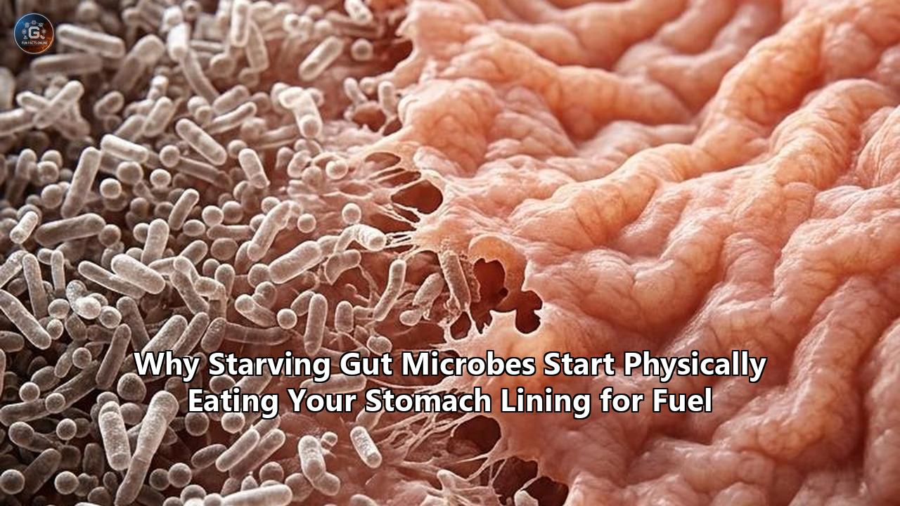
A series of multi-center human crossover trials and spatial metatranscriptomic studies have confirmed what was once observed only in isolated animal models: when dietary fiber is removed from the daily diet, commensal gut bacteria alter their enzymatic production within 24 to 48 hours, physically digesting the host’s protective mucus layer as an alternative food source. Conducted across research institutes including the Luxembourg Institute of Health, the University of Michigan Medical School, and partner clinical centers, these studies reveal that trillions of resident microbes possess a hardwired metabolic fallback switch. When deprived of complex, plant-derived polysaccharides, these microorganisms activate dormant genetic suites that produce specialized carbohydrate-active enzymes capable of dismantling the complex glycoproteins of the host mucosal barrier.
The physical degradation of what is colloquially termed the gut microbiome stomach lining—though anatomically extending through the gastric, small intestinal, and colonic mucosa—has direct clinical ramifications. The mucus gel layer, synthesized primarily by specialized goblet cells, serves as the primary physical and biochemical buffer separating the dense microbial biomass of the digestive lumen from the delicate, single-cell-thick epithelial wall.
The latest multi-omics data indicate that chronic or intermittent fiber starvation thins this protective mucus gel by over 70% in specific colonic segments. This structural breakdown grants commensal and opportunistic microbes direct physical access to the epithelial surface, triggering mucosal inflammation, tissue ulceration, and increased susceptibility to lethal enteric pathogens.
DIETARY FIBER PLENTY (Mutualistic State)
┌────────────────────────────────────────────────────────┐
│ Lumen: Intact Dietary Fiber (Pectins, Inulins, MACs)  │
│ Microbes: Bacteroides, Prevotella fermenting plant MACs│
├────────────────────────────────────────────────────────┤
│ Outer Mucus Layer: Basal turnover (A. muciniphila)    │
│ Inner Mucus Layer: DENSE, STERILE, IMPERMEABLE (~150µm)│
├────────────────────────────────────────────────────────┤
│ Epithelium: Intact tight junctions (Claudin, Occludin) │
│ Immune System: Basal surveillance, low inflammation   │
└────────────────────────────────────────────────────────┘

                         VS.

DIETARY FIBER STARVATION (Erosive State)
┌────────────────────────────────────────────────────────┐
│ Lumen: Depleted of plant glycans (Starvation state)    │
│ Microbes: CAZymes upregulated (Sialidases, Sulfatases) │
├────────────────────────────────────────────────────────┤
│ Outer Mucus Layer: Extensively degraded for O-glycans  │
│ Inner Mucus Layer: THINNED / PERFORATED (< 30µm)       │
├────────────────────────────────────────────────────────┤
│ Epithelium: Direct bacterial contact, TLR4 activation  │
│ Immune System: Cytokine cascade, colitis, leaky gut    │
└────────────────────────────────────────────────────────┘
This discovery has ignited debate among gastroenterologists, biochemists, and nutritional scientists. While some researchers advocate for aggressive dietary interventions centered on whole-food complex carbohydrate matrices, others are racing to commercialize targeted pharmaceutical countermeasures. These include synthetic decoy glycans, bacterial enzyme inhibitors, and engineered mucosal hydrogels designed to shield the intestinal lining without requiring massive changes in human eating behavior.
Comparing these divergent strategies reveals the complex biological trade-offs inherent in trying to control one of the most metabolically flexible ecosystems on the planet.
The Molecular Mechanism: How Starvation Flips the Bacterial Metabolic Switch
To understand why intestinal microbes turn on their host, researchers have mapped the precise genomic architecture of species like Bacteroides thetaiotaomicron, Akkermansia muciniphila, and Ruminococcus torques. The intestinal mucus barrier is largely composed of the polymeric mucin glycoprotein MUC2 (with MUC5AC and MUC6 predominating in the gastric compartment). These mucin backbones are heavily glycosylated, covered in complex carbohydrate branches termed O-glycans, which account for up to 80% of the molecule’s molecular weight.
        MUCIN POLYMER (MUC2 / MUC5AC) GLYCOPROTEIN BACKBONE
                            │
       ┌────────────────────┼────────────────────┐
       ▼                    ▼                    ▼
   [O-Glycan]           [O-Glycan]           [O-Glycan]
       │                    │                    │
 ┌─────┴─────┐        ┌─────┴─────┐        ┌─────┴─────┐
 ▼           ▼        ▼           ▼        ▼           ▼
Sialic     Fucose   Galactose   GlcNAc   GalNAc     Sulfate
 Acid       (Fuc)     (Gal)    (N-acetyl (N-acetyl  Groups
(Neu5Ac)                         glucos-   galactos- (-SO4)
                                 amine)    amine)
       │                    │                    │
       └────────────────────┼────────────────────┘
                            │
               BACTERIAL ENZYMATIC TARGETS
  ┌─────────────────────────┴─────────────────────────┐
  ▼                                                   ▼
Sialidases / Fucosidases (GH33, GH29)         Sulfatases & O-glycanases
[Cleave terminal protective caps]           [Expose and digest protein core]
These O-glycans consist of sugar monomers, including:
- N-acetylglucosamine (GlcNAc)
- N-acetylgalactosamine (GalNAc)
- Galactose
- Fucose
- Terminal sialic acid (N-acetylneuraminic acid)
- Chemically protective sulfate groups
Under normal physiological conditions with adequate dietary fiber—often classified as microbiota-accessible carbohydrates (MACs)—commensal bacteria prioritize plant-based polysaccharides such as arabinoxylans, cellulose, beta-glucans, and pectins. The bacteria utilize dedicated gene clusters known as Polysaccharide Utilization Loci (PULs) and Sus (Starch Utilization System) operons. These systems coordinate the binding, transport, and cleavage of plant carbohydrates into short-chain fatty acids (SCFAs) like acetate, propionate, and butyrate, which supply energy to human colonic cells and maintain immune balance.
                     DIETARY FIBER PRESENT
                               │
                Microbes express plant-targeting
                       PULs & Sus systems
                               │
                               ▼
               Production of short-chain fatty acids
                  (Acetate, Propionate, Butyrate)
                               │
                               ▼
                   Healthy Mucosal Homeostasis


                               VS.


                    DIETARY FIBER STARVATION
                               │
               Repression of plant PULs / Sus systems
                               │
                               ▼
             Upregulation of Mucin-Targeting CAZymes
            (GH20, GH29, GH33, GH95, Mucin Sulfatases)
                               │
                               ▼
               Enzymatic Stripping of Mucin O-Glycans
                               │
                               ▼
              Mucosal Thinning & Epithelial Barrier Loss
When plant fibers are missing from the digestive tract, this metabolic pathway shuts down. Metatranscriptomic profiling shows that fiber starvation triggers the repression of plant-targeting PULs and an immediate upregulation of gene clusters encoding Carbohydrate-Active Enzymes (CAZymes) targeting host glycans. These include:
- Glycoside Hydrolases (GHs): GH33 sialidases that remove terminal sialic acid caps; GH29 and GH95 fucosidases that cleave fucose residues; and GH20, GH84, and GH89 hexosaminidases that break down core GalNAc and GlcNAc bonds.
- Mucin Sulfatases: Specialized enzymes that remove ester-linked sulfate groups, unlocking the heavily protected inner core of the mucin polymer.
- Endo-acting O-glycanases and Mucinases: Enzymes (such as GH16 family members and metalloproteases) that cleave the central protein backbone once the protective sugar shield has been stripped away.
Microbiologists differentiate between two distinct groups of organisms driving this process: mucin specialists and mucin generalists.
Specialists, such as Akkermansia muciniphila and Barnesiella intestinihominis, possess an enzyme repertoire tailored for mucin degradation. They consistently survive on host mucus, even during healthy periods, providing low-level basal turnover.
Generalists, exemplified by Bacteroides thetaiotaomicron, Bacteroides caccae, and Mediterraneibacter gnavus, are opportunistic. They preferentially metabolize dietary fiber when available. When starved of fiber, these generalists dynamically redirect their transcriptional machinery toward host mucin. This sudden, population-wide shift turns billions of generalist microbes into active consumers of the host's physical mucus layer.
Competing Ecological Views: Essential Pruning vs. Pathological Destruction
The realization that gut bacteria feed directly on host biology has divided the scientific community into two main schools of thought regarding the ecological role of mucin degradation.
┌────────────────────────────────────────┬────────────────────────────────────────┐
│     PRUNING HYPOTHESIS (Beneficial)    │    EROSIVE STARVATION (Pathological)   │
├────────────────────────────────────────┼────────────────────────────────────────┤
│ • Controlled, basal mucin degradation  │ • Massive CAZyme upregulation          │
│ • Signals goblet cells to secrete MUC2 │ • Degradation outpaces synthesis rate  │
│ • Cross-feeds beneficial butyrate taxa │ • Eradicates sterile inner mucus layer │
│ • Prevents pathogen colonization       │ • Triggers TLR4-driven inflammatory    │
│ • Maintains dynamic mucosal turnover   │   cascades and epithelial barrier loss │
└────────────────────────────────────────┴────────────────────────────────────────┘
The Controlled Pruning Hypothesis
Proponents of the "Pruning Hypothesis" view mucin consumption not as an inherently destructive act, but as an evolved, mutualistic mechanism necessary for barrier maintenance. In healthy organisms receiving adequate nutrition, basal mucus degradation by specialists like Akkermansia muciniphila acts as a continuous biochemical signal.
When A. muciniphila hydrolyzes terminal sialic acids and fucose residues, it releases free monosaccharides, acetate, and propionate into the mucosal microenvironment. These fermentation products cross-feed secondary symbionts, such as the butyrate-producing Faecalibacterium prausnitzii and Anaerobutyricum hallii.
Butyrate fuels colonic epithelial cells (colonocytes), stimulating mitochondrial oxygen consumption. This maintains the hypoxia needed to preserve mucosal barrier proteins, including Claudin-1, Occludin, and Zonula Occludens-1 (ZO-1).
Furthermore, this controlled, low-level digestion of old, oxidized mucus stimulates neighboring goblet cells to upregulate MUC2 gene transcription and exocytose fresh, unoxidized mucin granules. Researchers supporting this model emphasize that without continuous microbial pruning, the intestinal mucus layer can become stagnant, heavily oxidized, and lose its antimicrobial efficacy.
                         CONTROLLED PRUNING
                                 │
                   Basal Mucin Cleavage (Akkermansia)
                                 │
                                 ▼
                     Acetate / Propionate Release
                                 │
                                 ▼
                   Cross-Feeding of Butyrate Taxa
                    (Faecalibacterium prausnitzii)
                                 │
                                 ▼
                   Colonocyte Oxygen Consumption
                        (Epithelial Hypoxia)
                                 │
                                 ▼
            Upregulated MUC2 Synthesis & Tight Junctions
The Erosive Starvation Model
In contrast, researchers supporting the "Erosive Starvation Model" point out that the equilibrium of the mucus layer depends entirely on the presence of luminal fiber. When fiber is absent, the system shifts from controlled pruning to runaway consumption.
In a typical healthy gut, the colonic mucus consists of two distinct zones:
- The outer mucus layer: A loosely organized, non-sterile matrix heavily colonized by microbes.
- The inner mucus layer: A dense, cross-linked, virtually sterile gel attached directly to the apical membrane of epithelial cells.
Under sustained fiber starvation, the consumption rate of O-glycans exceeds the maximum rate of goblet cell mucin exocytosis. The outer mucus layer is rapidly depleted, and both generalist and specialist bacteria migrate into the dense inner mucus layer.
As the inner layer thins from approximately 150 micrometers to less than 30 micrometers, microbial cell walls make direct contact with the epithelial microvilli.
                         EROSIVE STARVATION
                                 │
                    Luminal Fiber Deprivation
                                 │
                                 ▼
                  Massive Generalist CAZyme Switch
                     (Bacteroides, Ruminococcus)
                                 │
                                 ▼
                   Outer Mucus Rapidly Exhausted
                                 │
                                 ▼
             Microbial Infiltration of Inner Gel Layer
                                 │
                                 ▼
             Direct Epithelial Contact & TLR4 Cleavage
                                 │
                                 ▼
                   Inflammatory Barrier Collapse
This direct contact triggers Toll-Like Receptor 4 (TLR4) and Toll-Like Receptor 5 (TLR5) pathways, triggering the transcription factor NF-κB and releasing inflammatory cytokines, including IL-1β, IL-6, and TNF-α. The resulting inflammation damages epithelial tight junctions, inducing mucosal micro-ulcerations and creating a state of chronic barrier failure.
                     LUMINAL COMPARTMENT (COLON)
────────────────────────────────────────────────────────────────────────
 [Unrestricted Microbial Biomass: Bacteroides, Akkermansia, E. coli]
                           │       │       │
                           ▼       ▼       ▼
       [Pathological Secretion of CAZymes & Sulfatases]
                           │       │       │
                           ▼       ▼       ▼
════════════════════════════════════════════════════════════════════════
 OUTER MUCUS LAYER: ERODED AND LIQUEFIED
════════════════════════════════════════════════════════════════════════
                           │       │       │
                           ▼       ▼       ▼
────────────────────────────────────────────────────────────────────────
 INNER MUCUS LAYER: THINNED FROM ~150µm TO <30µm (PERFORATED)
────────────────────────────────────────────────────────────────────────
                           │       │       │
                           ▼       ▼       ▼
┌──────────────────────────────────────────────────────────────────────┐
│ EPITHELIAL CELL BORDER                                              │
│  [Epithelial Cell]  [Epithelial Cell]  [Epithelial Cell] (Apical)    │
│         │                  │                  │                      │
│         ▼ (TLR4 Activation)▼ (TLR4 Activation)▼ (TLR4 Activation)    │
│  [ Claudin / Occludin Tight Junction Disruption: LEAKY BARRIER ]     │
├──────────────────────────────────────────────────────────────────────┤
│ LAMINA PROPRIA & SYSTEMIC CIRCULATION                                │
│  Translocated LPS & Antigens ──> NF-κB Activation ──> IL-6 / TNF-α   │
└──────────────────────────────────────────────────────────────────────┘
Dietary Approaches: Whole Plants vs. Refined Supplements vs. Restrictive Diets
The dietary methods proposed to prevent microbial mucus degradation reflect fundamentally different philosophies regarding human nutrition, gut transit time, and microbial ecology.
┌────────────────────────────┬────────────────────────────┬────────────────────────────┐
│      WHOLE-FOOD MACs       │    ISOLATED PREBIOTICS     │   LOW-CARB / KETOGENIC     │
├────────────────────────────┼────────────────────────────┼────────────────────────────┤
│ • Intact plant cellular    │ • Purified powders         │ • Minimal carbohydrate     │
│   matrices                 │   (Inulin, FOS, Psyllium)  │   substrate for microbes   │
│ • Fermentation spans the   │ • Rapid proximal           │ • High mucin CAZyme        │
│   entire length of colon   │   fermentation in cecum    │   activation throughout    │
│ • Broad chemical diversity │ • Distal colon remains     │ • Significant thinning of  │
│   saturates varied PULs    │   vulnerable to starvation │   inner mucosal layer      │
│ • Higher volume required;  │ • Simple to standardize;   │ • Potential metabolic gains│
│   can cause mild gas/bloat │   can worsen IBS symptoms  │   at expense of gut barrier│
└────────────────────────────┴────────────────────────────┴────────────────────────────┘
Strategy 1: Complex Whole-Food MACs
The foundational nutritional strategy focuses on maximizing the intake of complex, intact plant matrices containing microbiota-accessible carbohydrates. Unlike simple sugars absorbed in the upper small intestine, complex plant cell walls—composed of intertwined networks of cellulose, hemicellulose, xyloglucans, and pectin—resist host enzymatic digestion.
- The Mechanism: The chemical diversity of natural plant fiber demands a wide variety of microbial enzymes. When a varied selection of root vegetables, legumes, whole grains, and alliums reaches the large intestine, the gut microbiota dedicates its PUL machinery entirely to processing these dietary substrates. This metabolic focus naturally suppresses the transcription of host-mucin-targeting CAZymes.
- The Transit Advantage: Whole-food matrices release their fermentable sugars slowly as the physical plant structure is broken down. This slow release ensures that fermentable carbon reaches the distal colon, the area most prone to ulcerative colitis and pathogen invasion.
- Trade-offs: Achieving protective MAC concentrations requires consuming 40 to 60 grams of diverse fiber daily—a level far above the standard Western intake of 12 to 15 grams. For patients with established inflammatory bowel disease (IBD) or irritable bowel syndrome (IBS), this high-fiber volume can cause abdominal distension, gas, and pain due to rapid fermentation kinetics and mechanical irritation of compromised tissue.
                 WHOLE-FOOD FIBER FERMENTATION PROFILE
Proximal Colon (Cecum)      Transverse Colon           Distal Colon (Sigmoid)
┌──────────────────────┐    ┌──────────────────────┐   ┌──────────────────────┐
│ Inulin & simple      │    │ Pectins & Resistance │   │ Insoluble Celluloses │
│ starches fermented   │───>│ Starches fermented   │──>│ & Lignified MACs     │
│ (Rapid SCFA spike)   │    │ (Sustained SCFA flux)│   │ (Protection to end)  │
└──────────────────────┘    └──────────────────────┘   └──────────────────────┘
 Result: Complete mucosal coverage from cecum to rectum.
Strategy 2: Isolated and Purified Prebiotic Supplements
To avoid the digestive discomfort and compliance challenges of high-volume diets, researchers have evaluated purified prebiotic supplements, such as isolated inulin, fructooligosaccharides (FOS), galactooligosaccharides (GOS), and psyllium husk.
- The Mechanism: These purified, soluble fibers provide concentrated doses of specific chemical linkages, selectively fueling beneficial genera like Bifidobacterium and Lactobacillus.
- The Proximal Trapping Problem: Unlike whole-food plant matrices, isolated soluble fibers (such as pure inulin or FOS) dissolve and ferment quickly upon entering the warm, aqueous environment of the cecum and ascending colon.
- The Trade-off: Metatranscriptomic mapping shows that while purified supplements effectively suppress mucin-degrading enzymes in the proximal colon, the substrate is often completely consumed within the first several inches of the large intestine. This rapid depletion leaves the transverse and descending colon starved of fermentable carbohydrates, causing bacteria in the distal bowel to ramp up mucus consumption. As a result, isolated supplements can leave downstream mucosal zones unprotected unless paired with slower-fermenting, complex substrates.
             ISOLATED PREBIOTIC FERMENTATION PROFILE (PROXIMAL TRAP)
Proximal Colon (Cecum)      Transverse Colon           Distal Colon (Sigmoid)
┌──────────────────────┐    ┌──────────────────────┐   ┌──────────────────────┐
│ 100% Inulin / FOS    │    │ Substrate Exhausted  │   │ Complete Carbohydrate│
│ Fermented Rapidly    │───>│ (Microbial Shift to  │──>│ Starvation (Hyper-   │
│ (High SCFA, Low pH)  │    │ Mucin Glycans Begins)│   │ degradation of MUC2) │
└──────────────────────┘    └──────────────────────┘   └──────────────────────┘
 Result: Distal colon remains unprotected; inner mucus barrier thins.
Strategy 3: Low-Carbohydrate, Ketogenic, and Carnivore Diets
The popularity of low-carbohydrate, ketogenic, and ultra-low-residue carnivore diets has created an unintentional physiological scenario: prolonged, total dietary fiber deprivation in the colon.
- The Trade-off: These dietary patterns can achieve short-term metabolic goals, such as improved glycemic control, reduced hepatic steatosis, and lower circulating triglycerides. However, they almost completely eliminate microbiota-accessible carbohydrates from entering the large bowel.
- Mucosal Impact: In the sustained absence of dietary carbohydrates, the gut microbiome adapts by shifting its metabolic machinery entirely to host-derived proteins and glycoproteins. Studies examining long-term fiber elimination show an increased abundance of mucin-degrading taxa, elevated levels of bacterial sulfatases, and a marked physical thinning of the colonic mucosal barrier. While elevated systemic ketone bodies can provide anti-inflammatory signals, the physical breakdown of the protective barrier increases vulnerability to bacterial translocation, endotoxemia, and acute enteric infections.
Biotechnology Interventions: Synthetic Glycan Decoys vs. CAZyme Inhibitors vs. Hydrogels
Because changing long-term human dietary habits can be challenging, biomedical researchers are developing targeted therapeutic technologies to prevent microbial degradation of the mucosal barrier.
┌─────────────────────────────────────────────────────────────────────────────┐
│                 BIOTECHNOLOGY PLATFORMS AT A GLANCE                         │
├────────────────────────────┬────────────────────────────┬───────────────────┤
│ PLATFORM                   │ PRIMARY MECHANISM          │ CORE CHALLENGE    │
├────────────────────────────┼────────────────────────────┼───────────────────┤
│ Synthetic Decoy Glycans    │ Diverts bacterial CAZymes  │ High synthesis    │
│ (Mucin Mimetics)           │ away from endogenous mucus │ cost; delivery    │
├────────────────────────────┼────────────────────────────┼───────────────────┤
│ Selective CAZyme &         │ Blocks catalytic sites     │ Risk of arresting │
│ Sulfatase Inhibitors       │ of sialidases & sulfatases │ necessary turnover│
├────────────────────────────┼────────────────────────────┼───────────────────┤
│ Engineered Mucosal         │ Mechanically shields       │ Transient mucosal │
│ Hydrogel Formulations      │ bare epithelial cells      │ clearance/erosion │
└────────────────────────────┴────────────────────────────┴───────────────────┘
Approach 1: Synthetic Mucin-Mimetic Decoy Glycans
One of the most active areas of therapeutic development involves synthesizing non-digestible, highly branched oligosaccharides that mirror the molecular structure of host O-glycans.
SYNTHETIC DECOY MECHANISM
[Bacterial CAZymes: Sialidases/Sulfatases]
         │
         ├─── EXOGENOUS DECOY GLYCAN (High Affinity Sponge) ──> [Enzyme Bound & Neutralized]
         │
└─── HOST MUCIN O-GLYCAN (Protected from enzymatic cleavage)- Mechanism: These synthetic structures are decorated with terminal sialic acid and sulfate groups, creating a "decoy sponge". When ingested, they pass unabsorbed through the stomach and small intestine into the colon, where they bind tightly to bacterial CAZyme catalytic sites. By offering a sacrificial glycan source, these compounds satisfy the enzymatic requirements of generalist bacteria like Bacteroides thetaiotaomicron, preserving the host's actual mucus layer.
- Evaluation: This approach avoids the gas, bloating, and fermentation spikes associated with large doses of prebiotic fiber. However, chemical synthesis of complex O-glycans with exact stereochemical arrangements remains costly and difficult to produce at commercial scale.
Approach 2: Small-Molecule Bacterial CAZyme and Sulfatase Inhibitors
Rather than providing decoy nutrients, pharmaceutical chemists are designing targeted small-molecule enzyme inhibitors that selectively block bacterial mucin-degrading enzymes without killing the bacteria themselves.
- Mechanism: By developing selective inhibitors against specific bacterial enzymes—such as the GH33 sialidases produced by Ruminococcus torques or specific colonic mucin sulfatases—these drugs block the removal of terminal protective sugars from the host mucus backbone. Without the ability to clip these initial bonds, downstream mucinases cannot access the core protein structure.
- Trade-offs: This strategy carries ecological risks. Completely halting mucin degradation can disrupt the baseline shedding and renewal of the mucosal barrier. Stagnant, non-degraded mucus can accumulate oxidized cellular debris and shed toxins, potentially impairing nutrient absorption and altering the natural balance of the microbiome. Additionally, designing an inhibitor specific to bacterial enzymes that leaves human epithelial glycosidases unaffected presents a significant molecular challenge.
       SELECTIVE CAZyme INHIBITION VS. NATURAL PATHWAY
Natural Breakdown Pathway:
[Host Mucin Layer] ──> (Bacterial Sialidase/Sulfatase) ──> [Stripped Core] ──> [Mucus Thinning]

Inhibitor-Blocked Pathway:
[Host Mucin Layer] ──> [ Small-Molecule Inhibitor ] ──X (Bacterial Sialidase Blocked)
                             │
                             ▼
              [Host Mucin Remains Structurally Intact]
Approach 3: Engineered Mucosal Hydrogels and Mucus Secretagogues
A third strategy bypasses microbial metabolism entirely, focusing on physically reinforcing the intestinal barrier.
- Mechanism: Biomaterial engineers have formulated cross-linked biopolymer hydrogels (using modified alginates, chitosan conjugates, and recombinant Trefoil Factor 3) that are delivered via pH-sensitive capsules directly to the distal gut. Upon release, these materials adhere to bare or inflamed epithelial cells, creating an artificial, bio-inert layer that blocks bacterial encroachment while native goblet cells regenerate the natural mucus lining.
- Trade-offs: These physical hydrogels offer direct protection against inflammation, making them promising candidates for active inflammatory bowel disease flares. However, their therapeutic effect is temporary. Gastrointestinal motility, peristalsis, and continuous cell turnover clear these hydrogels within 12 to 24 hours, requiring repeated dosing to maintain protection unless accompanied by dietary or microbial interventions.
Microbial Engineering: Live Biotherapeutics vs. Wild-Type Strain Manipulation
To establish long-term protection against mucus degradation, synthetic biologists are investigating ways to engineer the gut ecosystem directly.
┌────────────────────────────────────────┬────────────────────────────────────────┐
│     CRISPR-ENGINEERED BACTERIA (LBPs)  │     DEFINED MICROBIAL CONSORTIA        │
├────────────────────────────────────────┼────────────────────────────────────────┤
│ • Commensals with deleted mucin CAZymes│ • Multi-strain ecological networks     │
│ • Engineered to release trophic factors│ • Tightly coupled trophic chains       │
│   (IL-22, GLP-2 mimetics) in situ      │ • Eliminate reliance on host mucin via │
│ • Precise mechanism of action          │   efficient metabolite cross-feeding   │
│ • Regulatory hurdles & colonization    │ • Variable engraftment efficiency      │
│   resistance in complex native systems │   among diverse host genotypes         │
└────────────────────────────────────────┴────────────────────────────────────────┘
Live Biotherapeutic Products (LBPs) with Deleted CAZyme Suites
Researchers are using CRISPR-Cas systems to create gene-edited versions of common human gut commensals, such as Bacteroides ovatus or Bacteroides thetaiotaomicron, that lack the genetic operons for mucin degradation.
- How it Works: These engineered strains retain the ability to digest plant-based fibers and produce beneficial SCFAs, but are genetically incapable of switching to host O-glycans during periods of fiber starvation. Some versions are further engineered to continuously express mucosal repair peptides, such as Interleukin-22 (IL-22) or Glucagon-Like Peptide-2 (GLP-2) mimetics, directly at the epithelial boundary.
- The Ecological Barrier: The primary challenge with engineered single-strain probiotics is colonization resistance. The native gut microbiome forms a dense, highly competitive ecosystem. Introducing an engineered bacterium that has voluntarily surrendered a vital survival pathway (mucin foraging) places it at a fitness disadvantage compared to wild-type competitors during periods of dietary restriction, often leading to the strain being outcompeted and cleared from the gut.
           ECOLOGICAL COMPETITION IN FIBER-STARVED CONDITIONS
Native Wild-Type Generalist:
[Fiber Absent] ──> Upregulates CAZymes ──> Consumes Host Mucus ──> [Survives / Proliferates]

Engineered LBP (CAZyme-Deleted):
[Fiber Absent] ──> Cannot Cleave Mucus ──> Energy Starvation ──> [Outcompeted / Cleared]
Defined Multi-Species Microbial Consortia
As an alternative to single engineered strains, other researchers are developing multi-species ecological consortia. These communities are assembled using wild-type bacterial strains selected for their mutual metabolic dependencies.
- How it Works: In these consortia, primary fiber degraders break down complex carbohydrates into intermediate metabolites (such as lactate, acetate, and succinate). Secondary cross-feeders rapidly consume these intermediates to produce butyrate, creating a self-sustaining metabolic network.
- Evaluation: By building an efficient, cooperative metabolic web, these consortia reduce the selective pressure that drives individual generalist strains to consume host mucin. However, the success of this approach depends heavily on host-specific factors, including bile acid composition, intestinal transit time, and individual variations in host mucin glycosylation patterns.
Regional Dynamics: Gastric vs. Colonic Environments
The term gut microbiome stomach lining is frequently used in popular health discussions as a broad concept, but human digestive physiology varies markedly across different segments of the gastrointestinal tract.
The chemical and physical dynamics governing microbial mucus degradation change significantly as you move from the stomach to the distal colon.
GASTRIC MUCOSA (Stomach)
┌────────────────────────────────────────────────────────┐
│ • Environment: pH 1.5–3.5 (Extreme acid)               │
│ • Dominant Mucins: MUC5AC and MUC6                     │
│ • Biomass: 10¹–10³ CFU/g (Very Low)                    │
│ • Primary Degraders: Helicobacter pylori (Pathological)│
│ • Mechanism: Urease neutralization & mucinase cleavage │
└────────────────────────────────────────────────────────┘
                           │
                           ▼
SMALL INTESTINAL MUCOSA (Duodenum / Jejunum / Ileum)
┌────────────────────────────────────────────────────────┐
│ • Environment: Rapid transit, high antimicrobial load  │
│ • Dominant Mucins: Single unattached loose layer       │
│ • Biomass: 10³–10⁷ CFU/g (Moderate)                    │
│ • Antimicrobials: High RegIIIγ, Defensins, Secretory   │
│   IgA; bacterial-epithelial contact tightly restricted │
└────────────────────────────────────────────────────────┘
                           │
                           ▼
COLONIC MUCOSA (Cecum / Colon / Rectum)
┌────────────────────────────────────────────────────────┐
│ • Environment: Anaerobic, slow transit, neutral pH     │
│ • Dominant Mucins: MUC2 (Dense inner & loose outer)    │
│ • Biomass: 10¹¹–10¹² CFU/g (Massive)                   │
│ • Primary Degraders: Akkermansia, Bacteroides,         │
│   Ruminococcus species                                 │
│ • Mechanism: CAZyme/sulfatase-driven starvation foraging│
└────────────────────────────────────────────────────────┘
The Gastric Mucosa
The human stomach maintains an acidic environment (pH 1.5 to 3.5) designed to sterilize ingested food and initiate protein digestion. The gastric mucosa relies on specialized, surface-associated mucins—primarily MUC5AC secreted by superficial foveolar cells and MUC6 secreted by deeper glandular neck cells.
Because of the extreme acidity, the microbial population here is small (10¹ to 10³ cells per gram). Dietary fiber starvation does not cause general commensal foraging in the stomach because widespread microbial communities cannot survive there.
Instead, gastric mucosal degradation is driven by specific acid-tolerant organisms, particularly Helicobacter pylori. H. pylori utilizes the enzyme urease to generate an alkaline ammonia cloud around itself, while deploying specialized flagella and outer-membrane adhesins (BabA and SabA) that bind directly to Lewis b blood-group antigens on gastric mucin chains.
Once anchored, it secretes mucin-degrading proteases and phospholipases that break down the gastric mucus gel, exposing underlying parietal and chief cells to gastric hydrochloric acid and pepsin, leading to peptic ulcers.
       HELICOBACTER PYLORI GASTRIC MUCOSAL INVASION
1. Acid Neutralization:
   H. pylori ──> Urease enzyme: Urea + H2O ──> 2 NH3 + CO2 (Alkaline Cloud)

2. Mucin Attachment:
   BabA / SabA Adhesins ──> Binds to Lewis(b) Antigens on MUC5AC / MUC6

3. Enzymatic Digestion:
   Secreted Proteases & Phospholipases ──> Hydrolyzes Gel Structure

4. Acid Penetration:
   Luminal Gastric Acid (pH 2.0) ──> Breaches Damaged Mucosa ──> Peptic Ulceration
The Colonic Mucosa
In contrast, the large intestine contains the vast majority of the human microbiome (up to 10¹² cells per gram) operating in an anaerobic, neutral-to-mildly-acidic environment (pH 5.7 to 6.8) with slow transit times.
Here, the mucosal barrier is built on the large polymeric protein MUC2. It forms a two-layer defense system:
- An outer, loosely organized layer that houses billions of bacteria.
- An inner, dense, and sterile gel layer that physically blocks bacteria from reaching the underlying tissue.
Because of the massive bacterial population and its total reliance on dietary intake for fuel, the colonic mucosa is where dietary fiber starvation leads to rapid, widespread degradation of host mucus.
┌──────────────────────────────┬──────────────────────────────┬──────────────────────────────┐
│ CHARACTERISTIC               │ GASTRIC ENVIRONMENT         │ COLONIC ENVIRONMENT          │
├──────────────────────────────┼──────────────────────────────┼──────────────────────────────┤
│ Dominant Mucin Proteins      │ MUC5AC and MUC6              │ MUC2 (Polymerized network)   │
├──────────────────────────────┼──────────────────────────────┼──────────────────────────────┤
│ Microbial Density            │ 10¹ to 10³ CFU/g             │ 10¹¹ to 10¹² CFU/g           │
├──────────────────────────────┼──────────────────────────────┼──────────────────────────────┤
│ Primary Degradation Driver   │ Pathogen-specific enzymes    │ Broad microbial CAZyme switch│
│                              │ (Helicobacter pylori urease) │ driven by fiber starvation   │
├──────────────────────────────┼──────────────────────────────┼──────────────────────────────┤
│ Barrier Structure            │ Single, adherent mucus gel   │ Dual-layer: loose colonized  │
│                              │ protecting against acid      │ outer + dense sterile inner  │
├──────────────────────────────┼──────────────────────────────┼──────────────────────────────┤
│ Clinical Consequences of     │ Peptic ulcer disease,        │ Ulcerative colitis, systemic │
│ Barrier Failure              │ chronic gastritis, gastric CA│ endotoxemia, colorectal CA   │
└──────────────────────────────┴──────────────────────────────┴──────────────────────────────┘
Clinical Consequences: From Epithelial Encroachment to Systemic Disease
When the protective mucus barrier is degraded by starving microbes, the clinical impact extends far beyond local digestive discomfort. The loss of this barrier can trigger several progressive systemic pathologies.
               PATHOLOGICAL CASCADE OF MUCUS DEPLETION
                        Chronic Fiber Starvation
                                   │
                                   ▼
                   Microbial Mucin O-Glycan Digestion
                                   │
                                   ▼
                 Thinning & Perforation of Inner MUC2
                                   │
         ┌─────────────────────────┴─────────────────────────┐
         ▼                                                   ▼
 Direct Epithelial Access                            Epithelial Penetration
         │                                                   │
         ▼                                                   ▼
 TLR4 / NF-κB Pathway                             Tight Junction Disruption
         │                                                   │
         ▼                                                   ▼
 Local Chronic Inflammation                        Systemic Lipopolysaccharide
 (Colitis / Crohn's Flares)                       Translocation (Metabolic Leaky Gut)
         │                                                   │
         ▼                                                   ▼
 Epithelial DNA Damage / AOM                         Hepatic & Vascular Inflamm.
 (Colorectal Malignancy)                             (MASH / Atherosclerosis)
1. Inflammatory Bowel Disease (IBD) Flares
In both ulcerative colitis and Crohn’s disease, clinical biopsies routinely reveal a thinned or absent inner mucus layer accompanied by bacteria making direct contact with epithelial cells.
Multi-omics analyses of human stool and tissue samples show elevated expression of bacterial sialidases and sulfatases during active inflammatory flares.
When the inner mucus layer is breached, structural proteins like E-cadherin, Claudin-1, and Occludin are degraded by bacterial proteases. This breakdown permits continuous antigenic stimulation of the underlying lamina propria, causing sustained immune activation and tissue damage.
ULCERATIVE COLITIS CORRELATION
Healthy Mucosa:
[Thick MUC2 Layer] ──> [Shields Epithelial Cells] ──> [Immune Homeostasis]

Ulcerative Colitis Flare:
[Starved / Dysbiotic Flora] ──> [Degrades MUC2] ──> [Direct Epithelial Contact] ──> [Immune Infiltration & Ulceration]
2. Metabolic Endotoxemia and Low-Grade Systemic Inflammation
Mucosal barrier degradation allows Gram-negative bacterial fragments—particularly lipopolysaccharide (LPS), a potent endotoxin found in bacterial outer membranes—to leak across the intestinal lining into the portal bloodstream.
Once in systemic circulation, LPS binds to CD14 and TLR4 receptors on circulating macrophages, hepatic Kupffer cells, and vascular endothelial cells. This triggers low-grade, chronic systemic inflammation, which contributes to:
- Peripheral insulin resistance
- Non-Alcoholic Steatohepatitis (NASH/MASH)
- Vascular inflammation and accelerated atherosclerosis
                 METABOLIC ENDOTOXEMIA PATHWAY
 [Eroded Colonic Mucus Barrier]
              │
              ▼
 [LPS Translocation Across Epithelium]
              │
              ▼
 [Entry into Portal Circulation]
              │
              ▼
 [Binding to Hepatic Kupffer Cell TLR4]
              │
              ▼
 [Systemic Release of TNF-α, IL-6, and MCP-1]
              │
              ▼
 [Systemic Insulin Resistance & Hepatic Steatosis]
3. Heightened Vulnerability to Enteric Pathogens
A structurally intact mucus layer serves as the host's primary physical and biochemical line of defense against incoming pathogens. It traps pathogens and continuously flushes them out of the digestive tract through normal peristalsis.
When commensal bacteria consume the host's O-glycans during fiber starvation, this physical barrier is lost.
In vivo infectivity studies demonstrate that mice on a fiber-free diet experience rapid colonization by mucosal pathogens such as Citrobacter rodentium (an animal model for human enteropathogenic E. coli), leading to severe, fatal colitis from bacterial doses that a well-fed animal's mucosal barrier easily repels.
Pathogen Resistance in Fiber-Fed Host:
[Pathogen: E. coli / C. rodentium] ──> [Trapped in Thick Outer Mucus] ──> [Peristaltic Clearance]

Pathogen Susceptibility in Fiber-Starved Host:
[Pathogen: E. coli / C. rodentium] ──> [Reaches Bare Epithelium] ──> [Type III Secretion & Lethal Colitis]
4. Colorectal Carcinogenesis
Long-term epidemiological studies have linked low-fiber diets with an increased incidence of colorectal cancer.
Beyond the absence of protective butyrate, the physical degradation of the mucus barrier plays a direct mechanical role. When the inner mucus layer is eroded, bacteria expressing the pks genomic island (such as pks+ Escherichia coli) can attach directly to colonic stem cells located in the crypts of Lieberkühn.
These bacteria produce colibactin, an alkylating genotoxin that induces double-stranded DNA breaks and distinct mutational signatures in human epithelial cells, accelerating the development of adenomas and malignant tumors.
                    COLORECTAL ONCOGENESIS CASCADE
[Mucus Layer Depleted via Fiber Starvation]
              │
              ▼
[Direct Access to Crypt Stem Cells by pks+ E. coli]
              │
              ▼
[Colibactin Genotoxin Secretion onto Apical Membranes]
              │
              ▼
[Double-Stranded DNA Cleavage in Epithelial Stem Cells]
              │
              ▼
[Accumulation of Oncogenic Mutations (APC, KRAS, TP53)]
              │
              ▼
[Adenoma Formation and Malignant Transformation]
Comparative Assessment of Mucosal Defense Strategies
The following analysis compares the primary dietary, pharmacological, and biotechnological methods currently used or in development to protect the mucosal barrier against microbial degradation.
┌────────────────────────────────────────────────────────────────────────────────────────────────────────────────────────┐
│                                COMPREHENSIVE STRATEGY COMPARISON MATRIX                                                │
├──────────────────────┬───────────────────────────────┬───────────────────────────────┬─────────────────────────────────┤
│ INTERVENTION         │ MECHANISTIC PRINCIPLE         │ PRIMARY ADVANTAGES            │ KEY TRADEOFFS & LIMITATIONS     │
├──────────────────────┼───────────────────────────────┼───────────────────────────────┼─────────────────────────────────┤
│ Whole-Food MACs      │ Diverse plant structures      │ Sustained protection through  │ Requires high volume (40-60g);  │
│ (Dietary Matrix)     │ engage multiple microbial     │ entire colon; natural SCFA    │ can cause gas, distension, and  │
│                      │ PULs along the entire bowel   │ generation; cheap & scalable  │ poor compliance in active IBD   │
├──────────────────────┼───────────────────────────────┼───────────────────────────────┼─────────────────────────────────┤
│ Purified Prebiotics  │ Concentrated soluble single-  │ Simple to dose; selective     │ Rapid proximal fermentation     │
│ (Inulin, FOS, GOS)   │ linkage sugars (e.g., inulin) │ stimulation of Bifidobacteria │ leaves the distal colon starved │
│                      │ target specific taxa          │ without complex meal prep     │ and vulnerable to degradation   │
├──────────────────────┼───────────────────────────────┼───────────────────────────────┼─────────────────────────────────┤
│ Synthetic Decoy      │ Non-digestible engineered     │ Highly targeted; neutralizes  │ High chemical synthesis costs;  │
│ Glycans              │ oligosaccharides mimic host   │ mucin CAZymes without dietary │ lacks the metabolic cross-      │
│                      │ O-glycans to soak up enzymes  │ changes or large fiber volume │ feeding benefits of whole plants│
├──────────────────────┼───────────────────────────────┼───────────────────────────────┼─────────────────────────────────┤
│ Small-Molecule       │ Chemically inhibits active    │ Directly halts enzymatic      │ Risk of stopping normal mucus   │
│ CAZyme Inhibitors    │ sites of bacterial sulfatases │ degradation; drug-like dosing │ turnover; potential accumulation│
│                      │ and GH33 sialidases           │ suitable for active flares    │ of aged, oxidized mucus layers  │
├──────────────────────┼───────────────────────────────┼───────────────────────────────┼─────────────────────────────────┤
│ Engineered Mucosal   │ Cross-linked biopolymers      │ Immediate physical protection │ Transient residence time; wiped │
│ Hydrogels (Topical)  │ mechanically coat bare or     │ against bacterial contact;    │ out by peristalsis and cell     │
│                      │ ulcerated epithelial tissue   │ excellent for acute ulcers    │ turnover within 12 to 24 hours  │
├──────────────────────┼───────────────────────────────┼───────────────────────────────┼─────────────────────────────────┤
│ Gene-Edited Live     │ Commensals engineered to lack │ Permanent niche colonization; │ Low fitness in native microbial │
│ Biotherapeutics      │ mucin-degrading PUL suites;   │ constant SCFA production      │ ecosystems; outcompeted during  │
│                      │ secrete mucosal repair cues   │ without host mucus foraging   │ periods of dietary restriction  │
└──────────────────────┴───────────────────────────────┴───────────────────────────────┴─────────────────────────────────┤
1. Delivery Dynamics and Spatial Coverage
When comparing whole-food MACs to purified supplements, the most striking difference is where fermentation occurs within the colon.
Whole plant matrices—which combine insoluble cellulose with complex pectins and xylans—act as slow-release carriers. Because the plant cell walls must be broken down layer by layer, fermentable carbohydrates are released along the entire length of the large intestine, protecting the vulnerable distal colon.
In contrast, purified soluble prebiotics like inulin dissolve and ferment rapidly in the cecum. This proximal fermentation creates an unequal environment: the ascending colon receives an abundance of fuel, while the distal descending colon remains starved of carbohydrates, forcing downstream bacteria to feed on host mucin.
SPATIAL FERMENTATION PROFILES ACROSS INTESTINAL SEGMENTS
               Ascending Colon      Transverse Colon      Descending Colon
Whole-Food MACs:    [ HIGH ]              [ HIGH ]              [ HIGH ]
Purified Inulin:    [ MAXIMUM ]           [ LOW ]               [ EXHAUSTED ]
Synthetic Decoys:   [ CONTROLLED ]        [ CONTROLLED ]        [ CONTROLLED ]
2. Biological Trade-offs: Natural Diets vs. Targeted Pharmaceuticals
The comparison between dietary adjustments and pharmacological interventions reveals differing philosophies of care.
Dietary strategies using whole-food MACs are cost-effective, widely accessible, and provide diverse nutritional benefits, including the generation of health-promoting short-chain fatty acids. However, they require sustained patient adherence and can cause digestive symptoms—such as gas, bloating, and cramping—in individuals with existing gastrointestinal conditions or altered gut transit times.
Pharmaceutical approaches (such as small-molecule enzyme inhibitors and synthetic decoy glycans) offer targeted precision. They can rapidly stop bacterial degradation of the mucus layer during acute disease flares without requiring significant changes in patient diet.
Yet, these synthetic approaches carry distinct trade-offs: they can be expensive to produce, they do not provide the broad metabolic benefits of natural fiber fermentation (such as butyrate production), and completely blocking microbial enzymes risks halting the natural, healthy turnover of the gut's protective lining.
                             DECISION MATRIX
┌───────────────────────────────────────┬───────────────────────────────────────┐
│       WHOLE-FOOD INTERVENTIONS        │     PHARMACEUTICAL INTERVENTIONS      │
├───────────────────────────────────────┼───────────────────────────────────────┤
│ + High SCFA / Butyrate generation     │ + Immediate, targeted enzymatic block │
│ + Inexpensive and broadly accessible  │ + Independent of patient diet changes │
│ + Promotes broad microbial diversity  │ + Avoids fiber-induced gas & bloating │
│ - High volume required (40-60g daily) │ - High cost of drug development       │
│ - Poor compliance in modern lifestyles│ - Does not generate beneficial SCFAs  │
│ - Can trigger pain in active IBD      │ - Potential risks of stagnant mucus   │
└───────────────────────────────────────┴───────────────────────────────────────┘
Future Directions: Diagnostics, Personalization, and Clinical Milestones
As research into microbial mucin foraging continues to advance, several key clinical milestones and diagnostic technologies are emerging.
                         THE ROAD AHEAD
 ┌─────────────────────────────────────────────────────────┐
 │ 1. Non-Invasive Stool Glycan & Sialidase Biomarkers     │
 │    Mapping active CAZyme activity in human patients     │
 └────────────────────────────┬────────────────────────────┘
                              │
                              ▼
 ┌─────────────────────────────────────────────────────────┐
 │ 2. Host Secretor Phenotyping (FUT2 Genotype Screening) │
 │    Personalizing fiber requirements by mucosal genetics │
 └────────────────────────────┬────────────────────────────┘
                              │
                              ▼
 ┌─────────────────────────────────────────────────────────┐
 │ 3. Phase II/III Clinical Trials on Synthetic Decoys     │
 │    Validating engineered glycans in human IBD cohorts   │
 └─────────────────────────────────────────────────────────┘
1. Non-Invasive Stool Glycan Diagnostics
Current clinical practice lacks direct tools to measure whether a patient's gut microbiota is actively consuming their intestinal lining.
To address this gap, diagnostic laboratories are developing non-invasive stool assays that measure free cleaved mucin fragments, such as:
- Free N-acetylneuraminic acid (Neu5Ac)
- Specific O-glycan core structures (Core 1 through Core 4)
- Quantitative activity of fecal bacterial sulfatases and sialidases
These biomarkers aim to give gastroenterologists a direct readout of mucus barrier degradation before physical tissue damage and clinical symptoms appear.
PATIENT STOOL SAMPLE
         │
         ├─── Rapid Fluorometric Sialidase Assay ───> [High = Active Mucus Digestion]
         │
         └─── Core O-Glycan LC-MS Quantitation ──────> [Elevated Free Core 3 = Barrier Loss]
2. Personalized Mucosal Nutrition and Host Genetics
A major focus of ongoing research is understanding how host genetics influence bacterial mucus foraging. Human mucin glycosylation patterns are heavily shaped by host genes, particularly the FUT2 gene, which encodes the galactoside 2-alpha-L-fucosyltransferase 2 enzyme.
Individuals who carry non-functional FUT2 alleles—known as "non-secretors," representing roughly 20% of the global population—do not display ABO blood-group antigens and terminal fucose residues on their mucosal surfaces.
Because non-secretors produce a structurally distinct mucosal gel, their resident microbiota faces different selective pressures during fiber deprivation.
Emerging clinical protocols aim to integrate FUT2 genetic testing into dietary planning, tailoring specific fiber structures and prebiotic combinations to match each patient's individual mucosal biochemistry.
                   FUT2 GENETIC STRATIFICATION
                         Patient Genotype
                                │
         ┌──────────────────────┴──────────────────────┐
         ▼                                             ▼
  [FUT2 Secretor (80%)]                         [FUT2 Non-Secretor (20%)]
  • Mucus contains terminal fucose              • Mucus lacks terminal fucose
  • Diverse mucin-degrading flora               • Altered microbial flora
  • Standard complex MAC intervention           • Targeted sulfated glycans required
3. Upcoming Clinical Milestones
Over the next 24 to 36 months, several clinical trials will report outcomes testing these competing mucosal protection strategies:
- Human Synthetic Glycan Trials: Phase I/II human trials evaluating the safety and efficacy of synthetic decoy oligosaccharides in patients with quiescent ulcerative colitis to prevent disease relapses.
- Spatial Transcriptomic Mapping: Clinical trials using ingestible, location-specific sampling capsules to map the activation of bacterial CAZymes in real time across the human digestive tract during defined dietary shifts.
- Consortium-Based Therapeutics: Ongoing trials examining defined, multi-strain bacterial consortia designed to restore mucosal cross-feeding pathways and suppress mucin degradation in patients dependent on low-fiber enteral nutrition.
Understanding the mechanisms that drive gut microbes to feed on host mucus has fundamentally changed how researchers view the mucosal barrier. Rather than an inert, static lining, the mucosal interface is a dynamic, continuously contested boundary.
Whether through the deliberate consumption of complex whole-plant matrices, the deployment of engineered synthetic decoy sugars, or the use of targeted enzyme inhibitors, protecting this physical barrier remains one of the most critical frontiers in modern gastroenterology and human health.
Reference:
- https://www.michiganmedicine.org/health-lab/gut-bacteria-eat-colon-lining-when-starved-fiber
- https://pubmed.ncbi.nlm.nih.gov/27863247/
- https://microbiomepost.com/dietary-fiber-intake-shapes-gut-microbiota-activity-and-mucin-degradation-insights-from-a-human-dietary-intervention-study/
- https://pmc.ncbi.nlm.nih.gov/articles/PMC10154618/
- https://www.biorxiv.org/content/10.64898/2026.03.26.714468v1.full
- https://pubmed.ncbi.nlm.nih.gov/30792861/
- https://journals.asm.org/doi/10.1128/iai.00503-24
- https://www.tandfonline.com/doi/full/10.1080/19490976.2026.2614054
- https://pmc.ncbi.nlm.nih.gov/articles/PMC12721379/
- https://pmc.ncbi.nlm.nih.gov/articles/PMC5131798/
- https://www.mdpi.com/2072-6643/17/1/138
- https://www.ericmartenslab.org/project-1
- https://www.sciencedaily.com/releases/2026/08/260814235842.htm
- https://pubmed.ncbi.nlm.nih.gov/39796573/