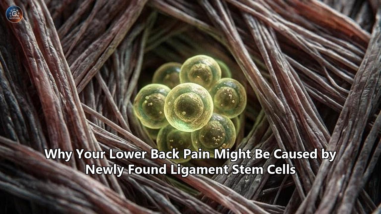
NEW YORK — Orthopedic surgeons and molecular biologists at Weill Cornell Medicine and Hospital for Special Surgery (HSS) have isolated the master stem cell responsible for creating and maintaining human tendons and ligaments. In doing so, they have exposed an unexpected biological culprit behind one of the most intractable orthopedic conditions: lumbar spinal stenosis.
Published in the journal Cell, the multi-year study reveals that lumbar spinal stenosis—a narrowing of the lower spinal canal that chokes passing nerves—is not merely the consequence of passive mechanical wear and aging. Instead, the condition is actively driven by a newly identified population of postnatal tendon and ligament stem cells (TLSCs) that malfunction, multiply excessively, and drive runaway tissue expansion inside the spinal column.
By analyzing human spinal ligament samples alongside genetically mapped animal models, the investigative team discovered that these progenitor cells become hyperactive through dysregulated intracellular calcium signaling. When that chemical signaling pathway surges, the cells churn out dense, aberrant fibrocartilaginous tissue. When researchers therapeutically blunted the same calcium cascade in the laboratory, the pathological ligament growth ceased entirely.
The discovery destabilizes long-held assumptions regarding the primary causes of lower back pain, challenging decades of clinical reliance on late-stage surgical bone removal and highlighting an avenue for early pharmacological intervention.
“Identifying these specialized stem cells unlocks a new area of research that allows us to address this disease much more mechanistically, rather than just waiting until a patient’s condition worsens and requires surgery to relieve the nerve compression,” said study co-corresponding author Dr. Sravisht Iyer, an associate professor of orthopedic surgery at Weill Cornell Medicine and an attending spine surgeon at HSS. “The findings are exciting for their potential to change the way we deliver spinal care.”
Dr. Matthew Greenblatt, co-corresponding author and associate professor of pathology and laboratory medicine at Weill Cornell Medicine, pointed out that the findings solve a decades-old anatomical riddle. “While previous studies had proposed several candidate stem cells, none had definitively shown that a single cell population could both self-renew and generate the full spectrum of tendon and ligament cell types,” Dr. Greenblatt said. “Though spinal stenosis is a complex condition, this really showed us that these cells are contributing to the pathology.”
The Clinical Challenge: Beyond the "Wear-and-Tear" Dogma
Lumbar spinal stenosis affects an estimated 103 million people globally, making it the leading indication for spinal surgery among adults aged 65 and older. For generations, textbook orthodoxy held that spinal stenosis was an unavoidable byproduct of mechanical senescence. The prevailing model assumed that as intervertebral discs desiccate and collapse with age, biomechanical loads shift onto adjacent facet joints and spinal ligaments, triggering a blunt, passive reaction characterized by friction, chronic inflammation, and scar tissue accumulation.
Under that model, the ligamentum flavum—a specialized band of elastic connective tissue that runs inside the posterior wall of the vertebral canal—gradually thickens, stiffens, and buckles inward. The encroaching tissue reduces the cross-sectional area of the central spinal canal and neural foramina, compressing the traversing nerve roots of the cauda equina.
Patients manifest symptoms through neurogenic claudication: a searing, aching, or cramping sensation in the lower back, buttocks, and thighs that intensifies during upright walking and standing, driving many to lean forward over shopping carts or walking frames to physically expand the canal and find transient relief.
Yet, the traditional model contained gaping clinical contradictions. If spinal stenosis were purely a mechanical consequence of disc collapse and vertebral friction:
- Why do many individuals with severe disc degeneration and high physical workloads never develop hypertrophic ligaments?
- Why do others with minimal skeletal alignment defects experience aggressive, disproportionate ligament expansion?
- Why does the hypertrophied ligamentum flavum display histologic features of hypercellularity and neo-vascularization, rather than the cell-sparse, acellular architecture of mature scar tissue?
The research spearheaded by Dr. Greenblatt, Dr. Iyer, and first author Dr. Lingling Hu answers these questions by reframing the condition as an active, stem-cell-driven proliferative disorder. The thickening of the ligament is not simple passive swelling or callous scar formation; it is an organized, abnormal cellular remodeling process fueled by an unmasked progenitor population.
This shifts the clinical understanding of the causes of lower back pain. While mechanical loading acts as an environmental catalyst, the true biological engine driving central canal compression is the dysregulated proliferative capacity of native connective tissue stem cells. For decades, medicine has treated the mechanical symptoms of this proliferation while remaining completely blind to the cellular machinery generating the problem.
Isolating the Elusive Master Cell: A Seven-Marker Breakthrough
Finding the cells that build and repair tendons and ligaments has long frustrated skeletal biologists. Unlike bone and bone marrow, which house well-mapped skeletal stem cells with distinct developmental trajectories, tendons and ligaments look deceptively uniform under standard light microscopy. They appear as seas of dense, aligned type-I collagen fibers populated by sparsely distributed, spindle-shaped cells collectively dismissed as generic "fibroblasts" or "tenocytes."
Dr. Greenblatt’s laboratory had previously identified specialized stem cell populations responsible for bone repair within the periosteum, as well as distinct progenitor lineages that construct the skull and vertebral bodies. Tendons and ligaments, however, posed a far more severe phenotypic camouflage. Mature tenocytes, fibroblastic stromal cells, and uncommitted progenitors share overlapping markers, obscuring functional hierarchies.
To isolate the elusive master cell, the Weill Cornell and HSS team used high-dimensional fluorescence-activated cell sorting (FACS) coupled with serial in vivo transplantation assays—the experimental gold standard for verifying stemness. They screened an extensive array of surface antigens on murine connective tissue before identifying a precise marker combination: cells negative for hematological and endothelial lineage markers, but uniquely defined by a distinct cluster of surface glycoproteins (Lin⁻ Thy1⁻ Sca1⁻ CD73⁺ CD140a⁻).
Lineage Progression of Tendon and Ligament Tissue:
[Postnatal TLSC] (Lin⁻ Thy1⁻ Sca1⁻ CD73⁺ CD140a⁻)
       │
       ▼ (Self-renewal via basal calcium maintenance)
[Committed Tenogenic Progenitor]
       │
       ▼ (Calcium signaling flux / Mechanotransduction)
[Pre-Tenocyte / Early Fibroblast]
       │
       ▼ (Collagen matrix assembly)
[Mature Tenocyte / Ligamentocyte]
When transplanted into animal models, these purified cells satisfied every operational requirement of adult stem cells:
- Self-Renewal: They could divide symmetrically to maintain a continuous reservoir of undifferentiated stem cells without exhausting their lineage pool.
- Multipotency within Lineage: They generated the full spectrum of cellular phenotypes necessary to assemble functional tendon and ligament tissue, forming organized tendon organoids in vivo.
- Hierarchy Apex: In serial transplantation models, isolated secondary daughter cells retained the capacity to reconstitute the original tissue architecture.
Once the murine stem cell was pinned down, the researchers partnered with Dr. Iyer to confirm its clinical relevance in human biology. Dr. Iyer collected hypertrophied ligamentum flavum tissue samples from patients undergoing surgical decompression for lumbar spinal stenosis, comparing them directly to control ligament samples excised from patients undergoing discectomy for acute disc herniations without any underlying canal stenosis.
The human equivalent of the stem cell was readily detected. Furthermore, the team found that this cell population was not confined to the spine. Dr. Greenblatt’s team mapped the exact same stem cell signature across disparate anatomical locations, including the human Achilles tendon and the patellar ligament.
“We looked in the kneecap ligament; we looked at the Achilles tendon; and everywhere we looked, we found this cell,” Dr. Greenblatt noted. The discovery established a unifying cellular progenitor for dense fibrous connective tissues throughout the human musculoskeletal system.
What Went Wrong: The Molecular Cascade of Pathological Overgrowth
The central problem exposed by the study is that in patients with spinal stenosis, these native stem cells undergo severe functional dysregulation.
When the researchers analyzed surgical specimens, the contrast was stark: ligaments harvested from individuals with lumbar spinal stenosis contained a dramatically expanded population of TLSCs compared to control tissues from herniated-disc patients. When these stenosis-derived human stem cells were isolated and xenografted into immunodeficient mice, they maintained their hyperactive state, churning out significantly higher volumes of dense connective tissue than cells derived from healthy ligaments.
The disease phenotype was hardwired into the stem cells themselves.
Pathological Proliferative Loop in Spinal Stenosis:

[Mechanical Instability / Shear Stress]
                  │
                  ▼
[Activation of Mechanosensitive Ion Channels (e.g., Piezo1, TRPV4)]
                  │
                  ▼
[Intracellular Calcium (Ca²⁺) Influx Surge]
                  │
                  ▼
[Hyperactivation of TLSCs (Lin⁻ Thy1⁻ Sca1⁻ CD73⁺ CD140a⁻)]
                  │
                  ▼
[Hyperplastic Expansion & Pathological Extracellular Matrix Deposition]
                  │
                  ▼
[Ligamentum Flavum Hypertrophy] ──► [Spinal Canal Narrowing / Nerve Compression]
To pinpoint what drives this hyperactivity, the researchers performed deep transcriptome profiling and functional signaling assays on stenosis-associated stem cells. The profiling uncovered a distinct biochemical anomaly: hyperactive calcium signaling.
Intracellular calcium (Ca²⁺) serves as a universal secondary messenger, translating extracellular physical cues into gene transcription. In healthy ligament stem cells, calcium flux operates under tight physiological constraints, triggering intermittent structural maintenance in response to physical movement. In stenosis-derived stem cells, however, the basal and stimulated calcium oscillations were massively elevated.
The team verified this mechanism through rigorous bidirectional testing:
- Inducing the Disease: Using genetic switches to artificially amplify calcium signaling within healthy ligament stem cells, the investigators watched the cells convert into a hyperplastic, destructive phenotype, reproducing the tissue thickening characteristic of spinal stenosis.
- Reversing the Disease: In animal models of injury-induced spinal stenosis, systematically dampening calcium signaling silenced the hyperactive stem cell response, arresting tissue overgrowth and preventing the development of spinal canal constriction.
This molecular insight clarifies why mechanical spinal problems frequently trigger catastrophic biological overgrowth. When an intervertebral disc degenerates or a motion segment becomes unstable, the biomechanical forces transmitted across the ligamentum flavum shift from uniform tension to turbulent shear stress. Mechanosensitive ion channels embedded within the TLSC membrane respond to this abnormal physical tension by opening their gates, flooding the cell interior with calcium ions.
In predisposed individuals, this mechanical signal fails to shut off. Instead, it locks the newly identified ligament stem cells into an endless regenerative loop, forcing them to construct more and more dense tissue inside a bony spinal canal that has zero room to expand.
Diagnostic Delays and the Limits of Current Medicine
The clinical fallout from this uncontrolled cellular growth is immense, largely because modern spine diagnostics are structured to detect macroscopic anatomical damage rather than underlying cellular pathology.
Patients suffering from stem-cell-mediated ligament overgrowth typically spend years bouncing between primary care clinics, physical therapy offices, and pain specialists before receiving an accurate diagnosis. In early stages, when ligament stem cells are actively multiplying and laying down excess collagen, conventional lumbar X-rays appear completely unremarkable or display routine, age-appropriate disc space narrowing.
As a result, patients are frequently told their discomfort stems from generic muscular strains, core weakness, or basic postural strain. They are advised to complete weeks of lumbar extension exercises—movements that compress the posterior spinal canal even further, directly squeezing the inflamed, hypertrophying ligament against vulnerable nerve roots.
By the time cross-sectional imaging, such as magnetic resonance imaging (MRI) or computed tomography (CT) myelography, is ordered, the disease has usually progressed to severe anatomical stenosis:
| Clinical Stage | Cellular & Molecular Status | Structural Presentation | Standard Clinical Intervention |
|---|---|---|---|
| Stage 1: Latent Induction | Mechanical instability activates mechanosensitive ion channels; Ca²⁺ signaling surges in TLSCs. | Ligamentum flavum thickness < 3.0 mm; central canal patent; X-rays show early disc desiccation. | Over-the-counter NSAIDs, generalized core physical therapy; frequently dismissed as mild strain. |
| Stage 2: Active Hyperplasia | TLSCs undergo accelerated self-renewal and matrix synthesis; micro-vascular proliferation. | Ligamentum flavum thickens (3.5–4.5 mm); early neural impingement; lateral recess narrowing. | Epidural steroid injections, activity modification, gabapentinoids; transient symptomatic relief only. |
| Stage 3: End-Stage Stenosis | Dense fibrous tissue maturation, elastic fiber degradation, occasional chondroid metaplasia. | Ligamentum flavum > 5.0 mm; severe central canal stenosis (< 100 mm² cross-sectional area); nerve bundling. | Surgical decompression (laminectomy, laminotomy), instrumentation, and interbody spinal fusion. |
Epidural corticosteroid injections, one of the most widely used nonsurgical interventions for spinal stenosis, highlight the limitations of current care. Corticosteroids act as blunt anti-inflammatory agents: they suppress local cytokine cascades, quiet vascular permeability, and temporarily calm irritated nerve endings.
They do not, however, alter the calcium signaling velocity within the underlying ligament stem cells. Consequently, injections provide temporary pain relief for weeks or months while the underlying stem cell population continues to produce excess tissue. Patients find themselves trapped in an exhausting cycle of recurring symptoms, returning for repeat injections until the drug's effectiveness wanes and the canal becomes structurally obstructed.
The Surgical Dilemma: Mechanical Solutions for Cellular Problems
When conservative measures fail and patients lose the capacity to walk more than a few yards without incapacitating leg pain, spine surgery becomes the standard recourse. More than 600,000 surgical decompressions for lumbar spinal stenosis are performed annually in the United States alone.
The dominant procedure is the decompressive laminectomy, during which a spine surgeon cuts through the paraspinal musculature, excises the bony lamina of the affected vertebrae, and physically carves out the hypertrophied ligamentum flavum to free the strangulated neural elements. In patients who display concurrent mechanical instability or spondylolisthesis, surgeons often add bilateral pedicle screws and interbody cages to fuse the motion segment solid.
While decompressive surgery can dramatically improve walking tolerance and reduce radicular pain, it represents an aggressive mechanical answer to what is fundamentally an uncontrolled cellular overgrowth. Relying on late-stage structural excision carries systemic risks, especially in the aging population most affected by stenosis:
- Perioperative Morbidity: Advanced age, cardiovascular disease, and metabolic comorbidities elevate the risks of general anesthesia, deep vein thrombosis, and perioperative infection during extensive spinal procedures.
- Iatrogenic Instability: Surgically removing bone and ligaments to decompress the spinal canal inevitably alters the spine's load-bearing mechanics. Destabilizing the posterior tension band frequently causes the treated level to become hypermobile, accelerating degeneration at adjacent vertebral segments.
- Post-Laminectomy Syndrome and Fibrosis: In 10% to 25% of open decompression cases, surgical trauma incites aggressive epidural scar tissue formation. This fibrous scarring can envelop the spinal cord and nerve roots, generating a secondary form of nerve tethering that is often more refractory to treatment than the original stenosis.
- Healthcare Expenditure: Lumbar spinal surgeries carry massive direct and indirect financial costs, running into billions of dollars annually for health systems and commercial insurers, driven by prolonged operating room times, post-acute inpatient rehabilitation, and hardware complications.
"Spine surgery is exceptional at mechanically unroofing a compressed nerve root when a patient can no longer walk," explained Dr. Iyer. "However, operating after the canal has already closed off means we have arrived at the final chapter of the disease process. The ultimate goal must be arresting the cellular machinery long before surgical decompression becomes unavoidable."
The Solution: Repurposing Drugs and Directing Molecular Therapies
The identification of the TLSC population and its calcium-dependent signaling cascade offers an opportunity to shift spinal stenosis treatment from reactive structural removal to targeted molecular prevention. Because Dr. Greenblatt and Dr. Iyer’s team showed that suppressing intracellular calcium flux arrested pathological tissue expansion in animal models, translational researchers are pursuing therapeutic strategies to control these rogue stem cells in humans.
Targeted Therapeutic Pathways for TLSC Inhibition:

[High Blood Pressure Therapies] ──► [Repurposed Calcium Channel Blockers]
                                                  │
                                                  ▼ (Suppression of Ca²⁺ Oscillation)
[Mechanotransduction Blockers] ───► [Inhibition of Piezo1 / TRPV4 Channels]
                                                  │
                                                  ▼
[TLSC-Targeted Biologics] ───────► [Local Sustained-Release Hydrogel Depots]
                                                  │
                                                  ▼
                       [Arrest of Ligamentum Flavum Hypertrophy]
                                                  │
                                                  ▼
                         [Preservation of Spinal Canal Diameter]
1. Repurposing Existing Calcium Channel Regulators
The most immediate clinical opportunity lies in repurposing pharmacological compounds that are already approved by regulatory authorities for other conditions. Calcium channel blockers (CCBs)—compounds like diltiazem, verapamil, amlodipine, and nifedipine—have been prescribed safely for decades to treat hypertension, cardiac arrhythmias, and vascular spasms.
On a cellular level, these drugs prevent extracellular calcium from crossing cell membranes through specialized voltage-gated or receptor-operated channels. Dr. Greenblatt’s group observed that dampening calcium channels blocks the hyper-proliferation of ligament stem cells.
Translating these findings into standard clinical practice, however, requires overcoming a primary pharmacological hurdle: systemic hemodynamics. Prescribing high-dose systemic oral calcium channel blockers to an elderly patient with normal or low blood pressure could provoke orthostatic hypotension, syncope, or dangerous cardiac conduction delays.
Consequently, researchers are formulating localized delivery systems:
- Injectable Bio-Resorbable Hydrogels: Biodegradable polymeric gels infused with micro-doses of calcium channel regulators, engineered for precise percutaneous injection directly adjacent to the ligamentum flavum under fluoroscopic or ultrasound guidance.
- Epidural Depot Microspheres: Controlled-release poly(lactic-co-glycolic acid) (PLGA) microspheres designed to elute sustained, sub-vasoactive concentrations of calcium signaling inhibitors over four to six months directly at the site of early ligamentous hypertrophy.
- Ion Channel-Specific Antagonists: Developing selective inhibitors targeting mechanosensitive ion channel families (such as Piezo1 and transient receptor potential vanilloid 4, or TRPV4), which serve as the primary mechanical transducers in connective tissue stem cells, avoiding the broad vascular effects of traditional L-type calcium blockers.
2. Monoclonal Antibodies and Cell-Surface Targeting
Beyond manipulating broad calcium channels, the distinct surface marker profile identified by the research team (Lin⁻ Thy1⁻ Sca1⁻ CD73⁺ CD140a⁻) provides a direct molecular address to identify and target these cells.
Translational bioengineers are exploring antibody-drug conjugates (ADCs) and ligand-functionalized nanoparticles engineered to bind specifically to CD73-positive, CD140a-negative ligament progenitors. By hitching intracellular signaling modulators or pro-apoptotic agents to these targeted delivery vehicles, therapeutics could selectively silence or normalize hyperactive TLSCs in the spinal canal without disrupting normal fibroblasts or neighboring osteoblasts in adjacent vertebral bone.
3. Modulating Calcineurin and the NFAT Pathway
Downstream of calcium influx, intracellular signaling cascades rely on the calcium-activated phosphatase calcineurin to dephosphorylate the nuclear factor of activated T-cells (NFAT), triggering gene transcription that drives cell proliferation.
In vitro investigations show that blocking calcineurin-NFAT transcriptional activity mirrors the therapeutic benefits of direct calcium channel blockade. Formulations of established calcineurin inhibitors, such as tacrolimus or cyclosporine A, modified for localized musculoskeletal delivery, are under evaluation in pre-clinical models to determine whether they can selectively halt tenogenic tissue growth while minimizing toxicity.
Expanding the Map: A Unified View of Lower Back Pain
The discovery of spinal ligament stem cells requires clinicians to rethink how they evaluate the diverse causes of lower back pain. Historically, musculoskeletal research has operated in anatomical silos. One faction of researchers focused strictly on the intervertebral disc, tracing pain to nucleus pulposus dehydration, annular tears, and discogenic inflammatory sensitization. Another faction concentrated on the facet joints, evaluating cartilage erosion, synovitis, and osteophyte formation. Ligaments were largely treated as passive structural straps, ignored until gross hypertrophy forced surgical removal.
The isolation of TLSCs reveals that the spine’s connective tissue acts as an active, biologically responsive cellular network. The tissues do not degenerate in isolation; they engage in continuous biomechanical and biochemical crosstalk:
Tri-Compartmental Model of Spinal Motion Segment Degeneration:

   [Intervertebral Disc]
      │ (Height Loss & Biochemical Degradation)
      ▼
   [Altered Kinematics & Shear Loads]
     ┌┴──────────────────────────┐
     ▼                           ▼
[Facet Joint Loading]     [Ligamentum Flavum Tension & Shear]
(Osteoarthritis & Bone)   (Activation of Mechanosensitive TLSCs)
     │                           │
     ▼                           ▼
[Joint Hypertrophy]       [Hyperplastic Tissue Overgrowth]
     └─────────────┬─────────────┘
                   ▼
[Central Canal & Foraminal Stenosis]- The Initiating Defect: Disc height loss reduces the vertical tension that normally keeps the ligamentum flavum taut.
- The Mechanical Shift: Instead of pure axial loading, the ligament begins experiencing abnormal cyclic shear, micro-buckling, and torsional forces during normal spinal flexion and extension.
- The Cellular Response: Mechanosensitive ion channels on native TLSCs register this abnormal mechanical stress, initiating an intracellular calcium surge.
- The Pathological Expansion: Rather than stabilizing the motion segment, hyperactive stem cells proliferate uncontrollably, transforming an elastic shock absorber into a rigid, space-occupying fibrous mass that impinges directly on descending nerve roots.
By understanding this dynamic, clinicians can better diagnose patient populations that have long slipped through diagnostic cracks.
For instance, younger patients exhibiting early, localized ligamentous hypertrophy on MRI can now be viewed as experiencing active cellular proliferation rather than irreversible wear. This provides an opportunity to deploy stabilizing physical interventions, motion-restricting bracing, and early targeted biologic agents to normalize the stem cell niche before structural nerve compression takes hold.
Diagnostic Evolution: Detecting Cellular Stress Before Mechanical Failure
If the future of spine medicine relies on halting stem cell overgrowth before structural stenosis develops, modern diagnostic imaging must advance in parallel. Today’s diagnostic standard—conventional structural MRI—is fundamentally reactive. It visualizes anatomical consequences, identifying the disease only after the ligamentum flavum has grown to 4, 5, or 6 millimeters in thickness and physically obliterated the epidural fat space surrounding the cauda equina.
To treat the biological driver rather than the structural aftermath, research institutions are developing molecular and advanced functional imaging techniques:
Molecular MRI and Sodium (²³Na) Imaging
Because hyperactive TLSCs radically alter extracellular matrix composition by shifting the balance from elastic fibers to dense, disorganized type-I/type-III collagen and glycosaminoglycans, ultra-high-field MRI can detect changes in tissue hydration and proteoglycan content long before gross physical thickening occurs.
T1rho (T1ρ) and chemical exchange saturation transfer (CEST) MRI techniques are being optimized to measure biochemical shifts within the ligamentum flavum, pinpointing the metabolic signatures of active cellular proliferation.
PET Radiotracers for Stem Cell Activation
Nuclear medicine investigators are screening radiotracers designed to track specific markers of cellular proliferation and mechanosensitive channel activation within spinal ligaments.
Positron emission tomography (PET) coupled with CT or MRI utilizing tracers that identify accelerated collagen synthesis, high fibroblast activation protein (FAP) expression, or elevated calcium-flux activity could illuminate active "hot spots" of stem cell hyperactivity in the lower spine. A patient presenting with indeterminate lower back pain could undergo molecular imaging to determine whether their spinal ligaments are actively expanding, allowing for targeted intervention years before nerve compression begins.
Mechanobiological Biomarker Panels
Researchers are evaluating blood- and cerebrospinal fluid-based biomarkers to identify systemically detectable products of pathological ligament remodeling.
Circulating fragments of specific extracellular matrix turnover products, unique microRNA (miRNA) transcripts associated with tenogenic stem cell differentiation, and inflammatory cytokines known to prime mechanosensitive channels are being evaluated in clinical cohorts. A biomarker panel identifying patients at high risk for aggressive ligamentous hypertrophy could transform routine screening for patients presenting with persistent lumbar complaints.
Biomechanical Realities: Preserving Stability While Halting Growth
A central challenge in translating these findings into clinical therapy is ensuring that halting stem cell activity does not compromise the spine's structural stability. Tendons and ligaments exist to absorb load, restrict excessive motion, and prevent vertebral translation. If a medical therapy shuts down stem cell self-renewal entirely, the tissue could lose its capacity to repair routine micro-damage, leading to progressive ligamentous laxity, joint instability, and secondary spondylolisthesis.
The goal of next-generation therapies is not to eradicate the newly discovered stem cells, but to recalibrate their signaling back to healthy baseline levels:
Targeting the Physiological Window of TLSC Homeostasis:

[Pathological Hyperactivity]  ──► (Current Stenosis: Uncontrolled Overgrowth, Canal Occlusion)
              ▲
              │   ◄── [Therapeutic Window: Targeted Calcium Normalization]
              ▼
[Physiological Homeostasis]   ──► (Healthy State: Baseline Repair, Matrix Balance, Stability)
              ▲
              │   ◄── [Danger Zone: Over-Inhibition / Complete Stem Cell Ablation]
              ▼
[Acellular Degeneration]      ──► (Ligamentous Rupture, Spinal Instability, Spondylolisthesis)
Pre-clinical experiments conducted by Dr. Greenblatt’s team indicate that this therapeutic balance is biologically achievable. When calcium signaling was dampened in animal models of spinal stenosis, the ligamentum flavum did not degenerate, atrophy, or rupture; it simply stopped expanding into the spinal canal. The stem cells returned to a quiescent, homeostatic maintenance state, preserving sufficient matrix synthesis to maintain tensile strength without driving pathological tissue overgrowth.
Achieving this balanced outcome in human patients will require careful titration of drug concentration, localized delivery kinetics, and comprehensive mechanical testing. Ongoing preclinical models at Hospital for Special Surgery are subjecting treated spinal motion segments to multidirectional biomechanical loading—measuring stiffness under flexion, extension, lateral bending, and axial rotation—to confirm that pharmacologically stabilizing ligament volume preserves native structural kinematics.
Next Milestones in Translational Spine Medicine
The identification of tendon and ligament stem cells as the primary cellular drivers of lumbar spinal stenosis marks a clear turning point in musculoskeletal medicine. For over a century, spine specialists treated spinal stenosis as an inevitable structural decay, addressing it with bone scalpels and mechanical screws. The work emerging from Weill Cornell Medicine and Hospital for Special Surgery establishes that this physical bottleneck is driven by an active, manageable cellular process.
Over the next three to five years, several critical milestones will determine how rapidly this laboratory discovery translates into clinical practice:
- Safety and Dosing Trials for Local Delivery: Academic medical centers are preparing Investigational New Drug (IND) applications to evaluate locally administered calcium signaling modulators in large-animal models of lumbar stenosis, testing long-term safety, local biocompatibility, and systemic pharmacokinetics.
- Human Biomarker Mapping: Longitudinal clinical studies are tracking patients with early-stage, asymptomatic ligamentum flavum thickening, utilizing advanced MRI protocols to correlate tissue growth rates with stem cell activation signatures and calcium signaling profiles.
- Investigation of Tendon Disorders Outside the Spine: Because the identical stem cell population was identified in the Achilles tendon and patellar ligament, researchers are testing whether dysregulated TLSC signaling underlies other difficult-to-treat fibroproliferative tendon conditions, such as calcific tendinopathy, chronic tendinosis, and heterotopic ossification.
- Evaluating Genetic and Epigenetic Predispositions: Ongoing genomic sequencing efforts aim to determine why specific individuals exhibit hyperactive calcium channels within their ligament stem cells, mapping candidate single-nucleotide polymorphisms (SNPs) that could identify patients genetically predisposed to stenosis decades before symptoms emerge.
The challenge of spinal stenosis is moving from late-stage mechanical rescue to targeted biological intervention. By exposing the stem cells that quietly drive canal narrowing, molecular medicine has opened the door to a future where lower back pain and spinal stenosis are identified at the cellular level and treated before a surgeon's scalpel is ever required.
Reference:
- https://news.weill.cornell.edu/news/2026/09/newly-discovered-stem-cell-reveals-potential-drug-target-for-spinal-stenosis
- https://www.ors.org/wp-content/uploads/AM24/Tendon/37.pdf
- https://scitechdaily.com/researchers-discover-what-may-be-fueling-one-of-the-worlds-most-common-spine-disorders/
- https://www.sciencedaily.com/releases/2026/09/260927225017.htm
- https://topics.consensus.app/news/research-indicates-increased-stem-cell-activity-linked-to-spinal-stenosis-evidence-review
- https://www.beckersspine.com/biologics/scientists-identify-stem-cells-behind-spinal-stenosis/
- https://www.lispine.com/blog/how-ligamentum-flavum-affects-spinal-stenosis-and-back-pain/
- https://nynjspine.com/blog/flaval-ligament-thickening-causes-symptoms-and-treatments/