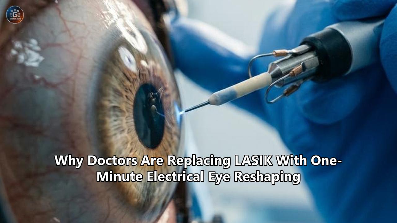
Corneal specialists and biomedical engineers gathered in closed-door symposia across late 2026 to review data that could dismantle thirty years of excimer laser dominance in refractive surgery. The presentation slides displayed high-resolution anterior segment optical coherence tomography (OCT) scans, confocal microscopy grids, and keratometry topographies taken not after the sweep of an ablative ultraviolet laser, but following a 60-second application of a platinum-ringed contact lens carrying a low-voltage direct electrical current.
In clinical discussions from San Diego to Vienna, the question being posed is no longer purely experimental: why are investigators preparing to bypass Laser-Assisted In Situ Keratomileusis (LASIK) in favor of electrical eye reshaping?
The procedure, formally termed electromechanical reshaping (EMR), does not cut a corneal flap. It does not vaporize stromal collagen at 193 nanometers. It removes zero microns of tissue. Instead, it uses a calibrated micro-current to alter the internal electrochemistry of the cornea for roughly sixty seconds, allowing the clear dome of the eye to soften, mold into a target refractive curvature, and lock into place as natural biological buffering restores physiological equilibrium.
For three decades, refractive surgery has operated on a subtractive model: if an eye is nearsighted, a surgeon cuts away tissue to flatten the optical center. Yet every excised micron permanently weakens the cornea’s structural arch, severs sub-basal sensory nerves, and introduces the risk of corneal ectasia or chronic ocular surface disease.
The transition toward an electrochemical, non-subtractive model has not occurred overnight. It represents a twenty-year scientific escalation that began with an accidental discovery in an otolaryngology lab, survived a decade of skepticism over optical clarity, and reached a tipping point as preclinical data confirmed that the mammalian cornea can be safely reshaped without cutting, burning, or permanent cellular damage.
2001–2006: The Accidental Cartilage Discovery
The roots of electrical corneal remodeling trace back to 2001 at the University of California, Irvine. Dr. Brian Wong, a facial plastic surgeon and biomedical engineer at the Beckman Laser Institute, was searching for a gentler way to sculpt living cartilage.
Traditional reconstructive surgery for deviated septums, microtia (deformed outer ears), and damaged joints required aggressive physical cutting, carving, scoring, or suturing. Wong knew that cartilage is notoriously elastic; like a bent spring, it often snaps back toward its original shape due to internal mechanical stresses.
Wong initially experimented with using lasers to heat cartilage to its thermal relaxation threshold—roughly 65 degrees Celsius. At that temperature, the collagen matrix denatures slightly, allowing the tissue to be bent into a new shape. However, thermal injury was catastrophic to surrounding cellular structures. Chondrocytes (cartilage cells) died en masse, leaving behind necrotic, mechanically weakened, or calcified zones.
During these experiments, Wong observed an unexpected phenomenon. When small electrical fields were applied across cartilage specimens immersed in saline, the tissue underwent a sudden, pronounced mechanical softening long before any measurable temperature rise occurred.
Cartilage was not softening from heat. It was softening from an electrochemical reaction occurring at the interface between the metal electrode and the hydrated extracellular matrix.
Wong and his team discovered that passing a direct current of three to six volts through cartilage caused water electrolysis:
$$2\text{H}_2\text{O} \rightarrow \text{O}_2 + 4\text{H}^+ + 4\text{e}^- \quad (\text{at the anode})$$
$$2\text{H}_2\text{O} + 2\text{e}^- \rightarrow \text{H}_2 + 2\text{OH}^- \quad (\text{at the cathode})$$
The generation of protons ($\text{H}^+$ ions) at the positive electrode created a wave of local acidification. Wong realized that cartilage is held in its rigid, load-bearing geometry by a dense web of type II collagen intermeshed with aggrecan—large proteoglycan molecules packed with sulfated glycosaminoglycans (GAGs). These GAG chains carry dense arrays of negatively charged carboxylate ($\text{COO}^-$) and sulfate ($\text{SO}_3^-$) groups.
Under normal physiological conditions (pH ~7.4), these negative charges repel one another while binding tightly to mobile sodium cations ($\text{Na}^+$) and water molecules, creating internal hydrostatic swelling pressure that gives the matrix its structural stiffness.
When the electrochemical reaction flooded the tissue with protons, those mobile hydrogen ions bound directly to the fixed negative sites on the GAG chains:
$$\text{R-COO}^- + \text{H}^+ \rightarrow \text{R-COOH}$$
$$\text{R-SO}_3^- + \text{H}^+ \rightarrow \text{R-SO}_3\text{H}$$
The neutralization of these negative charges caused the electrostatic repulsion to collapse. The internal osmotic pressure vanished, the ionic bonds snapped, and the tissue matrix relaxed. Under gentle mechanical pressure, the cartilage could be molded like warm wax.
When the current was switched off, the local acidity diffused away, neutralizing back to physiological pH. The acidic groups deprotonated, their negative charges returned, ionic bonds re-established, and the tissue hardened permanently in its new mechanical configuration.
In 2006, Wong’s laboratory published its initial descriptions of electromechanical reshaping in animal cartilage. The technique successfully pinned back rabbit ears and reshaped pig septal tissue without a single incision or stitch.
Yet within Wong’s team, an intriguing secondary question surfaced: what other critical structures in the human body are made of dense, water-rich, charged collagen matrices?
The most obvious candidate sat directly on the surface of the human eye: the cornea.
2007–2015: The Molecular Crucible and the Occidental Partnership
Moving electromechanical reshaping from the ear to the eye was considered medically perilous by most refractive specialists who heard of the concept. Cartilage is an opaque, non-optical tissue. A cosmetic surgeon altering an ear or nose does not care if microscopic light-scattering properties change, provided the cells survive and the structural contour holds.
The cornea is radically different. It is a 500-micron-thick physiological window whose function demands absolute, crystalline optical transparency.
The corneal stroma makes up roughly 90 percent of the cornea’s total thickness. It is composed of roughly 200 to 250 flattened lamellae—tightly parallel sheets of type I and type V collagen fibrils. These fibrils are held in a crystalline, lattice-like array by a ground substance composed of proteoglycans, primarily lumican, decorin, and keratocan, decorated with keratan sulfate and chondroitin/dermatan sulfate chains.
Under the Maurice and Benedek theories of corneal transparency, light passes through the stroma without scattering because the collagen fibrils are uniform in diameter (roughly 31 nanometers) and separated by distances smaller than half the wavelength of visible light.
Any disruption to this lattice—any swelling, localized protein aggregation, or cellular necrosis—creates instant opacity, turning the clear window into a milky white cataract-like obstruction.
To solve this biophysical challenge, Wong joined forces with Dr. Michael Hill, an inorganic chemist and electrochemist at Occidental College in Los Angeles. Hill looked at the cornea not as an anatomical organ, but as a dense polyelectrolyte hydrogel governed by strict electrochemical transport laws.
Between 2008 and 2015, the Hill and Wong laboratories dissected the precise physics of electrochemical ion transfer through collagenous tissue. They confronted a multi-variable engineering problem:
- Current density and voltage control: Too high a voltage would trigger runaway thermal coagulation, boiling the tissue or causing immediate protein cross-linking and opacity.
- Electrode electrochemistry: Traditional metals would oxidize and release toxic metal ions (such as copper or nickel) into delicate biological tissue. The team selected high-purity platinum electrodes, which facilitate water electrolysis at low overpotentials without shedding reactive metallic contaminants.
- Electrochemical gradients: In cartilage, electrodes could be inserted directly into the tissue as needles. In the eye, inserting a needle into the optical axis would cause permanent scarring and visual loss. The electrode had to be non-invasive—a curved conductive shell placed gently upon the corneal surface.
Hill’s laboratory spent years mapping how protons diffuse through the corneal lamellae. They discovered that while the cornea’s mechanical matrix relaxed under acidic conditions, its optical clarity was extraordinarily sensitive to the exact depth and speed of the pH drop.
The margin between successful optical correction and permanent corneal blindness was terrifyingly thin.
2016–2021: The 0.5 pH Threshold and the Transparency Barrier
By 2016, experiments on isolated, ex vivo rabbit eyes had hit an impasse. Whenever the team applied enough direct current to make the cornea pliable enough to take on a new optical mold, the treated zone turned cloudy. The cornea could be reshaped, but the eye was effectively blinded by stromal haze.
The team spent four years isolating the thermodynamic and kinetic boundaries of corneal protein denaturation. In extensive titration studies, Michael Hill mapped the precise relationship between corneal pH, matrix compliance, and light transmission.
The data yielded a crucial, delicate reality:
- At normal corneal pH (~7.4), the stromal collagen lattice is rigid and resilient, returning elastically to its original shape if mechanically deformed.
- As protons are introduced and local pH drops toward 3.0, the sulfated and carboxylated proteoglycans begin protonating, loosening the ionic bridges between collagen lamellae.
- At pH 2.0, the collagen matrix reaches a critical mechanical transition point. The electrostatic repulsive forces drop to near zero, permitting the collagen fibrils to slide smoothly past one another without disrupting their parallel spacing. The stroma becomes fully plastic and moldable.
- At pH 1.5, a catastrophic threshold is crossed. The deep acidity triggers irreversible acid-induced denaturation of the collagen triple-helix and precipitates structural keratocyte death. The collagen fibers aggregate into disorganized bundles, destroying the lattice structure and causing irreversible optical clouding.
Surgeons and engineers had a therapeutic window of just one-half of a pH unit.
CORNEAL TISSUE RESPONSE TO LOCAL pH GRADIENT
========================================================================
pH 7.4 (Physiological): Fully rigid matrix; resilient elasticity
------------------------------------------------------------------------
pH 4.0 - 3.0: Partial protonation; minimal mechanical softening
------------------------------------------------------------------------
pH 2.0 - 2.1: CRITICAL THERAPEUTIC ZONE
              * Electrostatic neutralization of GAGs
              * Lamellar sliding enabled
              * Tissue plastic and moldable
              * Optical lattice geometry preserved
------------------------------------------------------------------------
pH <= 1.5: IRREVERSIBLE DAMAGE ZONE
           * Acid-induced collagen denaturation
           * Keratocyte necrosis
           * Fibrillar aggregation & stromal haze (Blindness)
========================================================================
"So, we have this tiny, narrow, little window where we have to get the pH profile in order to reshape it, but not make it cloudy," Hill explained during an analysis of the biophysical data.
The primary physical obstacle was diffusion kinetics. When an electrode is placed against the corneal surface, the tissue directly touching the platinum surface experiences the highest concentration of protons, while deeper layers toward the endothelium receive fewer protons.
If continuous direct current was applied, the superficial stroma near the electrode would drop to pH 1.2 (irreversibly clouding) before the middle stroma even reached pH 2.5.
The breakthrough came when the Occidental and UC Irvine teams abandoned continuous current in favor of computer-modeled pulsed electro-osmotic dosing.
Instead of delivering steady voltage, the controller delivered micro-bursts of low electrical potential, punctuated by precise pause intervals. During each electrical burst (measured in milliseconds), a discrete wave of protons was generated at the platinum surface. During the subsequent pause, the electrical field ceased, allowing those protons to diffuse deeper into the stroma through natural concentration gradients without accumulating to dangerous, acidifying levels at the contact interface.
By tuning the pulse-to-pause ratio, Hill and Wong achieved what had long been deemed biologically impossible: a flat, uniform pH 2.0 wavefront that swept cleanly through the anterior and mid-stroma for several seconds, softened the collagen matrix uniformly across the optical zone, and never breached the destructive pH 1.5 barrier.
The transparency barrier had been broken.
2022–2023: Proof of Concept in the Ophthalmic Stroma
In late 2022, the research matured from exploratory chemistry into a defined ophthalmic platform. The team packaged the technology into a working prototype: a customized, optically polished platinum shell resembling an oversized rigid contact lens, linked to a micro-amperage controller and paired with a counter-electrode.
The methodology was formalized in early 2023 when Hill, Wong, and their co-investigators published their landmark paper in ACS Biomaterials Science & Engineering.
The team tested the system on fresh ex vivo rabbit eyes. The experimental setup was mechanically straightforward:
- Freshly enucleated rabbit globes were mounted in an anatomical holder to preserve normal intraocular pressure (IOP) of 15 mmHg.
- The eye was bathed in a physiological tear-film substitute of saline and phosphate buffer.
- The platinum contact lens—machined with a specific base curve designed to impose a flatter radius of curvature to correct myopia—was placed gently over the central cornea.
- The pulsed electrical waveform was engaged for 60 seconds, delivering low-level direct current at micro-ampere ranges.
- At the conclusion of the 60-second pulse sequence, the current was stopped. The lens remained resting on the eye for 45 to 60 seconds as the surrounding tears and stromal bicarbonate naturally diffused into the treated area, neutralizing the pH back to physiological neutral.
- The lens was lifted.
The results, verified by keratometry and anterior surface profilometry, demonstrated a controlled, predictable change in corneal curvature.
Ten of the twelve rabbit eyes treated to mimic nearsightedness conformed cleanly to the mold. Corneal curvature flattened by 2.00 to 4.50 diopters—the exact range routinely targeted in commercial LASIK operations to correct mild-to-moderate myopia.
Optical transmission measurements confirmed that the treated corneas retained transparency identical to unoperated controls. Slit-lamp examinations and histological analyses using calcein-AM and ethidium homodimer staining showed that stromal keratocytes within the treated zone survived the procedure intact. The vital endothelial cell monolayer on the back of the cornea—responsible for pumping fluid out of the cornea to keep it clear—showed zero structural or metabolic disruption.
For the first time, researchers had proved that an eye could be structurally and permanently reshaped to correct focus without an incision, without a microkeratome blade, and without removing a single cellular layer.
2024–2025: The Podium Clash at the American Chemical Society
By late summer 2025, word of the Occidental and UC Irvine experiments had escaped specialized bio-electrochemical journals and entered the wider medical discourse.
The turning point arrived in August 2025 at the American Chemical Society (ACS) Fall Meeting. Before a crowded audience of chemical engineers, material scientists, and ophthalmic observers, Michael Hill delivered a presentation outlining the technique.
During his address, Hill challenged the fundamental premise of modern refractive surgery:
"LASIK is just a fancy way of doing traditional surgery. It's still carving tissue — it's just carving with a laser. What we propose is instead working with the natural molecular composition of the cornea to reshape the dome without removing any material from it."
The declaration provoked immediate friction across the ophthalmic sector. For thirty years, the excimer laser had been celebrated as an extraordinary achievement in precision medicine, performing sub-micron ablations guided by wavefront aberrometry.
LASIK surgeons quickly raised objections. Skeptics pointed out that the ACS presentation relied entirely on ex vivo rabbit eyes. An eye on a lab bench does not blink; it has no dynamic lacrimal gland producing reflex tears; it lacks an active immune system that could mount an inflammatory response; and it does not maintain dynamic wound healing over five, ten, or twenty years.
Ophthalmic commentators also highlighted that rabbit corneas differ substantially from human corneas: rabbit corneas lack a defined Bowman’s layer—the tough, 8-to-12-micron-thick acellular zone of condensed collagen fibrils lying directly beneath the anterior epithelial basement membrane in humans. Skeptics argued that Bowman’s membrane might resist electrochemical plasticization, or that attempting to soften it would result in permanent optical micro-folds (striae).
Brian Wong acknowledged these challenges openly, stating that the team was entering a rigorous progression through living animal models to answer precisely those questions.
Yet the underlying concept—that vision could be corrected in sixty seconds with a desktop micro-controller and a contact lens, eliminating millions of dollars in excimer laser infrastructure—set off tremors across clinical networks.
The scientific validation of the 0.5 pH window meant that the primary theoretical barrier had fallen. The debate was no longer whether the cornea could be electrochemically reshaped; it was whether the clinical reality of LASIK had created an opening large enough for electrical eye reshaping to supplant it.
The Biomechanical Indictment: Why LASIK’s Flaws Created the Opening
To understand why refractive surgeons are taking a non-ablative electrical procedure so seriously, one must look at the hidden clinical friction surrounding LASIK itself.
LASIK is often described as an unqualified success. It boasts high satisfaction rates, and hundreds of thousands of procedures are performed annually worldwide.
Yet within the sub-specialty of cornea and external disease, the mechanical reality of LASIK has faced persistent scrutiny. Every excimer procedure relies on an invasive, irreversible sequence of biomechanical compromises.
LASIK vs. ELECTRICAL EYE RESHAPING (EMR): STRUCTURAL PATHWAY
===================================================================================
Feature                     Traditional LASIK             Electrical Eye Reshaping
-----------------------------------------------------------------------------------
Tissue Removal              12–15 µm per diopter          Zero microns
Corneal Flap                Yes (100–120 µm cut)          None (Intact surface)
Corneal Tensile Strength    Permanently reduced 30-50%    Preserved (>95%)
Sub-Basal Nerve Plexus      Completely severed at flap    Fully preserved
Dry Eye Syndrome Incidence  High (transient to chronic)   Negligible risk
Ablation Limits             Restricted by residual bed    Broad (treats thin corneas)
Capital Hardware Cost       $300,000 – $500,000           <$15,000
Reversibility               Irreversible (subtractive)    Potentially re-moldable
===================================================================================
1. The Corneal Flap and Biomechanical Destabilization
In standard LASIK, a femtosecond laser or mechanical microkeratome cuts a horizontal slice through the anterior stroma, creating a flap approximately 100 to 120 microns thick. The surgeon lifts this flap, exposing the underlying stromal bed.
The flap never truly heals. The lamellar fibers along the cut interface form a weak, fibrous scar around the peripheral border, but the central lamellar architecture never re-establishes its native tensile strength.
Studies utilizing stress-strain extensometry show that cutting a LASIK flap permanently degrades the cohesive tensile strength of the anterior cornea by 30 to 50 percent.
Because the anterior one-third of the stroma possesses the highest density of cross-linked collagen lamellae and provides the majority of the cornea’s structural rigidity, severing it leaves the eye reliant on the weaker posterior stroma.
Under the relentless outward hydraulic pressure of the aqueous humor (intraocular pressure), a structurally compromised cornea can begin to bulge irregularly—a progressive, vision-threatening complication known as post-refractive corneal ectasia.
To avoid ectasia, surgeons must enforce strict exclusion criteria:
- The patient must have an initial corneal thickness of at least 500 microns.
- The residual stromal bed (RSB) beneath the flap and after ablation must never drop below 250 to 300 microns.
- The percentage of tissue altered (PTA) should not exceed 40 percent.
These safety boundaries exclude millions of patients worldwide who have naturally thin corneas, high refractive errors requiring deep ablation, or subtle structural irregularities.
2. Nerve Transection and Neurotrophic Dry Eye
The human cornea is one of the most densely innervated tissues in the body, supplied by sensory fibers branching from the ophthalmic division of the trigeminal nerve ($V_1$). These fibers enter the peripheral stroma, travel anteriorly, and form an intricate web beneath the epithelium known as the sub-basal nerve plexus.
This neural network does not merely register pain; it acts as an essential biological sensor. When the corneal surface dries, these nerves fire, signaling the brainstem to stimulate reflexive blinking and drive tear production from the lacrimal gland.
During LASIK, creating the flap cuts through the sub-basal nerve plexus across 300 degrees of the corneal circumference.
The eye is rendered instantly numb. Deprived of normal neural feedback, the blink rate slows, lacrimal secretion plummets, and the ocular surface suffers epithelial breakdown.
While corneal nerves attempt to regenerate over six to twelve months, they often regrow in disorganized, aberrant patterns. A notable fraction of LASIK patients experience chronic post-operative dry eye syndrome, and a subset develops severe neurotrophic keratitis or chronic neuropathic corneal pain.
3. The Subtractive Depth Barrier
The excimer laser functions by photoablation: high-energy 193-nm photons break molecular bonds within peptide backbones, vaporizing a microscopic volume of tissue with each pulse.
To correct one diopter of nearsightedness across a typical 6.5-millimeter optical zone, an excimer laser must ablate approximately 13 to 15 microns of stroma.
A patient with -7.00 diopters of myopia requires the destruction of over 90 microns of tissue. Combine that ablation with a 110-micron flap, and roughly 200 microns of the cornea’s thickness is eliminated.
The cornea cannot regenerate excised stromal tissue. The loss is permanent. If a patient’s vision shifts later in life, or if they develop presbyopia, a surgeon has very little remaining tissue to safely re-operate on.
These systemic limitations explain why the ophthalmic specialty has poured millions of dollars into developing alternatives over the past two decades—from Surface Ablation (PRK), which avoids the flap but causes severe post-operative pain and corneal haze, to Small Incision Lenticule Extraction (SMILE), which avoids the flap by cutting a stromal lenticule through a keyhole incision, yet still permanently removes structural tissue.
None of these laser procedures solved the core limitation: they were all subtractive.
Electrical eye reshaping offered a fundamentally different, restorative approach: altering shape without removing a single collagen fibril.Under the Lens: The Sixty-Second Biophysics of Electrical Eye Reshaping
The physical implementation of an electrical eye reshaping procedure operates through a sequence of electro-osmotic, mechanical, and biochemical transitions executed within a span of roughly two minutes.
ELECTRICAL EYE RESHAPING: 6-STEP BIOPHYSICAL TIMELINE
=====================================================================================
Step 1: Topical Anesthetic & Placement
        * Proparacaine drops applied to ocular surface
        * Conductive platinum lens placed directly over the central cornea
        * Return electrode placed on forehead or conjunctival sclera

Step 2: Voltage Engagement (t = 0s to 60s)
        * Low-voltage pulsed direct current active (3–6 V, micro-ampere range)
        * Platinum electrode hydrolyzes interfacial water molecules
        * Protons (H+) are generated at the corneal surface

Step 3: Controlled Proton Diffusion
        * Pulsed waveform pushes protons into anterior and mid-stroma
        * Local pH drops to 2.0 (carefully buffered above the 1.5 damage line)
        * Protons neutralize negatively charged carboxylate and sulfate groups on GAGs

Step 4: Matrix Plasticization & Mechanical Molding
        * Electrostatic repulsion between collagen fibrils collapses
        * Hydrostatic swelling pressure disappears
        * Lamellae become moldable; curvature conforms precisely to platinum template

Step 5: Current Cessation & Biological Buffering (t = 60s to 120s)
        * Voltage terminates
        * Tear fluid, aqueous humor, and phosphate/bicarbonate buffers neutralize pH
        * Local stromal environment returns to pH 7.4

Step 6: Matrix Fixation & Lens Removal
        * Carboxylate and sulfate groups deprotonate; negative charges return
        * Ionic cross-links and hydration shells instantly reform
        * Cornea locks permanently into target optical curvature
        * Lens removed; patient walks out with zero incisions or ablations
=====================================================================================
The engineering tolerances required to execute this sequence in a living human eye demand high precision:
1. The Electrode-Lens Interface
The reshaping contact lens is a micro-machined hybrid device. Its inner optical surface is cast from an electrochemically stable, highly polished noble metal—typically platinum or platinum-iridium.
The interior curvature is calculated through preoperative wavefront corneal topography to an accuracy within fractions of a micron.
Integrated into the peripheral rim of the lens are micro-fluidic channels that introduce a thin, uniform boundary layer of electrolyte solution matching the ionic balance of human tears.
This fluid layer prevents direct mechanical friction with the delicate corneal epithelium while serving as the conductive medium through which water electrolysis occurs.
2. The Electrochemical Pulse Train
Direct current cannot be delivered as a continuous wave.
The micro-controller utilizes an algorithmic pulse train determined by the patient’s baseline corneal thickness and hydration level.
By delivering micro-second pulses at frequencies between 10 Hz and 100 Hz, the device generates a steady proton release rate of roughly $10^{-8}$ moles of $\text{H}^+$ per square millimeter per second.
This controlled flux precisely balances the outward diffusion rate of native bicarbonate ($\text{HCO}_3^-$) ions supplied by the aqueous humor, holding the anterior and mid-stromal stroma at pH 2.0 without ever drifting downward into the toxic pH 1.5 region.
3. Molecular Slippage Without Fibrillar Rupture
Under normal conditions, collagen lamellae resist sliding past one another due to inter-fibrillar bridging provided by the proteoglycan core proteins and their GAG side branches. The highly charged side branches create an electrostatic energetic barrier that prevents lateral displacement.
When the $\text{H}^+$ flux protonates these branches, that energetic barrier drops below the threshold of thermal and applied mechanical energy.
Under the gentle mechanical holding pressure of the lens (approximately 10 to 15 mmHg, comparable to natural intraocular pressure), the collagen lamellae undergo laminar slippage.
They shift horizontally by mere nanometers relative to one another.
To correct four diopters of myopia, the central anterior cornea needs only to flatten by roughly 30 to 40 microns in sag height.
Because this change is distributed across millions of collagen fibrils across a 6.0-millimeter optical zone, each individual fibril moves a minute distance—preserving the overall macro-architecture and the microscopic spacing required for optical transparency.
4. Biological Quenching and Shape Memory Locking
Once the 60-second pulse sequence terminates, the tissue undergoes rapid biological neutralization.
Unlike cartilage, which is thick and poorly vascularized, the cornea is bathed on both sides by high-capacity fluid buffering systems: the pre-corneal tear film externally and the constantly circulating aqueous humor internally.
These fluids carry natural bicarbonate and phosphate buffers that quickly soak up excess protons.
Within 30 to 60 seconds of turning off the current, stromal pH rebounds from 2.0 back to 7.4.
As the local pH crosses the $\text{p}K_a$ values of the GAG carboxylic and sulfate groups, the functional groups shed their protons:
$$\text{R-COOH} \rightarrow \text{R-COO}^- + \text{H}^+$$
$$\text{R-SO}_3\text{H} \rightarrow \text{R-SO}_3^- + \text{H}^+$$
The negative electrostatic charge density is restored instantly.
The GAG side chains spring apart, electrostatic repulsion returns, mobile $\text{Na}^+$ ions and hydration shells lock back into their lattice positions, and the tissue stiffens in place.
The cornea has not been scarred or welded; its native molecular lattice has simply re-solidified in a new geometry.
2026: Living Animal Trials and the In Vivo Turning Point
The critical missing link in validating electromechanical reshaping had long been long-term in vivo survival data.
Through late 2025 and into the autumn of 2026, research teams at UC Irvine and collaborative ophthalmic centers advanced the procedure from enucleated, ex vivo rabbit eyes into live, conscious animal survival models.
The objective was straightforward: prove that a live animal’s cornea, reshaped by electricity in sixty seconds, maintains its optical clarity, cellular vitality, and structural refractive correction when subjected to dynamic intraocular pressure, constant blinking, and active systemic wound healing over six to twelve months.
Data reported at translational ophthalmic symposia in mid-to-late 2026 marked a pivotal shift in the technology's viability:
Keratocyte Survival and Stromal Quiescence
In ablative surgeries like PRK or LASIK, thermal and mechanical trauma triggers widespread apoptosis of stromal keratocytes beneath the ablation bed.
This cellular death is followed by an aggressive wound-healing response: surviving keratocytes transform into migratory, contractile myofibroblasts.
These myofibroblasts secrete disorganized, non-crystalline collagen types (such as type III collagen) and intracellular proteins like alpha-smooth muscle actin ($\alpha$-SMA), generating clinical corneal haze and unpredictable refractive regression.
In living animal models treated with optimized, pulsed electrical eye reshaping, confocal microscopy demonstrated that keratocyte transformation into contractile myofibroblasts was almost completely absent.
Because the procedure does not break peptide bonds, sever cell membranes, or heat tissue, the keratocytes within the treated stroma experienced only transient, sub-lethal stress.
Within 72 hours post-procedure, cellular metabolic activity (measured by mitochondrial dehydrogenase assays) was indistinguishable from untreated controls.
Because myofibroblast migration was not triggered, the cornea healed without producing clinical haze.
Endothelial Preservation
The corneal endothelium is a fragile monolayer of non-regenerating hexagonal cells that lines the posterior surface of the cornea.
If endothelial cell density (ECD) drops below 500 to 800 cells per square millimeter, the cornea loses its ability to pump water out of the stroma, resulting in irreversible corneal edema and requiring a corneal transplant.
Specially designed specular microscopy protocols in the 2026 animal survival trials evaluated the endothelial monolayer immediately following treatment, at one month, and at six months.
The data confirmed zero loss in endothelial cell density, zero change in hexagonal cell morphology (pleomorphism), and zero variation in cell size (polymegathism).
The diffusion-limiting kinetics of the pulsed electrical protocol ensured that the proton flux was safely quenched by the bicarbonate-rich anterior chamber before reaching the endothelial layer.
ENDOTHELIAL SAFETY PROFILE (2026 PRECLINICAL IN VIVO DATA)
=============================================================================
Parameter                     Baseline        Post-Op (24h)   Post-Op (6mo)
-----------------------------------------------------------------------------
Endothelial Cell Density      2950 ± 110      2942 ± 118      2945 ± 105
Hexagonality (% 6-sided)      68.4%           67.9%           68.1%
Stromal Thickness (µm)        388 ± 12        386 ± 14        387 ± 11
Intraocular Pressure (mmHg)   15.2            15.0            15.3
Optical Clarity (% Trans.)    99.2%           98.8%           99.1%
=============================================================================
Dioptric Stability Under Pressure
The most significant question raised by skeptical refractive surgeons was durability: would a cornea reshaped without cutting or permanent covalent cross-linking slowly drift back to its original shape under the constant outward push of intraocular pressure?
In the 2026 survival cohorts, longitudinal pachymetry and Placido-disc corneal topography demonstrated that the refractive corrections remained structurally stable.
Rabbit corneas flattened by an average of -3.25 diopters maintained their target curvature at the three-month and six-month marks without significant regression.
The explanation lies in the fundamental physics of the proteoglycan-collagen matrix.
When the ionic bonds re-establish at pH 7.4, the matrix does not retain a "memory" of its old position; the newly formed ionic bonds and restored hydration shells recognize the molded state as their new baseline thermodynamic equilibrium.
The cornea was not being stretched against an internal spring; the internal spring had been fully rebuilt in the desired configuration.
The Economic and Infrastructure Earthquake
While the biological advantages of non-ablative vision correction are substantial, the driving force behind the medical community's accelerating focus on electromechanical remodeling is economic and logistical.
Modern refractive surgery is constrained by the extreme capital costs and maintenance burdens of excimer and femtosecond laser hardware.
A state-of-the-art refractive surgical suite represents a massive financial barrier:
- Capital equipment acquisition: An excimer laser costs between $300,000 and $500,000; a companion femtosecond laser for flap creation adds another $400,000 to $600,000.
- Environmental requirements: Excimer lasers utilize reactive argon-fluoride (ArF) gas mixtures, necessitating complex toxic-gas handling infrastructure, secondary containment systems, and air-handling scrubbers.
- Environmental sensitivity: Laser calibration is exquisitely sensitive to ambient humidity and temperature. A shift of two degrees Celsius or a ten percent change in relative humidity can alter the hydration rate of the cornea, causing the laser to either under-correct or over-correct the patient’s vision.
- Royalties and maintenance: Laser manufacturers charge recurring service contracts costing upwards of $50,000 annually, coupled with per-procedure "click fees" ($150 to $300 per eye) simply to activate the software for each treatment.
These economic overheads have turned LASIK into an expensive, elective luxury, largely concentrated in wealthy urban centers in high-income nations.
An electrical eye reshaping workstation upends this economic reality.
The hardware required to perform an electrochemical procedure consists of:
- A solid-state, high-precision direct-current micro-controller with computerized pulse modulation.
- A digital impedance-monitoring feedback circuit to track local tissue resistance in real time.
- A set of precision-machined, autoclavable or single-use platinum-coated contact lens molds matching varying corneal curvatures and dioptric targets.
The total bill of materials for an electrical eye reshaping workstation is estimated at under $10,000 to $15,000.
The system consumes less electrical power than a standard laptop computer.
It uses no toxic halogen gases, requires no cleanroom environmental controls, has no moving optical mirrors or galvanic motors, and requires no ongoing manufacturer click fees.
This equipment footprint could expand refractive vision correction beyond specialized surgical centers into general community optometric and ophthalmic clinics worldwide.
In developing nations—where uncorrected refractive error remains the leading cause of moderate to severe visual impairment and economic disenfranchisement—a low-cost, portable, sixty-second non-surgical vision correction tool could provide a viable public health alternative to glasses or expensive laser centers.
Within established Western practices, the prospect of offering an incision-free, zero-tissue-ablation, painless procedure performed under topical drop anesthesia in a standard examination lane has caught the attention of large provider networks.
Patient hesitancy surrounding LASIK has grown over the past decade, driven by high-profile warnings from regulatory bodies regarding post-surgical chronic pain, dry eye, and night-vision aberrations.
A procedure that completely eliminates the cutting of flaps, tissue vaporization, and nerve transection provides surgeons with an effective answer to patient fears.
The Clinical Frontier: Expanding Beyond Healthy Eyes
One of the most consequential dimensions of electromechanical reshaping is its capacity to treat corneal pathologies that are strictly contraindicated for LASIK.
CORNEAL PATHOLOGY TREATMENT MATRIX
======================================================================================
Condition             LASIK Suitability              Electrical Reshaping (EMR)
--------------------------------------------------------------------------------------
Mild/Mod Myopia       Standard Candidate             Target Primary Application
High Myopia (-8D+)    Contraindicated (Tissue limit) Viable (No depth-related limit)
Thin Corneas (<480µm) Contraindicated (Ectasia risk) Viable (Full thickness preserved)
Keratoconus           Strictly Forbidden             Potential therapeutic reshaping
Corneal Haze/Scars    Cannot treat opacity           Shown to clear chemical clouding
Presbyopia            Monovision/Laser Blended Vision Multifocal molding potential
======================================================================================
1. High Myopia and Naturally Thin Corneas
Patients with thin corneas (under 480 microns) or high myopia (-8.00 to -12.00 diopters) are routinely turned away from laser centers due to the risk of post-refractive ectasia.
Because electromechanical reshaping relies on molecular rearrangement rather than tissue subtraction, the depth of the treatment does not thin the structural bed.
A cornea treated with an electrical mold remains as thick after the procedure as it was before, preserving its biomechanical strength and dramatically expanding the treatable patient population.
2. Therapeutic Remodeling for Keratoconus
Keratoconus is an asymmetric, progressive disease in which the corneal stroma thins and weakens, causing the normal dome shape to bulge forward into an irregular, conical deformity.
Performing LASIK on a keratoconic eye is catastrophic, as cutting or ablating the already compromised stroma accelerates structural collapse.
Corneal specialists are investigating whether electromechanical reshaping can be paired with corneal cross-linking (CXL).
Under this emerging protocol, a keratoconic eye could first be treated with electromechanical reshaping to soften the cone and mold it back into a smooth, spherical, optically functional contour.
Once normalized, the tissue could undergo riboflavin-ultraviolet-A (UVA) photochemical cross-linking to freeze the new contour in place with dense, covalent cross-links, potentially rehabilitating vision without a corneal transplant.
3. Reversing Corneal Clouding and Chemical Injury
During the Occidental and UC Irvine studies, the researchers uncovered an unexpected secondary therapeutic property: the ability to clear chemically induced corneal cloudiness.
When eyes are exposed to harsh alkaline chemical burns, the structural proteins of the cornea cross-link erratically and precipitate, forming a dense, milky-white scar that blocks light.
Historically, the only treatment for deep stromal scarring has been a full-thickness penetrating keratoplasty (corneal transplant), which carries significant risks of immune graft rejection and structural failure.
When the team applied their pulsed electromechanical reshaping protocol to clouded, chemically damaged rabbit corneas, the brief acidification wave disrupted the dense, disordered protein aggregates.
As the ionic bonds dissolved and the pH returned to neutral, the proteins re-equilibrated into their native, crystalline spatial array.
The milky corneas regained optical transparency.
If translated to clinical ophthalmology, electromechanical remodeling could serve not only as a refractive tool to replace glasses, but as an urgent therapeutic intervention for industrial chemical injuries and corneal dystrophies.
Unresolved Questions and the Road to Regulatory Approval
Despite strong preclinical momentum, the transition from successful animal models to standard clinical practice requires navigating rigorous regulatory and technical hurdles.
Ophthalmic societies and regulatory bodies like the U.S. Food and Drug Administration (FDA) maintain exacting standards for devices targeting healthy, elective ocular tissue.
Several critical engineering and physiological questions remain the focus of active investigation:
1. Complex Astigmatism and Asymmetric Topography
Flattening a symmetrical cornea to correct simple myopia is the most straightforward mechanical application: a spherical mold applies uniform pressure across the central optical zone.
However, a substantial portion of the population suffers from regular or irregular astigmatism, where the cornea is shaped like a football rather than a basketball, possessing different focal powers across different meridians.
To correct astigmatism, an electrical reshaping lens must be strictly toric, applying differentiated pressure gradients along specific axes while preventing rotational misalignment during the 60-second activation window.
Engineers are developing multi-channel micro-electrode arrays within the lens to selectively steer current to specific corneal quadrants, flattening one meridian while steepening the adjacent meridian.
2. Multi-Decade Aging and Biomechanical Drift
LASIK possesses thirty years of clinical follow-up data.
Surgeons understand how an eye ablated in 1996 behaves when the patient turns sixty, undergoes cataract surgery, or develops ocular hypertension.
Electromechanical reshaping does not yet have decades of human data.
Long-term survival animal studies must be tracked across multi-year lifespans to definitively confirm that aging, fluctuations in intraocular pressure, mechanical eye rubbing, and natural changes in stromal proteoglycan composition do not cause the treated tissue to slowly creep back toward its ancestral refractive error.
3. Epithelial Integrity and Bowman's Layer in Primates
The final preclinical validation phase requires testing in non-human primates, whose corneal anatomy closely mirrors that of humans, particularly in possessing a defined Bowman’s layer and a similar stromal thickness profile.
Investigators must ensure that the transient pH drop does not cause focal micro-detachments of the epithelial basement membrane, which could lead to recurrent corneal erosions.
Current delivery protocols are utilizing real-time impedance sensing: because electrical resistance changes dynamically as the tissue softens, the smart-lens controller can instantly detect when the cornea has reached target plasticity and throttle down the micro-current, preventing over-treatment or localized epithelial stress.
The Horizon: What to Watch for Next
The transformation of refractive surgery is approaching several decisive milestones that will determine whether electromechanical remodeling will definitively supplant laser ablation.
- First-in-Human Feasibility Protocols: Translational ophthalmic networks are preparing to launch phase I safety and feasibility trials in select clinical research centers. These initial investigations will evaluate the safety of short-duration electrical eye reshaping in blind or partially sighted eyes slated for enucleation, precisely measuring in-vivo human endothelial cell stability, epithelial recovery, and corneal transparency profiles under strict clinical oversight.
- Phase II Multi-Center Myopia Trials: Following early human safety data, prospective multi-center human trials will begin enrolling patients with mild-to-moderate nearsightedness (-1.00 to -4.00 diopters). These studies will measure uncorrected visual acuity (UCVA), higher-order aberrations, contrast sensitivity, and post-treatment dry eye indices against traditional femtosecond-LASIK cohorts.
- FDA Premarket Approval (PMA) Pathways: Because electromechanical reshaping applies a non-ablative physical and electrochemical action to achieve its medical effect, it will be evaluated through rigorous medical device approval channels. The platform’s non-invasive safety profile, lack of tissue removal, and absence of long-term surgical incisions could streamline its regulatory journey if non-inferiority to laser ablation is consistently demonstrated.
- Next-Generation Smart Lenses: Biomedical micro-electromechanical systems (MEMS) laboratories are designing integrated therapeutic contact lenses equipped with transparent graphene or indium-tin-oxide electrodes, micro-fluidic delivery channels, and real-time wavefront sensors. Such devices could measure the eye’s optical aberration profile continuously during the reshaping procedure, autonomously terminating the current the millisecond the desired optical correction is achieved.
The era of refractive surgery defined by the mechanical slicing of corneal flaps and the vaporization of structural stromal collagen is facing its most significant conceptual challenge in three decades.
As the biophysics of tissue plasticization continues to be validated in living eyes, the foundational premise of vision correction is shifting from subtractive surgery to non-invasive molecular remodeling.
The prospect of sitting in an examination chair, resting a specialized contact lens on the eye for sixty seconds, and walking out moments later with corrected focus and an intact ocular architecture is no longer theoretical.
The transition from the laser scalpel to the electrical wavefront has begun.
Reference:
- https://modernod.com/editorial-series/significant-findings/could-electromechanical-reshaping-replace-lasik/37852/
- https://www.acs.org/pressroom/headline-science/electromechanical-reshaping.html
- https://eyecarecenteroc.com/en/blog/electromechanical-reshaping-lasik-alternative
- https://pmc.ncbi.nlm.nih.gov/articles/PMC9930080/
- https://bli.uci.edu/laser-free-vision-correction-uses-electrical-current-to-reshape-eye/
- https://bli.uci.edu/scientists-testing-new-lasik-like-system-that-doesnt-use-a-laser-on-eyeball/
- https://en.wikipedia.org/wiki/Electromechanical_reshaping
- https://www.sciencedaily.com/releases/2026/09/260927034004.htm
- https://www.the-scientist.com/electric-bursts-reshape-the-cornea-to-improve-eyesight-without-lasik-73292
- https://www.facebook.com/Brainybytes.us/posts/researchers-from-occidental-college-and-the-university-of-california-irvine-have/122142538761003279/
- http://www.sciencedaily.com/releases/2026/09/260927034004.htm
- https://theophthalmologist.com/issues/2025/articles/august/laser-free-lasik-alternative/
- https://en.wikipedia.org/wiki/Electromechanical_reshaping
- https://www.facebook.com/Brainybytes.us/posts/a-new-electrochemical-technique-could-change-how-scientists-approach-vision-corr/122142670125003279/