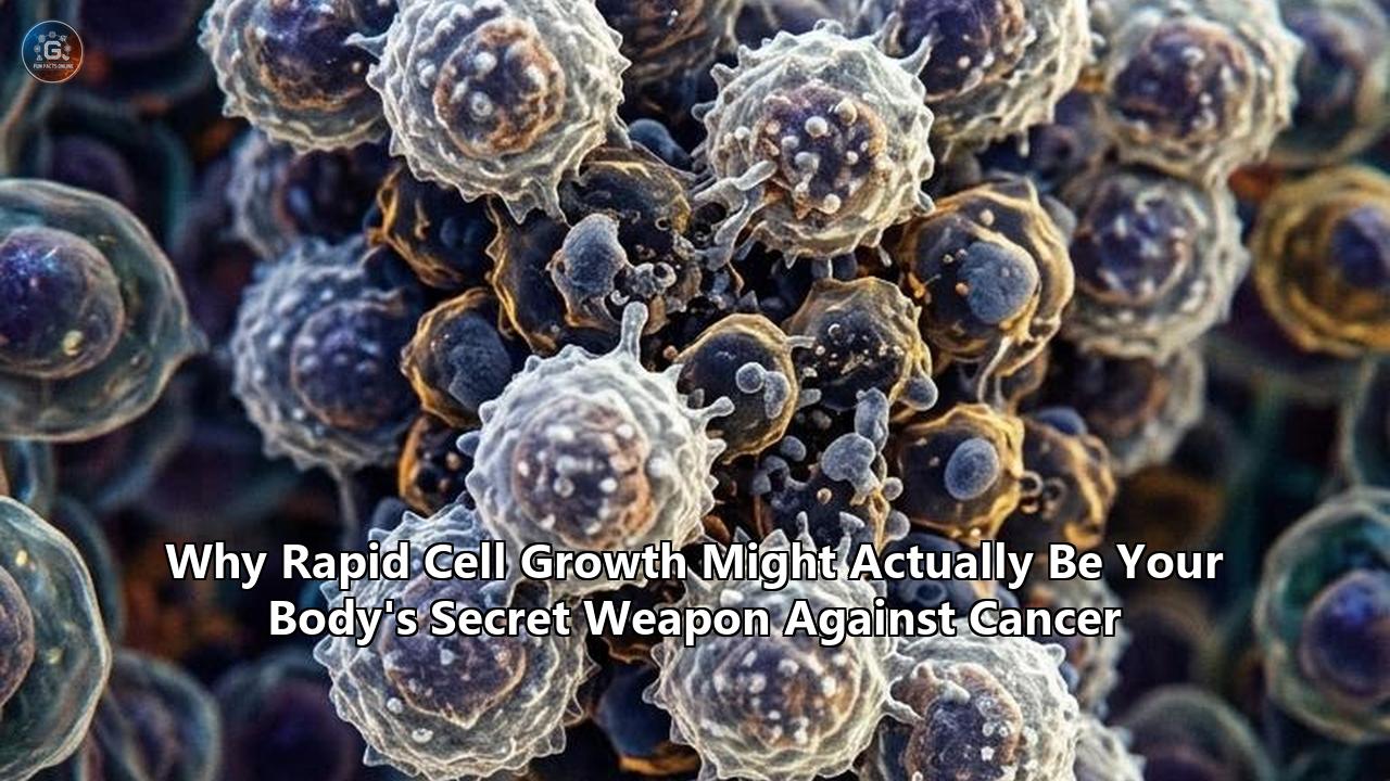
PASADENA, Calif. — In a study published in the Proceedings of the National Academy of Sciences, researchers at the California Institute of Technology demonstrated that high rates of cell division, long considered one of the primary drivers of tumor formation, act under specific biological conditions as an active defense mechanism that purges mutated cells before they can form malignancies.
The findings challenge an assumption that has anchored oncology for more than a century: that rapid cellular replication serves exclusively as a risk factor for malignancy. By combining engineering control theory with direct experiments in cancer-resistant marine organisms, the Caltech team proved that rapid cellular turnover acts as a biological "proofreading" system. When tissues divide rapidly and continuously, they can systematically detect, outcompete, and physically eliminate cells harboring dangerous mutations.
"Proofreading using cell turnover was a surprise to all of us," said Dr. Anish Sarma, lead author of the study and a resident physician in pediatrics at Boston Children’s Hospital and Boston Medical Center who conducted the research while completing his doctorate at Caltech. "We saw the possibility mathematically and thought, 'That can't be right,' and then experiments confirmed it".
The discovery reframes how biologists understand the relationship between proliferation and pathology. For decades, the conventional biological narrative warned that every single round of DNA duplication is an opportunity for genetic typos to slip past cellular proofreaders, eventually sparking malignancy. The new research demonstrates that without a swift, continuous engine of cellular generation and replacement, tissues lose their ability to flush out damaged cells, allowing precancerous mutations to take root.
========================================================================================
                          THE DUAL FACE OF CELL PROLIFERATION
========================================================================================

 TRADITIONAL PARADIGM:
 Cell Division = Mutation Gamble

 [ Normal Cell ] ──(Replication)──> [ DNA Copy Error ] ──(Accumulation)──> [ Tumor Growth ]
 * Assumption: More division directly multiplies cancer risk.

----------------------------------------------------------------------------------------

 NEW CONTROL-THEORY MODEL (Caltech / PNAS):
 Rapid Turnover = Active Quality Control Engine

                   ┌─── High-Speed Proliferation ───┐
                   │                                │
                   ▼                                ▼
 [ Somatic Tissue ] ──> [ Mechanical Competition ] ──> [ Apical Extrusion ] ──> [ Waste ]
                   ▲                                │
                   │                                ▼
                   └─── Selective Elimination ──────┴──> [ Clean, Healthy Tissue ]
 * Discovery: Halting cell division disables proofreading, allowing neoplasms to form.
========================================================================================
The Century-Old Assumption: Why Proliferation Was Branded the Culprit
To understand why the Caltech discovery disrupts established oncology, one must look at how science has viewed cell proliferation since the dawn of modern pathology.
In 1914, German biologist Theodor Boveri proposed that cancer arises from abnormal chromosomal constitutions within dividing cells. Over the subsequent century, that observation crystallized into a core principle: cell division is inherently dangerous. Every time a human cell divides, it must replicate 3.2 billion base pairs of DNA. Even with an astonishingly accurate replication machinery—which makes fewer than one error per billion bases copied—the sheer volume of cellular turnover across a human lifespan means trillions of mutations inevitably occur.
This mechanical reality formed the basis of the controversial "bad luck" hypothesis published in Science in 2015 by Cristian Tomasetti and Bert Vogelstein of Johns Hopkins University. Their statistical models demonstrated a strong positive correlation between the number of normal stem cell divisions in a given tissue and the lifetime risk of cancer in that tissue. Tissues that undergo continuous, rapid renewal—such as the epithelial lining of the small intestine, the colon, and the basal layer of the epidermis—account for a massive share of human carcinomas. In contrast, tissues that divide rarely or not at all, such as cerebral cortical neurons or cardiac myocytes, almost never generate primary malignancies in adulthood.
From this viewpoint, cell division has been treated as a form of biological debt. The faster the cell cycle turns, the higher the interest on genetic errors, and the quicker a tissue slides toward oncogenesis. In traditional frameworks, linking rapid cell growth cancer dynamics meant evaluating how runaway growth fuels tumor progression and therapy resistance.
Yet this statistical correlation has always harbored a major theoretical blind spot, known to evolutionary biologists as Peto’s Paradox.
========================================================================================
                       PETO'S PARADOX: THE CELLULAR ARITHMETIC
========================================================================================

  Organism       Lifespan        Body Mass (kg)     Estimated Cells    Lifetime Cancer Rate
  --------------------------------------------------------------------------------------
  House Mouse    2–3 years       0.025              ~10^10             ~20% - 30%
  Human          70–80 years     70                 ~3.7 x 10^13       ~30% - 40%
  Blue Whale     80–90 years     100,000            ~10^17             Near Zero (<1%)
  Jellyfish      Indefinite      Variable           Variable           Absolute Zero

  The Contradiction: If every cell division carries a fixed probability of oncogenic
  transformation, a blue whale or an elephant should develop thousands of lethal tumors
  before reaching reproductive maturity. Yet they do not.
========================================================================================
Formulated in 1977 by epidemiologist Richard Peto, the paradox observes that at the species level, there is no correlation between body size, lifespan, cell division count, and cancer incidence. An elephant possesses 100 times more cells than a human and lives for six decades, representing thousands of times more cumulative cell divisions, yet elephants rarely develop cancer.
Even more extreme are ancient, non-senescent organisms like cnidarians (jellyfish and hydra). Many cnidarian species maintain continuous, hyperactive stem cell divisions throughout their entire lifespans, constantly regenerating every tissue in their bodies every few weeks, yet they are biologically immune to neoplastic transformation.
The mathematical models long used to explain human oncogenesis could not resolve this contradiction without proposing increasingly complex and hypothetical tumor suppressor suites. The Caltech team decided to approach the problem from the opposite direction: What if rapid cell division is not the hazard, but the actual instrument of tumor defense?
Applying Control Theory: How Engineering Logic Decoded Biology
The breakthrough did not originate in a conventional oncology clinic. It emerged from an interdisciplinary partnership between Lea Goentoro, a professor of biology at Caltech, and John Doyle, the Jean-Lou Chameau Professor of Control and Dynamical Systems, Electrical Engineering, and Bioengineering, Emeritus.
┌───────────────────────────────────────────────────────────────────────────────────────┐
│                      CONTROL THEORY FEEDBACK IN CELLULAR TISSUES                     │
├───────────────────────────────────────────────────────────────────────────────────────┤
│                                                                                       │
│   Target Homeostasis (Constant Healthy Cell Density)                                  │
│         │                                                                             │
│         ▼                                                                             │
│   ┌─────────────┐       Dynamic Proliferation       ┌─────────────────┐               │
│   │ Controller  │ ────────────────────────────────> │ Tissue Effector │               │
│   └─────────────┘                                   └─────────────────┘               │
│         ▲                                                    │                        │
│         │              Kinetic Error Detection               ▼                        │
│         └────────── (Apoptosis & Cell Extrusion) ──── [ Sensor Array ]               │
│                                                                                       │
│   KEY ENGINEERING PRINCIPLE:                                                          │
│   In dynamical systems, higher bandwidth (faster cycle rates) enables tighter         │
│   feedback control and higher rejection rates of high-frequency disturbances.        │
│                                                                                       │
└───────────────────────────────────────────────────────────────────────────────────────┘
Doyle had spent decades analyzing robust control architectures in complex engineered systems—such as fly-by-wire jetliners, advanced power grids, and internet routing protocols. In all these systems, maintaining stability amidst constant external disruption requires rapid, continuous feedback loops.
If a system responds too slowly to an internal error, the error cascades and crashes the entire network. To maintain absolute stability, the error-correcting cycle must operate at high frequency.
When Sarma, Goentoro, and Doyle translated this engineering principle into tissue biology, they constructed a dynamical mathematical model tracking three coupled variables:
- The Rate of Proliferation ($\lambda_p$): The speed at which new cells are generated.
- The Rate of Somatic Mutation ($\mu$): The frequency with which environmental or replicative insults alter the genome.
- The Rate of Selective Clearance ($\delta_d$): The speed at which aberrant, damaged, or suboptimal cells are eliminated via apoptosis or physical extrusion.
In classical oncology models, $\lambda_p$ and $\mu$ are inextricably linked as drivers of disease. But when the Caltech researchers introduced the feedback principles of control theory, the mathematics produced an unexpected result.
If an organism maintains a high baseline rate of proliferation, it simultaneously generates the mechanical kinetic energy and biomass necessary to power continuous quality control. A tissue with rapid turnover functions like a swiftly flowing mountain stream: any sediment, debris, or mutant cell that drops into the current is swept downstream and cleared out before it can adhere to the riverbed.
Conversely, a tissue with a low proliferation rate is a stagnant pond. When a mutation occurs in a low-turnover environment, the damaged cell lingers in the tissue architecture for months or years, gaining a long temporal window to accumulate secondary genetic alterations, establish local vascular ties, and evolve invasiveness.
The mathematical simulations showed that tissues operating at high turnover rates could tolerate somatic mutation rates orders of magnitude higher than sluggish tissues, provided that the clearance feedback mechanism remained tightly coupled to cell division. The math was unambiguous: fast cell turnover was an active proofreading machine.
The Marine Proof: Forcing Neoplasms in Immortal Animals
To confirm that their mathematical equations matched living physiology, the researchers turned to an animal that had long baffled cancer biologists: the moon jellyfish (Aurelia).
Jellyfish possess an evolutionary design that makes them ideal for studying tissue dynamics. They are composed of simple, rapidly renewing epithelial layers governed by ancient, highly conserved genetic pathways identical to those found in human tissues. Despite being exposed in marine environments to severe ultraviolet radiation, chemical mutagens, and physical trauma, jellyfish in the wild do not develop tumors.
========================================================================================
                      CALTECH EXPERIMENTAL DESIGN & FINDINGS
========================================================================================

 EXPERIMENT 1: Standard Carcinogen Exposure
 [ Jellyfish ] + [ High-Dose Chemical Carcinogen ] ──> Zero Neoplasms Formed (100% Normal)
 * Result: Natural tissue defenses completely neutralized the mutational insult.

 EXPERIMENT 2: Apoptosis (Programmed Cell Death) Disabled
 [ Apoptosis Inhibited ] + [ Carcinogen ] ──> Severe Neoplasm Outbreaks
 * Result: Mutated cells could no longer be flagged for execution; tumors formed.

 EXPERIMENT 3: Cell Proliferation Arrested
 [ Proliferation Inhibited ] + [ Carcinogen ] ──> Severe Neoplasm Outbreaks
 * Result: Halting cell division crippled tissue proofreading; tumors formed.

 EXPERIMENT 4: Proliferation Restored
 [ Proliferation Reactivated ] + [ Carcinogen ] ──> Complete Neoplasm Elimination
 * Result: Resumed rapid turnover successfully flushed the mutant cells from tissue.
========================================================================================
The experimental protocol followed a clean four-step proof:
First, Sarma exposed wild-type moon jellyfish to potent chemical carcinogens known to cause widespread DNA damage. As anticipated, the animals suffered extensive somatic mutations but developed zero neoplastic growths. Their tissues continued normal morphogenesis and function.
Second, the researchers used pharmacological inhibitors to shut down the animals' programmed cell death machinery (apoptosis). When exposed to the same carcinogens, the animals could no longer instruct mutated cells to self-destruct. Neoplastic masses quickly formed throughout their bodies. This confirmed that selective cell clearance is mandatory for cancer resistance.
The critical test came in the third step. The team left the apoptotic machinery fully operational but administered targeted small-molecule inhibitors to selectively halt cell proliferation—slowing the animals’ cellular division rate to near zero.
If the century-old medical dogma was correct—that slowing cell division reduces cancer risk—these proliferation-arrested animals should have been well-protected from neoplasia.
The exact opposite happened.
When cell division was arrested, the carcinogen-exposed jellyfish developed extensive, aggressive neoplastic growths. Despite having fully functional apoptosis genes, the tissues were physically unable to eliminate the mutant cells. Without the mechanical push and rapid generation of new, healthy daughter cells, the mutated clones remained anchored in place, disorganized the surrounding tissue architecture, and expanded into tumors.
In the final step of the experiment, Sarma washed out the proliferation-inhibiting compounds, allowing the cells to resume their native, rapid rate of division.
Within days, the reactivated cellular engine restored tissue quality control. The newly proliferating normal cells crowded around the neoplastic lesions, physically displaced them from the tissue layers, and restored healthy, uniform morphology.
The experiment demonstrated that rapid cellular proliferation is not merely a downstream symptom of disease, but a biological prerequisite for keeping tissues free of tumors.
Mechanical Competition: The Physics of Cell Shedding
The Caltech discovery sheds light on a growing body of biomechanical research in mammalian systems: tissues do not rely solely on internal genetic switches to stop cancer; they use physical force.
In human biology, roughly 90 percent of all cancers arise in epithelial tissues—the sheets of cells that line the skin, the gastrointestinal tract, the lungs, the breast ducts, and the urogenital canals. These epithelial layers undergo continuous turnover. The human intestinal lining, for example, completely replaces its entire cellular surface every three to five days, shedding an estimated 10 billion cells every single day.
┌───────────────────────────────────────────────────────────────────────────────────────┐
│               THE BIOMECHANICS OF APICAL VS. BASAL CELL EXTRUSION                    │
├───────────────────────────────────────────────────────────────────────────────────────┤
│                                                                                       │
│   NORMAL QUALITY CONTROL: APICAL EXTRUSION                                            │
│   (Rapid proliferation pushes damaged cells out into the lumen)                      │
│                                                                                       │
│         [ Lumen / Gut Cavity / External Environment ]  <=== FLUSHED & DESTROYED       │
│                               ▲                                                       │
│                               │ (Sphingosine 1-Phosphate Signaling)                   │
│                        ┌─────────────┐                                                │
│                        │ Mutant Cell │  <=== Compressive Lateral Forces               │
│                  ┌─────┴─────────────┴─────┐                                          │
│                  │  Normal Dividing Cells  │                                          │
│   ═══════════════╧═════════════════════════╧═══════════════════════════════════════   │
│   [ Basement Membrane / Extracellular Matrix ]                                        │
│                                                                                       │
│   PATHOLOGICAL FAILURE: BASAL EXTRUSION                                               │
│   (Occurs when proliferation stalls or extrusion polarity is reversed)               │
│                                                                                       │
│   ═══════════════╤═════════════════════════╤═══════════════════════════════════════   │
│                  │  Normal Dividing Cells  │                                          │
│                  └─────┬─────────────┬─────┘                                          │
│                        │ Mutant Cell │  <=== Escapes into Interstitial Matrix         │
│                        └─────────────┘                                                │
│                               │                                                       │
│                               ▼                                                       │
│         [ Connective Tissue / Blood Vessels / Lymphatic Network ] ===> METASTASIS     │
│                                                                                       │
└───────────────────────────────────────────────────────────────────────────────────────┘
Research spearheaded by Dr. Jody Rosenblatt at the Huntsman Cancer Institute and the Dickson Poon School of Biomedical Sciences has elucidated how this mechanical defense operates. In a healthy epithelium, cell division and cell death are coupled through mechanical tension.
When cells divide, they crowd the epithelial sheet. This physical compression activates a mechanical sensor channel called Piezo1. Activation of Piezo1 triggers the synthesis and release of a lipid signaling molecule called sphingosine 1-phosphate (S1P).
S1P binds to the S1P2 receptor on neighboring cells, instructing an actomyosin ring—a microscopic muscular drawstring—to form around the base of the crowded or damaged cell.
The contracting ring squeezes the abnormal cell upward and out of the epithelial sheet in a process called apical extrusion. Once extruded into the lumen of the gut, lung, or skin surface, the cell is entirely detached from its survival signals, undergoing a specialized form of cell suicide known as anoikis (Greek for "homelessness") before being harmlessly excreted as waste.
A parallel line of research led by Dr. Shunsuke Kon at the Tokyo University of Science has shown that this mechanical extrusion is the primary mechanism of cell competition. When a normal mammalian epithelial cell acquires a classic oncogenic driver mutation—such as an activating mutation in the Ras or Src oncogenes—it does not instantly form a tumor.
Instead, the surrounding normal, rapidly dividing cells recognize the mutant cell as an aberrant neighbor. The rapidly dividing healthy cells generate lateral compressive forces that physically crush the Ras-mutant cell, forcing it out of the epithelial layer into the apical lumen.
However, this mechanical defense functions only as long as the surrounding healthy tissue maintains a high rate of cell proliferation and turnover.
If healthy cell turnover slows down due to aging, chronic inflammation, or tissue exhaustion, the mechanical forces drop below the threshold required for apical extrusion.
Under these compromised conditions, the mutant cell experiences basal extrusion: instead of being pushed out into the digestive cavity to die, it is extruded downwards through the basement membrane into the underlying connective tissue, where it gains direct access to blood vessels, lymphatic ducts, and the pathway to lethal metastatic spread.
Understanding the protective mechanics of continuous cell turnover explains why a temporary acceleration of cellular turnover is the body's native defense against carcinogens.
When tissues encounter a toxic compound, an infectious pathogen, or physical abrasion, their immediate homeostatic reaction is hyper-proliferation: dividing at maximum velocity to accelerate the conveyor belt of apical shedding, flushing the compromised cells out of the body before they can settle into the deep tissue layers.
The Proliferation Trap: Intrinsic Suicide Switches
Beyond the mechanical forces of cell competition, rapid cell division protects the body through an entirely separate, cell-intrinsic mechanism: it acts as a tripwire that forces precancerous cells to trigger their own destruction.
When an aberrant cell mutates an oncogene—such as c-Myc, Cyclin D1, or K-Ras—it attempts to force itself into rapid, deregulated replication.
Healthy cells divide only when given precise, paracrine growth factor signals from their neighbors. Precancerous cells ignore these stop signs, attempting to run their DNA replication machinery at full throttle.
========================================================================================
             HOW ONCOGENIC HYPER-PROLIFERATION TRIGGERS SELF-DESTRUCTION
========================================================================================

    [ Oncogene Activation ] (Myc / Ras / Cyclin E Overexpression)
              │
              ▼
    [ Hyper-Accelerated Cell Cycle Entry ]
              │
              ├─── (1) Nucleotide Pool Depletion
              ├─── (2) Replication Fork Collisions (Transcription vs. Replication Machinery)
              └─── (3) Super-Enhancer DNA Double-Strand Breaks
              │
              ▼
    [ SEVERE REPLICATION STRESS ]
              │
              ▼
    [ ATM / ATR Kinase Cascade Activation ]
              │
              ▼
    [ Phosphorylation of p53 & Inactivation of MDM2 ]
              │
         ┌────┴─────────────────────────────┐
         ▼                                  ▼
    [ Irreversible Senescence ]        [ BAX / PUMA Activation ]
    (Permanent Metabolic Freeze)       (Mitochondrial Apoptosis / Cell Death)
========================================================================================
The human cell cycle has evolved so that pushing replication past safe biological speeds causes catastrophic structural damage to the DNA itself—a safety mechanism termed oncogene-induced replication stress.
For a cell to duplicate its DNA safely, it requires:
- Adequate pools of balanced deoxynucleotide triphosphates (dNTPs, the chemical building blocks of DNA).
- Precise spatial coordination between RNA polymerases (which read genes to make proteins) and DNA polymerases (which replicate the chromosomes).
- Strictly timed licensing of replication origins across all 46 chromosomes.
When an oncogene forces hyper-proliferation, the cell enters the DNA synthesis phase (S phase) without sufficient nucleotide pools.
The DNA replication forks—the molecular engines copying the double helix—run out of fuel and stall.
Stalled replication forks collapse, creating jagged, lethal double-strand DNA breaks.
Furthermore, as demonstrated in a study from the Hebrew University of Jerusalem published in Science Advances, hyper-proliferating cells run their transcriptional super-enhancers at maximum capacity, creating violent physical collisions between the machinery transcribing RNA and the machinery copying DNA.
These collisions trigger a biochemical alarm system:
- The sensor kinases ATR (Ataxia Telangiectasia and Rad3-related) and ATM (Ataxia Telangiectasia Mutated) detect the stalled forks and broken DNA strands.
- ATM and ATR rapidly phosphorylate the tumor suppressor master switch, p53.
- Under normal conditions, p53 is kept at low levels by its negative regulator, MDM2. Research from the University of Oxford’s Department of Biochemistry published in Nature Communications showed that MDM2 functions as a biological countdown timer. If cell division proceeds normally, MDM2 levels remain sufficient. But if a damaged, hyper-proliferating cell experiences replication delay and takes too long to complete division, MDM2 degrades completely.
- The unchecked p53 immediately halts the cell cycle and transcribes pro-apoptotic executioners like BAX and PUMA, puncturing the mitochondrial membrane, releasing cytochrome c, and destroying the cell from within.
If the apoptotic pathways are partially compromised, the replication stress triggers an alternative defense: oncogene-induced senescence (OIS).
The cell permanently locks its own cell cycle machinery, entering a dormant state where it can never divide again.
Through these mechanisms, the body uses the metabolic demands of rapid proliferation to create an inescapable trap: any cell attempting to run faster than the physiological speed limit triggers its own destruction.
The Immune Beacon: How Fast Growth Exposes Concealed Cells
For a developing cancer to become a lethal clinical tumor, it must remain invisible to the immune system.
The adaptive immune system—specifically CD8+ cytotoxic T lymphocytes and natural killer (NK) cells—patrols every tissue in the body, conducting surveillance to destroy cells that express abnormal antigens or display cellular distress markers.
One of the primary shields used by cancer cells is dormancy and slow cycling.
When precancerous cells divide slowly, their metabolic output is low, they process few abnormal proteins, and they display minimal stress signals on their outer membranes. They fly entirely beneath the radar of immune surveillance.
┌───────────────────────────────────────────────────────────────────────────────────────┐
│              IMMUNOLOGICAL VISIBILITY: FAST-DIVIDING VS. SLOW-DIVIDING               │
├───────────────────────────────────────────────────────────────────────────────────────┤
│                                                                                       │
│   SLOW-DIVIDING / DORMANT PREMALIGNANT CELL (Immune Evasion)                         │
│                                                                                       │
│      [ Low Metabolism ] ──> Few Neoantigens ──> No Surface Stress Markers             │
│                                                       │                               │
│                                                       ▼                               │
│                                          [ CD8+ T Cell Passes By ]                    │
│                                          (Cell Remains Undetected)                    │
│                                                                                       │
│   RAPIDLY DIVIDING PREMALIGNANT CELL (Immune Activation)                              │
│                                                                                       │
│      [ Hyper-Metabolism ] ──> Endogenous Retroelements (Z-RNA) ──> ZBP1 Sensor        │
│      [ High Protein Turnover ] ──> Abundant MHC-I Neoantigen Presentation            │
│      [ DNA Damage Response ] ──> MICA / MICB Surface Expression                      │
│                                                       │                               │
│                                                       ▼                               │
│                                       [ NK & CD8+ T Cells Engage ]                    │
│                                       (Perforin & Granzyme Destruction)               │
│                                                                                       │
└───────────────────────────────────────────────────────────────────────────────────────┘
When a cell undergoes rapid proliferation, it strips away this invisibility cloak.
Rapidly dividing cells require massive increases in transcription, translation, and metabolic flux.
This high-speed biochemical assembly line generates three distinct immune triggers:
1. Defective Ribosomal Products (DRiPs) and Neoantigen Loading
In hyper-proliferating cells, ribosomes operate at near-maximum translation capacity to generate the biomass required for cell doubling.
Under this intense pace, up to 30 percent of newly synthesized polypeptides are misfolded or prematurely terminated.
The cell’s proteasomes rapidly chop these defective proteins into short peptides, which are loaded onto MHC Class I molecules and pushed to the cell surface.
If the cell carries even subtle somatic mutations, the high rate of protein turnover floods the cell surface with mutant peptide flags (neoantigens), providing CD8+ T cells with clear targets for targeted cytotoxicity.
2. Induction of MICA/MICB Stress Ligands
The DNA damage and replication stress caused by rapid division activate the ATM/ATR signaling pathways, which directly induce the transcription and surface expression of MICA and MICB (MHC Class I Polypeptide-Related Sequence A and B).
MICA and MICB are unambiguous cellular distress beacons.
They bind directly to the NKG2D receptor expressed on natural killer (NK) cells and gamma-delta ($\gamma\delta$) T cells.
The moment an NK cell encounters a cell displaying high levels of MICA/MICB, it forms an immunological synapse, releasing cytotoxic granules containing perforin and granzyme B, blowing holes in the target membrane and inducing rapid lysis.
3. Z-RNA Generation and ZBP1-Mediated Immune Suicide
A discovery published in Nature by researchers at Fox Chase Cancer Center revealed that rapidly dividing, stressed cells generate their own internal alarm signals using ancient, integrated viral remnants in the human genome.
When cells experience severe transcriptional and replicative stress, they accidentally transcribe non-coding repetitive DNA elements called endogenous retroelements.
These retroelements fold into a rare, left-handed double-helix RNA configuration known as Z-RNA.
A specialized intracellular sensor protein called ZBP1 (Z-DNA Binding Protein 1) immediately detects this host-generated Z-RNA.
Once activated, ZBP1 triggers necroptosis—an inflammatory form of programmed cell death.
As the cell lyses, it spills damage-associated molecular patterns (DAMPs) and inflammatory cytokines into the microenvironment.
This chemical alarm summons mature dendritic cells and macrophages, transforming an isolated mutant cell into an inflammatory target that the adaptive immune system recognizes and eliminates.
Rapid division, therefore, acts as a biological amplifier: it turns subtle, sub-microscopic genetic flaws into broad, highly immunogenic targets that the immune system can identify and eliminate.
The Double-Edged Sword: When Protection Crosses into Pathology
If rapid cell turnover provides all these layers of tumor defense—mechanical shedding, replication stress suicide, and immune exposure—how does clinical cancer ever emerge?
The answer lies in the breakdown of the clearance feedback loop.
As the Caltech control-theory models demonstrated, rapid proliferation is protective if and only if the tissue retains its capacity for selective clearance and structural polarity.
Malignancy represents the specific failure mode where a cell acquires the ability to proliferate rapidly while simultaneously disabling every single clearance switch.
========================================================================================
                      THE STEPWISE COLLAPSE OF TISSUE DEFENSE
========================================================================================

 STAGE 1: HEALTHY PROLIFERATIVE HOMEOSTASIS
 Rapid Proliferation + Intact Apoptosis + Active Extrusion = ZERO CANCER (Immunity)

 STAGE 2: LOSS OF INTRINSIC SENSORS
 Cell acquires TP53 or ATM mutation. Replication stress no longer triggers apoptosis.
 * Tissue Defense Status: COMPROMISED, but mechanical competition still extrudes mutant cells.

 STAGE 3: LOSS OF STRUCTURAL POLARITY
 Cell acquires APC or E-Cadherin mutation. Actomyosin ring fails; apical extrusion halts.
 * Tissue Defense Status: FAILING. Mutant cells undergo basal extrusion into stroma.

 STAGE 4: IMMUNE EVASION
 Cell upregulates PD-L1 and sheds surface MICA/B (via ADAM proteases).
 * Tissue Defense Status: TOTAL COLLAPSE. Uncontrolled rapid cell growth cancer establishes.
========================================================================================
Clinical oncogenesis is not caused by rapid proliferation alone; it is caused by the uncoupling of rapid division from homeostatic clearance.
A critical example is found in the development of human colorectal cancer.
In normal colonic crypts, cells proliferate rapidly at the base, migrate up the crypt walls, and are extruded at the surface into the lumen.
When a cell sustains an initial mutation in the APC (Adenomatous Polyposis Coli) gene, it hyperactivates the Wnt signaling pathway.
As shown by Dr. Kon’s team in Nature Communications, Wnt hyperactivation specifically disrupts the directionality of cell competition.
When these APC-mutated cells subsequently acquire a Ras mutation, the neighboring normal cells no longer squeeze them upward into the intestinal cavity.
Instead, the polarity of the actomyosin ring reverses, causing the mutated cells to invade downward into the interstitial matrix.
Similarly, aggressive tumors evolve mechanisms to disarm the immune system’s stress sensors.
Many solid tumors produce matrix metalloproteinases (such as ADAM10 and ADAM17) that cleave MICA and MICB off their outer cell membranes.
By shedding these distress ligands into the bloodstream, the tumor cell strips its surface bare, blinding circulating NK cells and cytotoxic T lymphocytes.
At this evolutionary transition point, the biological process that originally protected the tissue is co-opted: the malignant clone uses high-speed proliferation exclusively for biomass expansion, having systematically dismantled every self-destruct mechanism that nature engineered into the cell cycle.
Weaponizing Proliferation: New Clinical Strategies
The realization that rapid cell division is an inherently fragile, high-stress state has spurred a shift in cancer therapeutics.
Historically, systemic cancer therapies relied on broad chemotherapeutic poisons (such as cisplatin, doxorubicin, or paclitaxel) designed to kill any cell caught in the act of division.
While effective at shrinking bulky tumors, this blunt-force approach inflicts heavy collateral damage on healthy high-turnover tissues—causing hair loss, severe gut mucositis, and bone marrow suppression.
Worse, traditional chemotherapy frequently spares the most dangerous cells in a tumor: slow-cycling, quiescent cancer stem cells.
These dormant cells survive chemotherapy, linger in tissues for years, and eventually awaken to drive lethal metastatic relapse.
Modern clinical strategies are turning this dynamic on its head. Instead of attempting to shut down proliferation, oncologists are developing therapies that exploit the vulnerabilities of fast-dividing cells, forcing them over the edge into metabolic catastrophe.
========================================================================================
                  EMERGING THERAPEUTIC STRATEGIES EXPLOITING TURNOVER
========================================================================================

 THERAPEUTIC CLASS           TARGET / MECHANISM                     CLINICAL OBJECTIVE
 ---------------------------------------------------------------------------------------
 Synthetic Lethality         ATR (Ceralasertib) &                   Forces hyper-dividing
 Kinase Inhibitors           WEE1 (Adavosertib) Inhibitors          cells past DNA repair
                                                                    checkpoints into mitotic
                                                                    catastrophe.

 Metabolic Overload          Polyamine Synthesis Inhibitors         Depletes iron-storage
 Combinations               + GPX4 Inhibitors                      polyamines, inducing
                                                                    lethal lipid ferroptosis.

 Dormancy Awakening         APC/C Cyclosome Modulators             Wakes quiescent stem cells,
 Combinations                                                      forcing them into division
                                                                    where they become vulnerable
                                                                    to targeted therapy.

 Epigenetic Retroviral      DNA Methyltransferase Inhibitors       Forces production of Z-RNA
 Mimicry                     (Decitabine / 5-Azacytidine)           and retroelements, turning
                                                                    tumors visibly "infected."
========================================================================================
1. Synthetic Lethality via Checkpoint Abrogation
Because cancer cells divide with pre-existing replication stress and DNA damage, they rely heavily on the remaining cell cycle checkpoints—specifically the G2/M checkpoint—to pause and repair their fragmented genomes before attempting chromosome segregation.
Small-molecule inhibitors targeting WEE1 (such as adavosertib) and ATR (such as ceralasertib) dismantle this final checkpoint.
When a rapidly dividing cancer cell is treated with a WEE1 inhibitor, it is prematurely forced into mitosis with hundreds of shattered chromosomes.
The cell attempts to pull fragmented chromosomes apart on the mitotic spindle, resulting in complete mechanical shattering of the genome—a lethal event termed mitotic catastrophe.
Healthy cells, which have intact G1/S checkpoints and low baseline replication stress, pass through this therapeutic window unharmed.
2. Exploiting Metabolic Bottlenecks: The Polyamine-Iron Trap
A study from the Whitehead Institute for Biomedical Research and the Massachusetts Institute of Technology, published in Cell, uncovered a direct metabolic vulnerability created by rapid cellular proliferation.
Rapidly dividing cells require high concentrations of small, positively charged organic molecules called polyamines (such as spermine and spermidine).
For decades, polyamines were assumed to function solely as stabilizers of nucleic acids.
The Whitehead team discovered that polyamines serve as metabolic storage lockers for reactive iron.
Rapidly growing cells require vast quantities of iron to run their mitochondrial respiration and synthesize DNA, but free ionic iron ($Fe^{2+}$) is toxic, reacting with oxygen to generate destructive hydroxyl radicals via the Fenton reaction.
Polyamines safely bind free iron in a non-reactive state.
When researchers treated rapidly dividing tumors with drugs that lower polyamine levels, the cells lost their iron buffer.
The sudden release of free iron catalyzed lipid peroxidation, rendering the cells susceptible to ferroptosis—an iron-dependent form of non-apoptotic cell death.
By combining polyamine depletion with inhibitors of GPX4 (glutathione peroxidase 4, the primary cellular defense against lipid oxidation), the researchers selectively triggered ferroptosis across fast-dividing cancer cells while leaving slow-dividing normal tissues unaffected.
┌───────────────────────────────────────────────────────────────────────────────────────┐
│               THE POLYAMINE-IRON FERROPTOSIS VULNERABILITY MECHANISM                 │
├───────────────────────────────────────────────────────────────────────────────────────┤
│                                                                                       │
│   RAPIDLY DIVIDING CANCER CELL                                                        │
│   High-speed growth creates massive demand for Iron (Fe2+) and Polyamines             │
│                                                                                       │
│      [ High Polyamines ] ──(Safely Locks)──> [ Labile Iron Pool (Fe2+) ]             │
│                                                          │                            │
│   THERAPEUTIC INTERVENTION:                               │                           │
│   (1) Polyamine Synthesis Inhibitor (e.g., DFMO)         │                           │
│   (2) GPX4 Lipid Antioxidant Inhibitor                   │                           │
│                                                          ▼                            │
│   Polyamine Buffer Collapses ──> Massive Burst of Free, Reactive Ionic Iron           │
│                                                          │                            │
│                                                          ▼                            │
│                                       [ Violent Fenton Chemistry ]                    │
│                                                          │                            │
│                                                          ▼                            │
│                                       [ Unchecked Lipid Peroxidation ]                │
│                                                          │                            │
│                                                          ▼                            │
│                                       [ LETHAL FERROPTOTIC CELL LYSIS ]               │
│                                                                                       │
└───────────────────────────────────────────────────────────────────────────────────────┘
3. Awakening Dormant Cells to Clear Residual Disease
At the National Cancer Institute's Center for Cancer Research, a team led by Dr. Steven D. Cappell revealed how the enzyme APC/C (anaphase-promoting complex/cyclosome) operates as a dynamic metabolic and replicative switch, cycling on and off as cells emerge from quiescence.
By manipulating the APC/C switch, researchers can force dormant, therapy-resistant cancer stem cells out of their quiet hiding spots and into rapid proliferation.
Once forced into the cell cycle, these newly awakened cells lose their chemotherapy resistance, express surface neoantigens and stress ligands, and can be cleared using standard chemotherapies or immune checkpoint inhibitors.
The Pediatric Puzzle: High Proliferation with Minimal Carcinoma
The implications of the Caltech findings extend into clinical pediatrics.
One of medicine's most profound epidemiological mysteries is the distinct pattern of childhood cancers.
Children undergo the most rapid somatic cell proliferation in human life.
Between conception and early childhood, trillions of cell divisions build organs, expand bones, and coat vast epithelial surfaces.
If cell division were purely a risk factor for cancer, children should suffer high rates of the epithelial carcinomas that afflict older adults—such as colon, skin, lung, and stomach cancers.
========================================================================================
                      PEDIATRIC VS. ADULT ONCOLOGICAL DYNAMICS
========================================================================================

  Parameter                 Early Childhood Development       Adult Aging Biology
  --------------------------------------------------------------------------------------
  Tissue Proliferation Rate  Extremely High (Continuous Growth) Low to Moderate (Maintenance)
  Dominant Cancer Types      Embryonal / Liquid / Blastomas    Epithelial Carcinomas (>85%)
                             (Neuroblastoma, ALL, Wilms)       (Colon, Lung, Breast, Skin)
  Primary Cause of Malignancy Developmental Arrest / Chimeric   Cumulative Mutational Wear /
                             Oncogenic Fusion Translocations   Loss of Clearance Feedback
  Epithelial Proofreading    MAXIMUM INTENSITY                 COMPROMISED / EXHAUSTED
  Capacity                   (Instant Extrusion & Clearance)   (Basal Extrusion & Invasion)
========================================================================================
Yet primary epithelial carcinomas in young children are exceptionally rare.
Childhood cancers represent less than 1 percent of all cancer diagnoses, and they are almost entirely restricted to liquid leukemias or blastomas—cancers of embryonal development where cells become trapped in an immature, non-differentiated state.
Dr. Sarma’s transition from the Caltech biophysics laboratory to a clinical residency in pediatrics at Boston Children’s Hospital and Boston Medical Center was guided by this contrast.
"I am particularly interested in how these control models apply to early cancer development and pediatric biology," Sarma explained.
In a developing infant, high baseline proliferation is tightly coupled with healthy clearance mechanisms.
During embryogenesis and early growth, tissues use high-speed cell turnover to conduct constant quality control, proofreading developing organs, carving out functional tissue architectures, and eliminating defective cells via apoptosis and apical shedding.
Only later in life, when cellular senescence, chronic lifestyle inflammation, and cumulative environmental damage degrade tissue polarity and clearance feedback, does cellular turnover slow down, allowing mutated clones to evade mechanical competition and form adult carcinomas.
The Next Horizon: What to Watch in Evolutionary Oncology
The discovery that rapid cellular turnover can serve as a tissue defense is reshaping modern oncology, systems biology, and drug development.
As research groups worldwide build upon the Caltech mathematical models and experimental findings, several milestones and open questions will define the field in the coming years:
- Mapping Human Proliferation-Clearance Thresholds: Clinical research consortia are working to identify the precise mathematical tipping point where cellular turnover transitions from an active defense to a pathological vulnerability. By utilizing high-resolution single-cell spatial transcriptomics, pathologists aim to quantify the ratio of apical to basal extrusion in premalignant lesions (such as colonic polyps or cervical intraepithelial neoplasia), providing an objective biomarker for early cancer progression.
- Next-Generation Extrusion Therapeutics: Rather than focusing solely on killing cancer cells, biotechnology programs are investigating compounds that enhance mechanical tissue competition. Developing agonists that activate the Piezo1 mechanical channel or upregulate the S1P2 receptor signaling cascade could restore apical extrusion to aging epithelial sheets, enabling normal tissues to mechanically eject premalignant clones into the lumen before they breach the basement membrane.
- Replication-Stress Synthetic Lethality in the Clinic: Phase II and Phase III clinical trials evaluating combinations of ATR, WEE1, and DNA-PK inhibitors alongside standard-of-care immunotherapy are approaching completion. These trials will confirm whether pharmacologically driving fast-dividing cancer cells past their replication stress tolerance can turn immunologically "cold" tumors "hot" by triggering internal Z-RNA cascades and retroelement-driven immune clearance.
- Decoupling Tissue Regeneration from Oncogenic Risk: For regenerative medicine and stem cell therapeutics, understanding that rapid turnover provides proofreading capability will help tissue engineers design synthetic stem cell grafts that replace damaged organs without increasing the lifetime risk of malignancy.
The longstanding view that cell division is merely a dangerous biological roll of the dice is giving way to a more dynamic understanding.
When properly balanced by selective clearance and structural tissue polarity, the rapid generation of new cells is an evolutionary mechanism designed to keep complex, multicellular bodies clean, functional, and cancer-free.
Reference:
- https://www.caltech.edu/about/news/jellyfish-enable-surprising-new-findings-about-cancer
- https://www.tus.ac.jp/en/mediarelations/archive/20231122_4682.html
- https://www.sflorg.com/2026/08/bio08182601.html
- https://pmc.ncbi.nlm.nih.gov/articles/PMC9027054/
- https://www.eurekalert.org/news-releases/1139601
- https://pmc.ncbi.nlm.nih.gov/articles/PMC3843373/
- https://thesis.caltech.edu/18448/02/CondiotteThesisFinal.pdf
- https://www.sciencedaily.com/releases/2017/02/170215131543.htm
- https://news.cancerresearchuk.org/2021/09/17/how-cancer-hijacks-cell-death-a-new-view-of-metastasis/
- https://www.youtube.com/watch?v=ZEVuy5zpzEk
- https://www.foxchase.org/publications-research/study-reveals-hidden-immune-defense-mechanism-could-be-used-fight-cancer
- https://pmc.ncbi.nlm.nih.gov/articles/PMC4898971/
- https://www.uclahealth.org/news/release/suppressing-protein-vital-cell-division-stops-certain-cancer
- https://fablab.web.ox.ac.uk/article/cancer-beats-clock
- https://www.abbviescience.com/en/r&d-focus/tumor-intrinsic.html
- https://lulab.ucsf.edu/research/cancer-intrinsic-mechanisms-mediate-immune-evasion-brain-cancer
- https://www.probiologists.com/article/mechanisms-of-cancer-cell-rescue-against-pancreatic-cancer-therapeutics-intrinsic-and-acquired-resistance
- https://scienceblog.cincinnatichildrens.org/cell-cycle-length-emerges-as-a-critical-cancer-driver/