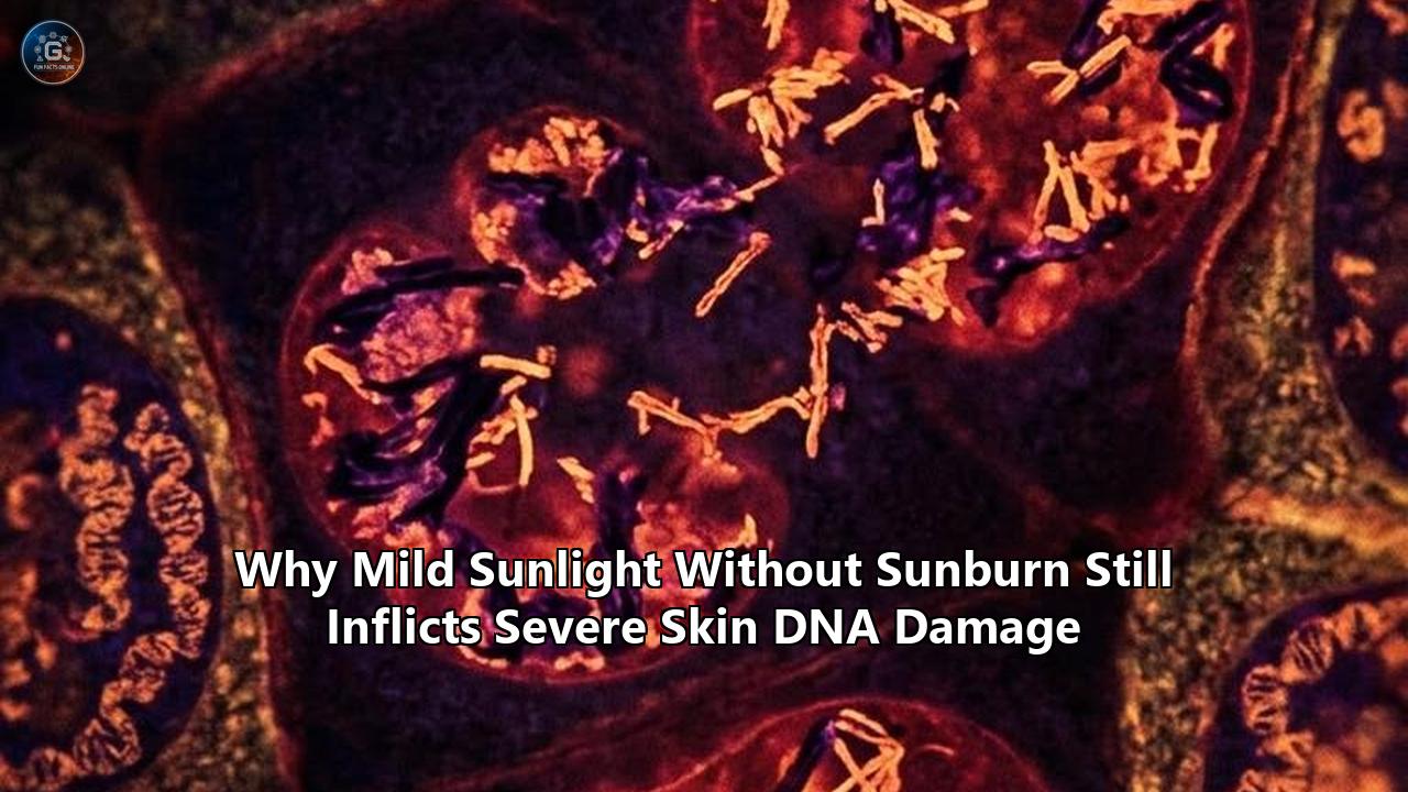
The findings, published by the QIMR Berghofer Medical Research Institute in Brisbane, dismantle one of the most resilient dogmas in public health photobiology: the belief that skin remains biologically protected so long as it avoids erythema—the vascular inflammatory reaction commonly recognized as a sunburn.
In a controlled clinical trial named the Low-Dose UV Study, lead investigators Professor Rachel Neale and Professor David Whiteman AM examined 58 adult volunteers with light to olive skin tones (Fitzpatrick skin phototypes I through IV). Researchers delivered measured quantities of solar-simulated ultraviolet radiation (UVR) directly to test patches on the participants’ backs across multiple consecutive days. The exposures mimicked low-intensity ambient light—such as the weak solar flux found in mid-morning or late afternoon—as well as condensed, higher-intensity intervals equivalent to midday sun. Crucially, the cumulative UV doses were kept deliberately sub-erythemal, meaning not a single participant developed pink or inflamed skin.
Subsequent full-thickness punch biopsies and genomic profiling revealed that the participants' basal keratinocytes and melanocytes sustained severe, measurable DNA lesions regardless of exposure intensity. The biological damage was governed not by how fast the solar energy was delivered, but by the absolute number of photons absorbed.
"You can get the same dose of UV radiation in a short time in the middle of the day or a longer time earlier or later in the day. Our research has shown it is the total dose that is important—it doesn't matter how long it takes to get it," explained Professor Neale upon releasing the trial results. "People potentially get lulled into a sense of security when the intensity of sunlight is weak and they spend too long outdoors without adequate protection. We have shown that is a problem for skin damage."
Professor Whiteman underscored that while an individual episode of sub-erythemal exposure does not instantly produce a malignancy, its molecular footprint is indelible. "Our lab results show these small, incremental doses of UV have caused some damage to the DNA in the skin cells, enough for the cells to then respond to the damage," Whiteman said. "Over months and years, these episodes of incremental, incidental UV exposure will have a cumulative impact on the skin and can lead to mutations that will initiate skin cancer."
The trial acts as an incisive case study into a systemic blind spot in preventative medicine. For more than half a century, clinical dermatology, sun-safety campaigns, and international regulatory standards for sunscreens have relied on skin redness as their primary biological proxy for injury. The QIMR Berghofer data, arriving alongside newly illuminated biochemical mechanisms from laboratories at the University of South Florida and the University of Copenhagen, prove that erythema is not a reliable gauge of genomic security. Instead, silent, sub-erythemal sunlight dna damage occurs continually at solar intensities long considered harmless by the general public.
  Solar UV Exposure
         │
         ├───► High Intensity / Acute Dose ──► RNA Damage / Ribotoxic Stress (ZAKα) ──► Erythema (Sunburn)
         │                                                                                 ▲
         │                                                                                 │ (DECOUPLED)
         │                                                                                 ▼
         └───► Low Intensity / Chronic Dose ──► Direct CPDs + Dark Chemiexcitation ──► Somatic DNA Mutations
Deconstructing the Erythema Fallacy: Inside the Low-Dose UV Study
The QIMR Berghofer trial was designed to strip away the confounding variables that have historically compromised observational photodermatology. Prior population-level surveys frequently relied on self-reported sun habits, which tend to conflate outdoor leisure duration with personal recollections of burning. By contrasting discrete delivery rates of UV radiation in a regulated clinical environment, the Brisbane team isolated photon kinetics from thermal perception.
Each subject was exposed to precise doses of UV radiation equivalent to 0.25 to 0.5 of their individual Minimal Erythema Dose (MED)—the benchmark metric defined as the lowest radiant exposure necessary to produce the first perceptible, uniform redness 24 hours post-exposure. One group received this sub-erythemal allotment compressed into brief, higher-flux sessions, simulating unshaded midday conditions. Another group received an identical total radiant energy spread across prolonged, attenuated sessions resembling early morning gardening, walking the dog, or sitting beneath a shaded patio.
When the investigators analyzed tissue specimens collected 24 to 48 hours following exposure, the histological data were unequivocal:
- Equalized Photoproduct Burden: Both the high-rate and low-rate cohorts exhibited virtually identical densities of cyclobutane pyrimidine dimers (CPDs)—the primary molecular lesion caused by ultraviolet radiation. The rate of photon arrival did not cushion the genome against injury.
- Transcriptional Stress Responses: Skin tissue demonstrated pronounced upregulation of the tumor-suppressor protein p53, along with downstream transcriptional arrests indicating the cells had halted their replication cycles to manage structural chromosomal faults.
- Absence of Clinical Warnings: Despite this severe cellular trauma, the skin retained its baseline pigmentation and tone without clinical inflammation, edema, or microvascular engorgement.
For decades, the public has operated on the intuitive assumption that thermal heat and erythema are synonymous with cellular harm. If the skin is cool, or if the light feels gentle on an autumnal morning, the conscious mind registers zero threat. The Low-Dose UV Study exposes this heuristic as a physiological trap. Sunburn is an acute emergency alarm, but its silence does not signify cellular safety.
The Molecular Decoupling: Why Sunburn and DNA Mutation Follow Different Pathways
To understand why mild sunlight inflicts severe genomic alterations without causing a burn, it is necessary to examine how skin cells process ultraviolet photons. The field of photobiology has undergone a structural revision regarding why human skin turns red in the first place.
Historically, academic literature presumed that erythema was the direct consequence of extensive DNA damage. The long-standing hypothesis posited that as keratinocytes suffered massive DNA double-strand breaks and nucleotide lesions, their apoptosis prompted a wholesale release of pro-inflammatory cytokines, initiating local microvascular dilation.
Investigations led by Dr. Anna Constance Vind and Professor Simon Bekker-Jensen at the University of Copenhagen, published in tandem with collaborators at Nanyang Technological University, revealed that visible sunburn is driven primarily by ribotoxic stress rather than direct genomic breaks. UV radiation strikes and cleaves cytosolic messenger and ribosomal RNA. This damage causes translating ribosomes to collide and stall during protein synthesis.
The ribosome stall is subsequently detected by a specialized sentinel kinase known as ZAK-alpha (ZAKα). When ZAKα senses the arrested translation machinery, it triggers the ribotoxic stress response, activating downstream p38 and JNK mitogen-activated protein kinase cascades. These pathways drive the swift synthesis of prostaglandins, leukotrienes, and interleukins that force nearby dermal capillaries to dilate, generating classic erythema, localized heat, and swelling.
  [UV Photons] ──► Dermal Cleavage of mRNA/rRNA ──► Ribosome Stalling
                                                           │
                                                           ▼
                                                    ZAKα Kinase Alarm
                                                           │
                                                           ▼
                                               p38/JNK Signaling Cascade
                                                           │
                                                           ▼
                                              Prostaglandin Release & Erythema
This discovery provides the missing mechanistic explanation for the QIMR Berghofer findings. The ribotoxic threshold required to trigger ZAKα-mediated sunburn is relatively high. It requires an acute, dense flux of shorter-wavelength UVB photons to destabilize ribosomes at a scale that trips the body's vascular fire alarm.
Nuclear DNA, however, possesses no such high-barrier alarm system. Direct cyclobutane dimer formation occurs upon the absorption of individual, single photons by adjacent pyrimidine bases (thymine or cytosine). There is no minimum threshold or molecular firewall required to initiate sunlight dna damage. A solitary photon possessing sufficient energy can induce covalent cross-links between neighboring bases instantly, bending the DNA helix by approximately 30 degrees and stalling replication forks.
Because the DNA damage response does not inherently initiate rapid, widespread prostaglandin-driven vasodilation, the cell undergoes extensive mutagenesis in complete silence. Erythema and genotoxicity are biochemically decoupled. The absence of a sunburn simply indicates that the ribotoxic stress response was avoided, while the nuclear genome may have absorbed thousands of mutagenic hits.
Expanding the Action Spectrum: Dark CPDs and the UVA Threat
The vulnerability of human skin to weak sunlight is further compounded by the optical composition of ambient solar radiation. Terrestrial sunlight that strikes the Earth’s surface during early morning, late afternoon, or through high-altitude cloud cover is not evenly distributed across the ultraviolet spectrum.
| Spectral Band | Wavelength Range | Environmental Proportion | Direct Biological Target | Penetration Depth | Erythema Contribution | Primary Mutation Mode |
|---|---|---|---|---|---|---|
| UVB | 290 – 320 nm | ~5% at solar noon; <1% in low sun | DNA, RNA, Keratin | Stratum Corneum & Epidermis | Very High (Major driver of sunburn) | Direct absorption photoproducts (CPDs, 6-4PPs) |
| UVA-II | 320 – 340 nm | Substantial throughout daylight | Endogenous photosensitizers, Melanin | Basal Epidermis & Upper Papillary Dermis | Moderate to Low | Mixed: Direct CPDs & radical-driven oxidation |
| UVA-I | 340 – 400 nm | >90% of all terrestrial UV | Melanin, Porphyrins, Flavins | Deep Basal Layer, Reticular Dermis | Minimal | Indirect ROS, Chemiewcitation, "Dark CPDs" |
As demonstrated by the table above, low-angle or "weak" sunlight is dramatically depleted of short-wavelength UVB because those rays are scattered and absorbed by the oblique traversal of the atmosphere. Instead, sub-erythemal ambient light consists almost entirely of UVA radiation.
For decades, basic dermatological pedagogy held that UVA posed primarily an aesthetic risk—photoaging, elastin degeneration, and matrix metalloproteinase induction—while UVB was branded the sole oncogenic culprit. A landmark study published by Dr. Marcus Cooke of the University of South Florida and Dr. Sanjay Premi of the Moffitt Cancer Center radically revised this model.
The USF and Moffitt research team demonstrated that the biological action spectrum for UV genotoxicity is far wider than traditional public health models claim. Historically, the action spectrum was thought to mimic the direct physical absorption curve of purified DNA, which peaks sharply in the deep UVB range and collapses to nearly zero across the UVA spectrum. Cooke and Premi revealed that cells generate extensive DNA-damaging excited states through complex, long-wavelength redox chemistry.
  UVA Photons (Weak / Low-Angle Sun)
         │
         ▼
  Melanin & Endogenous Chromophore Activation
         │
         ▼
  Induction of Nitric Oxide Synthase (iNOS) & NADPH Oxidase (NOX)
         │
         ▼
  Generation of Superoxide (O₂•⁻) + Nitric Oxide (•NO) ──► Peroxynitrite (ONOO⁻)
                                                                 │
                                                                 ▼
                                                  Excitation of Melanin Fragments
                                                                 │
                                                                 ▼
                                                Triplet State Dioxetane Energy Transfer
                                                                 │
                                                                 ▼
                                                 CPD Formation in the Dark ("Dark CPDs")
This phenomenon centers on what is known as chemiexcitation. When lower-energy UVA photons, ubiquitous in gentle early-morning sunlight, penetrate deep into the skin's basal layer, they excite electrons inside melanin polymers and cellular chromophores. This excitation triggers enzymes including inducible nitric oxide synthase (iNOS) and NADPH oxidase, generating superoxide radicals ($O_2^{\bullet-}$) and nitric oxide ($\text{NO}$).
These radicals instantly combine to form peroxynitrite ($\text{ONOO}^-$), an exceptionally potent oxidant. The peroxynitrite cleaves melanin macromolecular complexes into high-energy dioxetane intermediates. As these unstable rings degrade, they do not release their energy as harmless fluorescence or heat. Instead, via an electron-exchange mechanism, they transfer radiationless triplet-state excitation energy directly onto nuclear DNA.
The devastating biological consequence is that cyclobutane pyrimidine dimers—the classic mutations previously thought to arise only from direct UVB absorption—continue to assemble inside skin cells for more than three to four hours after an individual has completely left the sun.
"Over half of a person's DNA damage arises in the car on the way home from the beach or after returning to the office," observed Dr. Douglas Brash, clinical professor of therapeutic radiology and dermatology at Yale School of Medicine, who pioneered early work on chemiexcitation with Premi. Melanin, long championed as the ultimate physiological shield against cutaneous malignancy, functions as a double-edged sword. In mild, sub-erythemal sunlight, eumelanin and pheomelanin absorb abundant UVA photons and subsequently channel that stored energy into the cell's genetic core via chemiexcited intermediates, creating "dark CPDs" long into the evening.
Public Health Blind Spots: The Proxy Metric Trap
The societal persistence of the belief that gentle sun is harmless provides an instructive study in how public health policies can inadvertently engineer collective vulnerabilities. When health agencies distilled complex photobiology into actionable consumer guidance in the late twentieth century, they required quantifiable, easily recognizable metrics. They selected two primary anchors:
- The Minimal Erythema Dose (MED): Used as the foundational biological unit for all photoprotection evaluations.
- The Sun Protection Factor (SPF): An in vivo clinical index defined explicitly as the ratio between the UV dose required to produce erythema on sunscreen-protected skin compared to unprotected skin:
$$\text{SPF} = \frac{\text{MED}_{\text{protected}}}{\text{MED}_{\text{unprotected}}}$$
By yoking both consumer warnings and regulatory testing to the endpoint of visible vascular inflammation, health frameworks established an unwritten, erroneous syllogism in the public consciousness:
$$\text{Sunburn causes cancer} \implies \text{No sunburn} = \text{No damage}$$
This simplification created profound secondary failures across health communication, environmental policy, and individual behavior.
  Traditional Assumption:
  [UV Exposure] ──────► [Visible Redness / Erythema] ──────► [Somatic DNA Damage]
                         (Assumed Universal Indicator)

  Actual Biological Reality:
  [UV Exposure] ──┬───► [Direct CPDs + Oxidative Chemiexcitation] ──► [Mutations] (Silent)
                  │
                  └───► [ZAKα Ribosomal Cleavage] ─────────────► [Erythema] (Lagged Alarm)
The UV Index "Free Pass"
World Health Organization (WHO) and regional meteorological guidelines universally advise the public to apply sun protection when the global solar Ultraviolet Index (UVI) reaches 3 or higher. Conversely, when the UVI is reported between 0 and 2—a common reading throughout autumn, winter, early morning, and late afternoon across temperate zones—the official guidance universally dictates that "no protection is required."
The QIMR Berghofer findings expose this blanket threshold as mathematically and biologically inaccurate. The UV Index is structurally weighted against the McKinely-Diffey Erythema Action Spectrum, an internationally accepted formula that measures how efficiently specific wavelengths turn skin red.
Because the erythema action spectrum drops by a factor of 1,000 between 300 nm (UVB) and 360 nm (UVA), the index severely discounts the massive UVA flux present during early mornings, overcast skies, and shoulder seasons. A UVI rating of 2 delivered over three hours of continuous outdoor activity can deliver an aggregate photon load capable of causing significant sunlight dna damage, even while registering as zero on a standard erythema scale.
  Solar Wavelength Sensitivity:

  Erythema Action Spectrum (Index Weighting):
  300 nm (UVB) | ████████████████████████████████████ (Factor 1.0)
  360 nm (UVA) | █ (Factor 0.001)  <-- Vastly discounted by UVI!

  True Mutagenic Action Spectrum (CPDs + Chemiexcitation):
  300 nm (UVB) | ████████████████████████████████████ (Direct CPDs)
  360 nm (UVA) | ████████████████████ (Significant "Dark CPDs" via Melanin)
The Illusion of Thermal Warning
Humans possess no biological sensory receptors for ultraviolet photons. Our conscious thermal sensations are mediated entirely by transient receptor potential (TRP) channels that react to infrared radiation (heat) and visible light.
When outdoor temperatures feel pleasant—such as 21°C (70°F) on a breezy spring morning—individuals experience zero thermal discomfort. Because human psychology intuitively relies on discomfort to gauge physical hazard, people stay outdoors for prolonged durations without photoprotective garments or topical barriers, remaining entirely unaware that the radiant flux is inflicting billions of base-pair alterations throughout their epidermises.
The Behavioral Compensation of "Safe Hours"
Public health campaigns routinely urge people to "avoid the midday sun between 10:00 AM and 4:00 PM." While this advice effectively caps peak exposure to short-wavelength UVB, it has caused an adverse compensatory behavior: individuals intentionally schedule outdoor sports, yard work, and social engagements outside those hours under the conviction that the light is entirely benign.
The QIMR Berghofer study demonstrated that spending two hours in mild 9:00 AM sunlight can deliver an identical sub-erythemal genotoxic dose to spending twenty minutes in direct 1:00 PM sun. By shifting the temporal window without deploying broad-spectrum protection, millions of people unknowingly substitute a rapid, noticeable burn for a protracted, invisible shower of mutagenic photons.
Stem Cell Reservoirs and the Proliferation of p53 Clones
The clinical consequence of repeated, sub-erythemal sunlight dna damage is the gradual, clonal transformation of human skin. Skin is not a static canvas; it is a rapid-turnover organ whose entire cellular surface is renewed every 28 to 40 days through the mitotic division of long-lived, self-renewing epidermal stem cells anchored within the basal layer.
When mild sunlight strikes the skin, high-energy photons bypass the upper, dead stratum corneum and deposit energy directly into this basal niche. When uncorrected by nucleotide excision repair (NER) pathways, the resultant cyclobutane pyrimidine dimers provoke distinctive genetic alterations known as "UV signature mutations": cytosine-to-thymine ($C \rightarrow T$) and tandem double cytosine-to-double thymine ($CC \rightarrow TT$) base transitions.
  Basal Keratinocyte Stem Cell
         │
         ├───► Sub-Erythemal Photon Influx (No Burn, No Alarm)
         │
         ├───► CPD Cross-Link Unrepaired by NER
         │
         ▼
  DNA Polymerase η (eta) Translesion Error during Replication
         │
         ▼
  C ──► T or CC ──► TT Transition Mutation in TP53 Gene
         │
         ▼
  Loss of Apoptosis Signaling
         │
         ▼
  Competitive Clonal Expansion over Normal Cells
         │
         ▼
  Macroscopic "Patch" of Mutated Keratinocytes (Actinic Keratosis / Field Cancerization)
The primary genomic casualty of this process is the TP53 tumor suppressor gene. Known as the "guardian of the genome," TP53 transcribes a master transcription factor that arrests the cell cycle when DNA is compromised, either directing enzymatic repair or executing programmed cell death (apoptosis) if the chromosome is irreparably shattered.
When sub-erythemal exposures inflict a $C \rightarrow T$ transition within key DNA-binding domains of TP53, the stem cell becomes functionally crippled. It loses the capacity to commit apoptosis when genetically compromised. Liberated from normal growth checkpoints, this mutated stem cell outcompetes its normal, unmutated wild-type neighbors. It expands laterally across the basal lamina, seeding an invisible colony of pre-malignant cells known as a p53 clone.
Deep single-cell genomic sequencing has demonstrated that clinically pristine, normal-appearing skin in individuals over forty years of age contains thousands of these distinct mutated clonal patches. Each patch can encompass millions of epidermal cells.
Subsequent mild sun exposures do not kill these clones. Instead, they provide a positive selection pressure. The mild UV insults selectively eliminate vulnerable wild-type cells via baseline pathways while sparing the apoptosis-resistant TP53-mutated clones, allowing them to expand further. Over decades, this process of field cancerization yields the visible precursors of cutaneous oncology: actinic keratoses, basal cell carcinomas (BCC), and invasive cutaneous squamous cell carcinomas (cSCC).
The critical epidemiological realization extracted from the QIMR Berghofer data is that this clonal architecture is not built solely during dramatic, blistering sunburns suffered on tropical vacations. It is systematically assembled, stone by stone, through decades of daily incidental transit, morning dog walks, and outdoor gardening under low-intensity sunlight.
The Regulatory Crisis: Structural Flaws in Modern Photoprotection
The realization that low-intensity sunlight silently drives skin carcinogenesis exposes systemic flaws in international photoprotection standards. For decades, the global personal care sector and drug regulators have prioritized metrics that shield against acute vascular response rather than silent genotoxicity.
  Sunscreen Formulation Priority Disconnect:

  Market Standard (Current Regulation):
  [High UVB Filtration] ──► Extinguishes Erythema (High SPF Number) ──► Consumer Assumes Safety
  [Minimal Broad-Spectrum] ─► Lets Sub-Erythemal UVA Through ───────► DNA Damage Continues Unchecked

  Required Biological Standard:
  [Broad-Spectrum UV Balance] ──► Eliminates Direct CPDs
  [Topical Radical Quenchers] ─► Blocks Dark Chemiexcitation
  [DNA Repair Enzymes] ────────► Accelerates Removal of Incidental Lesions
The Limitations of the SPF Formula
Because SPF is defined entirely by the suppression of erythema, a sunscreen with an SPF rating of 50 is legally formulated to delay visible skin reddening by a factor of 50. In practical terms, it attenuates UVB wavelengths with extraordinary efficacy. However, unless formulated to meet the highest international broad-spectrum standards (such as the European Commission's mandatory 1:3 UVA-to-UVB protection factor ratio), an SPF 50 lotion may allow substantial levels of long-wavelength UVA to penetrate the basal layer unobstructed.
When users apply such formulations, they experience zero sunburn, enabling them to remain outdoors during gentle, low-angle sunlight for prolonged periods under the assumption that they are entirely insulated from risk. In reality, while their skin remains visually pale, the uninterrupted influx of sub-erythemal UVA photons drives relentless, uninterrupted dark CPD production via chemiexcited melanin intermediates.
The Measurement Void for Cellular Protection
Currently, consumer protection bodies possess no standardized, commercially deployed metric for measuring a product’s Biological DNA Protection Factor (DPF). While the persistent pigment darkening (PPD) assay and the critical wavelength standard ($\lambda_c \ge 370\text{ nm}$) measure broad-spectrum attenuation, neither monitors real-time suppression of cyclobutane dimers, 8-oxo-deoxyguanosine (8-oxo-dG) base oxidations, or post-exposure radical cascades.
Dermatological chemists are now arguing that photoprotection must extend beyond simple reflective or absorbing filters to include:
- Chemiexcitation Quenchers: Molecules capable of safely neutralizing high-energy triplet-state dioxetane intermediates before their energy can transfer to DNA. Compounds such as ethyl sorbate and specialized alpha-tocopherol derivatives have exhibited the capacity to interrupt this post-sun chemical cascade.
- Enzymatic Repair Accelerators: Topical delivery of encapsulated bacterial and algal DNA repair enzymes—most notably T4 endonuclease V and CPD-photolyases—that directly target and excise pyrimidine dimers before transcription stalls can trigger somatic mutations.
- Endogenous Antioxidant Up-regulators: Agents that stimulate nuclear factor erythroid 2-related factor 2 (Nrf2) pathways to augment intrinsic intracellular defenses against peroxynitrite-driven stress.
The Public Health Paradox: Vitamin D and Cardiovascular Optimization
The realization that mild sunlight inflicts severe genomic alterations forces a direct, contentious confrontation with widespread clinical guidelines regarding vitamin D synthesis and systemic health.
Over the past two decades, bone health and immunology research communities have issued widespread public recommendations urging populations to seek 10 to 20 minutes of daily unprotected sun exposure during non-peak hours (early morning or late afternoon). The biological rationale appeared sound: brief, low-intensity exposure was believed to facilitate cutaneous cholecalciferol synthesis via the photolysis of 7-dehydrocholesterol without incurring the tissue damage or carcinogenic risk associated with a painful sunburn.
       The Incidental Exposure Dilemma:

       Advised Exposure Window: Mild Sun (Low-Angle / Non-Peak)
                          │
         ┌────────────────┴────────────────┐
         ▼                                 ▼
   Vitamin D Synthesis               Genomic Reality
  - Photolysis of 7-DHC             - Direct Pyrimidine Dimers
  - Systemic Nitric Oxide Release   - Chemiexcitation "Dark CPDs"
  - Trace calcitriol conversion     - Basal TP53 Mutations
                                    - Long-Term Skin Cancer Risk
The clinical findings from QIMR Berghofer, paired with the USF/Moffitt data, severely complicate this recommendation:
- Inefficient Vitamin D Wavelengths in Low-Angle Light: The synthesis of previtamin D3 occurs almost exclusively within a razor-thin optical window of UVB radiation, peaking sharply between 295 nm and 303 nm. In mild, low-angle sunlight, this precise narrow band is largely filtered out by the Earth’s atmosphere. Consequently, seeking "gentle" sunlight for vitamin D synthesis is an inefficient enterprise: the individual absorbs substantial doses of mutagenic UVA, which drives oxidative and chemiexcited sunlight dna damage, while receiving negligible quantities of the exact UVB photons required to synthesize cholecalciferol.
- Cardiovascular Nitric Oxide Release: Photodermatologists have demonstrated that UVA light mobilizes stored bioactive nitric oxide metabolites (nitrite and S-nitrosothiols) from dermal stores into systemic circulation, promoting vasodilation, lowering resting arterial blood pressure, and reducing cardiovascular mortality. This sets up a profound biological conflict: the same sub-erythemal photons that trigger chemiexcitation and induce DNA lesions appear to confer measurable cardiovascular benefits.
Given the irreversible nature of somatic chromosomal damage, preventative health models are reconsidering this trade-off. Nutritional delivery of vitamin D via oral supplementation bypasses cutaneous mutagenic risk entirely. Conversely, substituting dietary interventions for sunlight-induced cardiovascular improvements remains a complex metabolic challenge, reinforcing the necessity for sunscreen formulations that preserve beneficial systemic biochemical dynamics while neutralizing mutagenic photon cascades.
Actionable Photobiological Guidance Under the New Model
The collapse of the "burn-first" risk paradigm requires a structural pivot in how individuals manage real-world solar exposure. Clinical photobiologists emphasize several operational protocols:
  Traditional Guidance (Flawed)         Updated Biological Protocol
  ─────────────────────────────────     ──────────────────────────────────────────
  - Seek shade only if turning pink     - Treat any direct daylight as mutagenic
  - Rely on UV Index ≥ 3 warnings       - Use physical shade at all daytime UVI
  - Unprotected morning sun for vit D   - Oral vitamin D; eliminate unprotected sun
  - SPF numbers define safety           - Prioritize high-UVA/broad-spectrum ratios
- Cease protection when out of sun - Post-exposure antioxidant applications- Rethink the Shading Baseline: Do not treat shade, cloud cover, or early daylight hours as an all-clear signal. If an environment provides sufficient optical illumination to cast a perceptible shadow, it contains adequate radiant energy to initiate cyclobutane dimer synthesis within cutaneous stem cells.
- Enforce Broad-Spectrum Balance Over Raw SPF Numbers: When utilizing topical photoprotection, high SPF values (e.g., SPF 100) must not be treated as a license to extend time outdoors, particularly in mild, non-burning light. Formulations must specifically feature verified broad-spectrum ratings—such as the European Union's UVA Circle icon, the UK Boots 5-Star metric, or an explicit critical wavelength $\ge 370\text{ nm}$—to ensure that the long-wavelength rays responsible for dark CPD generation are mitigated.
- Account for Post-Exposure Reaction Windows: Acknowledging that chemiexcited genomic damage persists for several hours after retreating indoors necessitates the deployment of evening-after photoprotection. Topically applied antioxidant regimens containing high-potency radical quenchers, lipid-soluble vitamins, and DNA repair liposomes should be utilized after sunlight exposure ends to disrupt the persistent peroxynitrite-melanin cascade.
- Adopt Structural Shielding as the Primary Defense: Because chemical and mineral filters suffer from uneven user application, degradation, and perspiration wash-off, physical barriers—specifically clothing woven to verified Ultraviolet Protection Factor (UPF) standards, wide-brimmed headwear, and optical-grade eyewear—represent the only intervention that uniformly blocks both the ribotoxic wavelengths that cause sunburn and the longer, ambient rays that silently degrade cellular DNA.
The Horizon: Scientific Frontiers and Public Policy Redesign
The QIMR Berghofer clinical trial marks the beginning of an overhaul in how science approaches the boundaries of photodamage. As research laboratories worldwide digest the data from Brisbane, USF, and Copenhagen, the dermatology landscape faces an aggressive slate of scientific, regulatory, and epidemiological questions.
  Near-Term Frontiers in Photobiology:

  1. Clinical Metrics       ──► Transition from MED to "DNA Protection Factor" (DPF)
  2. Public Policy          ──► Overhaul of the Global UV Index & "Safe Hours" Messaging
  3. Formulation Science    ──► Integration of Triplet-State Quenchers & CPD Repair Enzymes
  4. Population Genetics    ──► Evaluating Chemiexcitation Dynamics across Fitzpatrick I-VI
Developing In Vivo DNA Biosensors
Clinical trials require non-invasive diagnostic methodologies to evaluate genomic trauma in real time. The invasive full-thickness punch biopsies used in the QIMR Berghofer study are impractical for wide-scale consumer safety assessments.
Teams across biomedical engineering are developing non-invasive tape-stripping assays coupled with ultra-sensitive droplet digital PCR (ddPCR) to measure the immediate shedding of cyclobutane pyrimidine dimers and 6-4 photoproducts from the surface of the stratum corneum. This technology promises to enable real-time, consumer-accessible tracking of silent cellular trauma, definitively retiring reliance on the visual onset of erythema.
Revising Global Meteorological Warning Systems
The revelation that low-level UV exposures accumulate to produce high-grade mutagenic burdens forces a necessary recalculation of the World Health Organization’s Global Solar UV Index.
Working groups within photobiology and atmospheric chemistry are drafting revisions to replace the singular, erythema-weighted curve with a dual-reporting framework:
- The Erythemal Metric: Warning populations of acute sunburn risk, ribotoxic stress, and immediate ocular injury.
- The Cumulative Genotoxic Index: Reporting absolute photon energy across the UVA-UVB spectrum to inform populations of silent, mutational risks irrespective of temperature, cloud cover, or solar zenith angle.
Investigating Variable Ethnic Photobiology
A major unresolved frontier is the precise kinetics of chemiexcitation and sub-erythemal injury across diverse skin phototypes. While melanin provides significant photoprotective filtration against direct UVB-induced pyrimidine dimerization, its role as a persistent reservoir for post-exposure chemiexcitation means that darker skin types (Fitzpatrick IV through VI) experience distinct, long-lasting radical cascades.
Pilot studies from institutions like the Charité - Universitätsmedizin Berlin reveal that while dark skin exhibits initial melanin-mediated shielding against acute damage, it displays delayed DNA repair kinetics and substantial residual lesions 24 hours post-exposure due to melanin photosensitization. Establishing how different ratios of eumelanin to pheomelanin affect dark CPD formation in non-burning light remains one of the most critical objectives in modern cellular photobiology.
The Low-Dose UV Study delivers a final, unvarnished biological principle: human skin does not possess a "free pass" in the sun. The biological boundary between safety and damage does not align with the threshold between pale and red skin. Erythema is merely an inflammatory reaction to collapsed ribosomes, while the cellular genome degrades quietly, photon by individual photon, in the gentlest light nature provides.
As photoprotective medicine rewires its assumptions, the imperative for clinical practice, regulatory agencies, and the broader public is clear: sun protection can no longer be treated as an emergency protocol reserved for the scorching midday beach, but as an ongoing physiological defense against the invisible mutagenicity of daylight itself.
Reference:
- https://www.sciencedaily.com/releases/2026/09/260930020324.htm
- https://www.usf.edu/arts-sciences/news/2026/sept-26/dna-damage-continues-long-after-sun-exposure-usf-researchers-say-protective-measures-need-to-catch-up.aspx
- https://www.techexplorist.com/new-study-reveals-surprising-cause-sunburn-not-dna/96194/
- https://pubmed.ncbi.nlm.nih.gov/40617063/
- https://pourmoiskincare.com/blogs/beautyforecast/sunburn-reimagined-how-a-new-study-flips-everything-we-know-about-uv-damage
- https://news.yale.edu/2015/02/19/sunlight-continues-damage-skin-dark
- https://medicine.yale.edu/news-article/sun-damage-occurs-even-after-sunset/
- https://www.oncology-central.com/study-indicates-that-sunlight-damages-skin-for-hours-after-exposure-and-in-the-dark/
- https://www.cbsnews.com/news/sun-damage-to-skin-continues-after-dark/