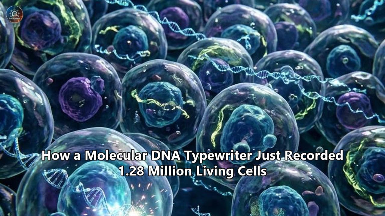
On October 8, 2026, researchers at the Seattle Hub for Synthetic Biology, the University of Washington School of Medicine, and Dartmouth College unveiled a cellular family tree that developmental biologists had sought for half a century. Writing in Science, a team led by genomicists Jay Shendure, Junhong Choi, and Chengxiang Qiu reported the dense lineage reconstruction of 1,281,141 living cells inside a mammalian embryo, tracing every single one of them back to the fertilized egg.
The scale of the dataset is unprecedented in mammalian biology. By deploying an engineered molecular recorder directly into a single-cell mouse zygote, the researchers logged embryonic cell divisions continuously across 13.5 days of gestation—roughly two-thirds of the animal’s pregnancy. When the embryo was harvested at late organogenesis, its heart was beating, its neural tube had closed into distinct brain ventricles, and its limb buds were forming digits. Tucked inside the nucleus of more than 1.28 million of those embryonic cells was a physical, readable diary stamped directly into the double helix: an engineered molecular DNA typewriter that had typed sequential genetic symbols across engineered synthetic tapes every time a cell divided.
“Think of it like an actual typewriter, except it types onto a cell’s own DNA instead of paper,” explained Haedong Kim, a postdoctoral researcher at the University of Washington and co-first author of the paper. “Every time a cell divides, it strikes one new character onto the next blank line, always in order, never overwriting what’s already there”.
The resulting map reveals foundational rules of mammalian life that had previously remained inaccessible. It proves that the very first cleavage of the fertilized egg creates an asymmetric legacy: one founding daughter cell generates roughly 57 percent of the animal’s body, while the sister cell accounts for 42 percent. Yet despite this numerical imbalance, both blastomeres retain identical potential, contributing equally to all primary germ layers and 138 differentiated cell types. Furthermore, the molecular clockwork proved that cellular fates do not lock into place simultaneously: while red blood cells and retinal precursors commit to their specialized identities early in development, the outer layers of the skin remain developmentally flexible until late organogenesis.
Decades of developmental biology led to this point. Mammalian embryogenesis occurs inside an opaque uterus, obscuring billions of migrating, dividing, and differentiating cells from direct visual inspection. Reconstructing this living history required converting the tools of genome editing from molecular scissors into an indelible, sequential recording apparatus.
1974–2015: The Invariable Worm and the Mammalian Black Box
The dream of reconstructing an animal’s entire cellular genealogy began in a laboratory basement in Cambridge, England. Between 1974 and 1983, British biologist John Sulston sat hunched over a differential interference contrast microscope, tracking cell divisions in the transparent roundworm Caenorhabditis elegans. Sulston sketched each division by hand in notebooks, following cells through mitosis, migration, and apoptosis until he had charted every branch of the worm’s 959 somatic cells.
Sulston’s triumph earned him a Nobel Prize, but it also highlighted a frustrating limitation: C. elegans is an evolutionary anomaly. Its development is rigidly deterministic. Every single wild-type worm develops along an invariant script, with the exact same cell division generating the exact same tissue at the exact same minute.
Mammals obey no such script.
A mammalian embryo develops through stochastic, regulative biology. If an experimenter destroys half the cells of an early mammalian embryo, the remaining cells compensate, adjusting their signaling gradients and yielding a complete, viable organism. Furthermore, mammalian embryos are opaque and develop inside a maternal womb. A mouse begins as a single cell roughly 80 micrometers in diameter; two weeks later, it has expanded into tens of millions of cells folded into intricate three-dimensional organ structures. By adulthood, that number swells past several billion.
For decades, biologists attempted to peer through this opacity using optical and biochemical labels:
- Fluorescent Dyes: Lipophilic dyes such as DiI were injected into embryonic regions under a microscope. While useful for short-term tracking, the dyes dilute by 50 percent with every mitotic division, fading to invisibility after five or six cycles.
- Retroviral Barcodes: In the late 1980s and 1990s, teams used defective retroviruses to deliver unique genetic markers into early blastomeres. While permanently integrated into the host genome, retroviral tagging suffered from an intrinsic limitation: it provided a static snapshot. It marked the ancestor, but could not record subsequent branching events within the developing clone.
- The Brainbow Strategy: In 2007, researchers developed combinatorial fluorescent protein cassettes that assigned distinct hues to individual cells via Cre-lox recombination. Brainbow revolutionized neuroanatomy, but spectral overlap capped its discriminative resolution at a few dozen colors—orders of magnitude below what is required to track an entire organ system.
Developmental biologists realized that optics could never solve the mammalian problem. Light scatters; deep tissues remain dark; and observing a living mammalian fetus continuously at cellular resolution across weeks in utero was biophysically impossible. If an embryo’s history was to be recovered, the cells would have to record that history themselves.
2016–2019: The Double-Strand Dilemma and the Scramble for Digital Marks
The development of CRISPR-Cas9 genome editing in the early 2010s transformed the field. Biologists recognized that if Cas9 could be directed to cut synthetic target sites engineered into an organism’s genome, the cell’s error-prone repair pathways would patch the wound with insertions and deletions (indels). These indels, inherited by all subsequent daughter cells, could function as permanent genetic scars—a molecular paper trail.
In 2016, Jay Shendure and his colleagues at the University of Washington published GESTALT (Genome Editing of Synthetic Target Arrays for Lineage Tracing). By introducing Cas9 into zebrafish embryos containing an array of CRISPR target sites, GESTALT demonstrated that accumulating indels could reconstruct lineages across early embryonic tissues. Similar systems emerged in rapid succession: MEMOIR, ScarTrace, and LINNAEUS.
Yet as researchers attempted to push Cas9 scar-based recording from simple aquatic organisms into mammals, the strategy hit a biological wall.
The primary mechanism of standard CRISPR-based lineage recording relied on double-strand breaks (DSBs) repaired by non-homologous end joining (NHEJ). When deployed across long developmental timescales in complex tissues, DSB-based scars created catastrophic data corruption:
- Large Deletions and Information Loss: When multiple guide RNAs targeted adjacent sites in a recording array, Cas9 often cut several loci simultaneously. Instead of producing tidy, sequential point indels, the cellular repair machinery bridged the distal cuts, excising the intervening sequence entirely. These deletions erased previous recording marks, destroying the very lineage data researchers were trying to preserve.
- Burnout and Premature Saturation: Standard Cas9 target sites possess a narrow temporal window. Because the enzyme cuts rapidly, the majority of editable sites in an engineered array were consumed within the first few cell divisions. By the time the embryo initiated gastrulation, the recording array was “burned out,” leaving late-stage organogenesis completely unmonitored.
- Loss of Temporal Order: Standard indels are stochastic and unordered. If a cell carried a 3-base-pair deletion at site A and a 2-base-pair insertion at site B, there was no physical way to determine which mutation occurred first. Temporal order had to be reconstructed through probabilistic inference, introducing substantial phylogenetic noise.
- Cellular Toxicity: Repeatedly severing both strands of the mammalian genome activates p53-dependent DNA damage checkpoints. In early embryos, frequent double-strand breaks induce cell cycle arrest, trigger apoptosis, or select for clones with impaired DNA-repair pathways, fundamentally skewing normal development.
Biologists found themselves with an imperfect recorder. To map an entire mammalian embryo, they needed an entirely different physical mechanism: an editor that could write new information without severing the DNA backbone, advance across a recording tape in a strict chronological sequence, and operate steadily without exhausting its substrate.
2019–2022: The Birth of the Typehead Mechanism
The solution arrived through a synthesis of synthetic biology and precision genome editing. In late 2019, David Liu’s laboratory at the Broad Institute published prime editing, an engineered system that combined an engineered Cas9 nickase (H840A) with an engineered reverse transcriptase. Instead of breaking both DNA strands, prime editing nicks a single strand and uses an extended prime editing guide RNA (pegRNA) to directly reverse-transcribe new genetic sequences into the target locus without requiring double-strand breaks or donor DNA templates.
At the University of Washington, Jay Shendure and postdoctoral fellow Junhong Choi saw the prime editor not merely as a therapeutic gene repair tool, but as a potential mechanical write-head.
Prime editing operates with surgical precision. It does not induce widespread indels, and it allows researchers to specify the exact nucleotide string being inserted. Choi and Shendure set out to design a molecular engine where the insertion of a DNA sequence at one site physically created the target for the next edit, shifting the write-head down a repetitive DNA register like the carriage of an early twentieth-century typewriter.
The architecture they engineered, formally introduced in Nature in July 2022, was the molecular DNA typewriter.
THE DNA TYPEWRITER RECORDING LOGIC
─────────────────────────────────────────────────────────────────────────────
Step 1: Inactive Tape
[5' KEY ]---[ Monomer 1: Active Typehead ]---[ Monomer 2: Truncated ]---[ Monomer 3: Truncated ]

Step 2: Keystroke Event (Prime Editor + pegRNA)
           ▼
[5' KEY ]---[ Symbol Inserted: NNNGGA ]
                     └─ Disables Monomer 1 Typehead
                     └─ Supplies missing 'GGA' Key to Monomer 2

Step 3: Carriage Shifted
[ Monomer 1 (Spent) ]---[ Monomer 2: NEW Active Typehead ]---[ Monomer 3: Truncated ]
─────────────────────────────────────────────────────────────────────────────
The system relied on three interlocking synthetic components:
- The DNA Tape: An engineered genomic cassette consisting of tandem repeats of a 14-base-pair monomer. Crucially, all the monomers in the array except the first were truncated at their 5′ end by three base pairs, rendering them completely invisible and inaccessible to the prime editor. The first monomer was preceded by a 3-base-pair "key" sequence (GGA), making it the sole functional protospacer—the active "typehead".
- The Prime Editor (PEmax): An enhanced prime editor enzyme engineered for maximum mammalian activity while maintaining strict single-strand nicking fidelity.
- The Symbolic Guide RNAs (epegRNAs): Engineered guide RNAs that specified a 6-nucleotide insertion consisting of a variable 3-nucleotide symbol followed by the 3-nucleotide key (NNNGGA).
The mechanical elegance of the design lay in its sequential irreversibility. When the prime editor complex engaged the active typehead at Monomer 1, it nicked the strand and reverse-transcribed the 6-base-pair symbol into the locus. This single event had two physical consequences:
- It altered the sequence of Monomer 1, permanently destroying its typehead so it could never be recognized or edited again.
- Because the inserted symbol ended with the three-base-pair "GGA" key, its physical presence completed the truncated sequence of Monomer 2 immediately downstream.
Monomer 2 was now an active typehead. The carriage had advanced one space. Monomers could only be written 5′ to 3′, strictly one after another. Because double-strand breaks were avoided, adjacent records were never deleted.
The physical order of the letters on the DNA tape directly mirrored the temporal order of events inside the living cell.
2022–2024: From In Vitro Proof to the Multicellular Wall
In their initial 2022 proof-of-concept experiments, Choi, Shendure, and their collaborators demonstrated that the molecular DNA typewriter could record synthetic messages inside human HEK293T cells. They programmed cells to record thousands of sequential transfections and assembled lineage trees spanning several thousand cells cultured in standard plastic dishes.
Later, in 2024, the team published standardized protocols and demonstrated recording across monoclonal gastruloids—aggregates of embryonic stem cells that mimic early post-implantation development—building phylogenies of up to 50,000 cells. That same year, in a study published in Nature, they adapted the typewriter to record environmental signals, linking specific guide RNA expressions to Wnt and BMP signaling pathways.
Yet moving from 50,000 cells in a flat culture well to more than a million cells in a living, breathing mammalian fetus was considered by many in the field to be nearly impossible.
The developmental barriers were formidable. In vitro, researchers could flood cells with transgenes, maintain steady drug selections, and harvest cells at uniform densities. In an embryo, an engineered recorder faced biological realities:
THE IN VIVO SCALING BOTTLENECK
┌───────────────────────────────┬───────────────────────────────────────────────┐
│ In Vitro Gastruloids (2024)   │ In Vivo Mouse Embryo (2026 Target)            │
├───────────────────────────────┼───────────────────────────────────────────────┤
│ ~50,000 cells                 │ >1,000,000 cells                              │
│ Artificial synthetic media    │ Maternal-fetal metabolic gradients            │
│ Uniform plasmid transfection  │ Single pronuclear injection into 1-cell zygote│
│ High tolerability for dropout │ Unbiased capture across 100+ divergent tissues│
│ Low mechanical tissue shear   │ Massive cell migration, morphogenetic folding │
└───────────────────────────────┴───────────────────────────────────────────────┘
The UW team identified three specific failure modes that had ruined earlier attempts at whole-mammal lineage recording:
- Delivery Exhaustion: All components—the prime editor, the guide RNAs, the transposase, and multiple copies of the DNA tape array—had to enter the zygote within minutes of fertilization. If the recorder expressed too late, early development would go unrecorded. If it integrated erratically, different tissues would inherit different sets of tapes, destroying the global tree.
- Signal-to-Noise Retrieval: Developing embryos rapidly differentiate into rigid musculoskeletal matrices, dense neural clusters, and delicate endothelial sheets. Trying to dissociate a late-stage embryo into intact single cells destroys fragile cell types and leaves durable ones behind. The single-cell readout had to occur at the level of individual nuclei. Yet nuclei contain only a tiny fraction of the RNA transcripts present in the whole cytoplasm, making it difficult to recover synthetic barcodes from single nuclei without losing the cell’s transcriptional identity.
- Combinatorial Saturation: If an embryo carried only one or two tapes, the system would run out of recording capacity within a few generations. To record across millions of cells, each cell needed to harbor multiple, independently editing tapes, generating a combinatorial diversity large enough to prevent identical twin lineages from being misclassified as the same cell.
2024–2025: Engineering the Zygote Delivery Architecture
To bridge the gap between cell culture and developing organisms, the Seattle Hub team completely redesigned the physical tape and delivery chassis. Postdoctoral fellow Haedong Kim and senior computational biologist Chengxiang Qiu spent two years re-architecting the system for mouse pronuclear injection (PNI).
First, they engineered a circularized RNA recording format designated circTAPE.
Instead of relying on fragile linear messenger RNAs or transient nuclear transcripts, the researchers drove the expression of the DNA Tape using a Polymerase III promoter engineered to circularize the transcript. Circular RNA lacks free 5′ and 3′ ends, rendering it extraordinarily resistant to cellular exonucleases. This design ensured that throughout the rapid cell cycles of early embryogenesis, stable, high-copy circular RNA transcripts of the tape accumulated within the nucleus, ready to be captured during downstream single-nucleus combinatorial indexing.
Second, they solved the integration challenge using a piggyBac transposase system. They co-injected two plasmids alongside piggyBac transposase mRNA into the male pronucleus of fertilized C57BL/6 mouse zygotes:
- Construct 1: Encoded constitutively expressed PEmax driven by a ubiquitous mammalian promoter.
- Construct 2: Contained an engineered expression cassette bearing an NNNGGA-encoding epegRNA library, a unique 12-base-pair integration barcode (tape-BC), and a 6-monomer TAPE-1 array.
SCHEMATIC: THE IN VIVO PNI CONSTRUCT
─────────────────────────────────────────────────────────────────────────────
[ piggyBac ITR ]──[ Pol-III ]──[ epegRNA Library (NNNGGA) ]
                 ──[ 12-bp Tape-BC ]──[ 6x TAPE-1 Monomers (circTAPE) ]──[ piggyBac ITR ]
─────────────────────────────────────────────────────────────────────────────
This configuration meant that as the transposase mobilized the cassette, each integration landed at a distinct genomic locus, marked by its own permanent 12-base-pair integration barcode.
The mathematical power of this multiplexed arrangement was staggering. Each 6-monomer tape could accept six sequential keystrokes. With multiple distinct symbols available at each position, an individual tape could adopt over 260,000 distinct configurations ($8^6$).
When multiple tapes were integrated into a single genome, their combinatorial permutations multiplied. For a cell harboring 11 independently operating tape integrations, the theoretical diversity of possible barcode patterns reached $8^{(6 \times 11)}$—approximately $10^{59}$ unique recording states.
This capacity eliminated the risk of barcode saturation. Every time a cell divided anywhere in the embryo, prime editing events on any one of the 11 tapes would create a unique molecular signature, inherited permanently by that cell’s progeny. The stage was set for the primary trial.
Late 2025–October 2026: Embryo #3 and the 1.28-Million Cleavage Event
In the vivarium suites of the Seattle Hub for Synthetic Biology, the team injected 100 fertilized mouse eggs with the refined molecular DNA typewriter machinery. The zygotes were implanted into pseudo-pregnant surrogate mothers and allowed to develop.
Mouse gestation lasts roughly 20 days. The researchers targeted embryonic day 13.5 (E13.5) for harvest. E13.5 represents an ideal developmental nexus: gastrulation is complete, organogenesis is fully underway, the primary vascular network is transporting erythrocytes throughout the fetus, and major neural systems are structurally established. Yet at this stage, the embryo is still small enough to allow single-nucleus recovery across its entire anatomy.
Of the 100 injected zygotes, 10 successfully reached E13.5 with confirmed genomic integrations and detectable recording activity. Bulk amplicon sequencing quickly revealed that one specimen stood far above the rest: Embryo #3.
Embryo #3 had integrated 11 distinct single-copy TAPE-1 cassettes into its genome without disrupting essential organogenic loci. Transcriptomic profiling confirmed that its cell-type distributions were identical to wild-type mice, matching the single-cell developmental reference atlas with a Spearman correlation of $\rho = 0.95$ ($p < 10^{-81}$). The molecular typewriter had not hindered normal development.
RECONSTRUCTION SUMMARY: EMBRYO #3 AT E13.5
─────────────────────────────────────────────────────────────────────────────
• Harvest Window:                Embryonic Day 13.5 (E13.5)
• Total Single Nuclei Profiled:  1,753,895 nuclei
• High-Confidence Tree Tips:    1,281,141 cells
• Internal Ancestral Nodes:      1,142,588 dated ancestors
• Active Genomic Tapes:          11 independent integrations
• Transcriptomic Coverage:       138 validated cell types across 6 germ layers
• Total Illumina Sequencing:     8.5 Billion (RNA) + 1.6 Billion (circTAPE) reads
─────────────────────────────────────────────────────────────────────────────
To extract the lineage records, the researchers deployed three-round single-nucleus combinatorial indexing (sci-RNA-seq3). They harvested 1,753,895 single nuclei, preserving both the cellular mRNA transcripts and the circTAPE arrays.
Sequencing on Illumina NovaSeq systems yielded 8.5 billion transcriptome reads and 1.6 billion circTAPE reads. After removing PCR duplicates, low-quality nuclei, and doublets, the team locked onto a pristine core dataset: 1,281,141 transcriptionally profiled single cells with high-confidence, paired lineage recordings.
When the computational algorithms began assembling the trees, the data delivered an immediate analytical breakthrough.
In Embryo #3, the molecular DNA typewriter had initiated a burst of editing immediately following pronuclear injection, right at the single-cell stage. As the fertilized egg executed its very first mitotic division—splitting into two daughter blastomeres—the typewriter stamped distinct, permanent, independent marks into the tapes of each daughter cell.
This meant that every single one of the 1.28 million cells harvested two weeks later carried an unalterable tag identifying which of the two original founding blastomeres was its ancestor. Nature had provided an internal experimental replicate inside a single living mammal.
What 1.28 Million Cells Revealed: Fate, Timing, and Gastrulation Hegemony
The tree reconstructed from Embryo #3, made accessible through the open-source NextCell browser developed by the Seattle team, is ultrametric: every leaf node terminates at E13.5, while every internal branch point represents a mathematically dated ancestral cell.
               THE TWO-CELL DIVISION ASYMMETRY
                       Fertilized Egg (E0)
                               │
            ┌──────────────────┴──────────────────┐
     First Cleavage                        First Cleavage
     Blastomere A                          Blastomere B
     (57.3% of Embryo)                     (42.7% of Embryo)
            │                                     │
   ┌────────┴────────┐                   ┌────────┴────────┐
Ecto      Meso      Endo                Ecto      Meso      Endo
(33.1%)   (48.2%)   (18.7%)             (33.4%)   (47.9%)   (18.7%)
By cross-referencing each cell's developmental ancestry against its active transcriptome, the researchers answered foundational questions about mammalian lineage architecture.
1. The Asymmetric Founding Cleavage
For over a century, embryologists debated whether the first two cells of a mammalian embryo contribute equally to the mature organism. The molecular DNA typewriter answered with mathematical finality.
Descendants of Blastomere A accounted for 57.3 percent of the profiled embryonic cells, while Blastomere B accounted for 42.7 percent. The split was decidedly unequal. Yet when the team examined the germ-layer and cell-type distributions of those descendants, the proportions were identical. Blastomere A yielded 33.1 percent ectoderm, 48.2 percent mesoderm, and 18.7 percent endoderm; Blastomere B yielded 33.4 percent ectoderm, 47.9 percent mesoderm, and 18.7 percent endoderm.
Even at the granular level of all 138 classified cell types—from Purkinje neurons to cardiomyocytes—both founding blastomeres contributed descendants in near-perfect equilibrium. Numerical dominance in early cleavage does not alter cellular multipotency.
2. Uncoupling Commitment Clocks
Classic embryology often treated cell-fate commitment as a synchronized, step-wise cascade linked to embryonic staging. The 1.28-million-cell tree demonstrated that mammalian tissues run on wildly asynchronous commitment clocks:
- Early-Locking Lineages (Hematopoiesis and Retina): Erythroid (red blood) cells, primitive macrophages, and neural retina cells separated into dedicated, monophyletic clades exceptionally early in development. Once a lineage branch acquired these developmental markers, its descendants almost never contributed to other tissue classes. Their developmental choices were locked in.
- Late-Plasticity Lineages (The Epidermis): In stark contrast, the cells destined to form the surface ectoderm—the outermost layer of the skin—remained phylogenetically intertwined with neural crest and mesenchymal precursors until late in organogenesis. Cells contributing to the skin did not separate into dedicated clades until after embryonic day 9.
Development does not flip a single switch. Some cell lineages freeze their trajectories while the embryo is an amorphous disk, whereas others maintain broad plastic potential well into organ construction.
DEVELOPMENTAL TIMELINE OF FATE LOCK-IN
─────────────────────────────────────────────────────────────────────────────
Embryonic Age   Developmental Event               Lineage Status
─────────────────────────────────────────────────────────────────────────────
E0.0            Zygote Injection (PNI)            Typewriter begins logging
E1.5            2-cell Cleavage                   Blastomeres A & B permanently tagged
E6.5 - E7.5     Gastrulation                      Massive clonal bottleneck:
                                                  Pre-gastrulation founders take over
E8.0 - E9.0     Early Organogenesis               Blood & retinal lineages lock in
E10.0 - E12.0   Mid Organogenesis                 Organ-specific dominance phases
E13.5           Embryo Harvest                    Surface ectoderm (skin) finally locks
─────────────────────────────────────────────────────────────────────────────
3. Gastrulation Bottlenecks and Clonal Dominance
One of the most striking phenomena captured in Embryo #3 was what Shendure’s team termed "clonal hegemony".
When analyzing ancestral lineages dated to the onset of gastrulation (E7.5), the researchers discovered that a tiny cohort of pre-gastrulation epiblast cells expanded aggressively to populate the entire somatic body. Lineage inequality spiked dramatically within just one to two cell cycles of founder allocation. A small fraction of early cells effectively won the developmental race, contributing dozens of times more descendants than their neighboring counterparts.
Yet this dominance was not driven by genetic superiority or altered cell fate: these hyper-proliferative founder clones remained completely multipotent, seeding cells into every organ system in the mouse.
Algorithmic Architecture: Scaling to a Million Leaves
Building a phylogenetic tree of 1,281,141 cells presented a monumental computational challenge.
Traditional phylogenetic software (such as RAxML, MEGA, or standard maximum parsimony algorithms) was designed to analyze evolutionary relationships among dozens or hundreds of species, or at most a few thousand single cells. Confronted with an $N$ of $1.28 \times 10^6$, classical tree-building algorithms grind to a halt. The search space of possible tree topologies for 1.28 million taxa exceeds the number of fundamental particles in the observable universe.
To solve this, Qiu, Kim, and Shendure constructed a multi-tiered algorithmic pipeline rooted in the physical logic of the typewriter:
TREE RECONSTRUCTION ALGORITHM
┌──────────────────────────────────────────────────────────────┐
│  Phase 1: Parsimony-Supported Backbone (640,012 Cells)       │
│  - Filter for cells with near-complete tape recovery         │
│  - Exploit strict 5'->3' sequential monomer ordering         │
│  - Build non-conflicting, high-depth ancestral skeleton     │
└──────────────────────────────┬───────────────────────────────┘
                               │
                               ▼
┌──────────────────────────────────────────────────────────────┐
│  Phase 2: Distance-Based Placement (641,129 Cells)           │
│  - Project remaining cells with partial tape dropouts        │
│  - Assign to closest ancestral nodes via Hamming distance    │
└──────────────────────────────┬───────────────────────────────┘
                               │
                               ▼
┌──────────────────────────────────────────────────────────────┐
│  Phase 3: Transcriptomic Co-Embedding Validation             │
│  - Cross-reference with 11.4M-cell mouse developmental atlas │
│  - Map tip phenotypes to 138 cell types via kNN (k=20)       │
└──────────────────────────────────────────────────────────────┘
The algorithm leveraged a core physical advantage: sequential monotonicity.
Because prime editing insertions on TAPE-1 only occur 5′ to 3′, backwards transitions are impossible. If Cell X carries the edit string Symbol-3 at Monomer 1, Symbol-7 at Monomer 2, and Symbol-12 at Monomer 3, it is physically impossible for its parent to have lacked Symbol-3 or Symbol-7.
This unidirectional physical constraint eliminated millions of invalid branch permutations. The algorithm constructed an initial, high-confidence, parsimony-supported backbone tree comprising 640,012 cells. Once the developmental backbone was solidified, the remaining cells—which possessed partial tape dropouts due to sequencing depth constraints—were placed onto the branches using distance-based parsimony algorithms, yielding the complete 1,281,141-tip phylogeny.
The completed tree was rendered in NextCell, allowing any developmental biologist to zoom into any internal node, query its estimated embryonic timestamp, and examine the transcriptomic identities of its descendants across all 14 days of development.
The Companion Horizon: Organoids and Parallel Frontiers
The publication of the 1.28-million-cell mouse phylogeny in Science was accompanied by a companion study in Cell led by Junhong Choi at Memorial Sloan Kettering Cancer Center in collaboration with Shendure’s laboratory.
While the Science paper solved the in vivo developmental challenge in whole mouse embryos, the Cell paper demonstrated the technology's power in controlled synthetic models: mouse embryonic stem cell-derived organoids.
Using the exact same molecular DNA typewriter framework, Choi mapped how unorganized, pluripotency-maintained stem cells break symmetry in vitro. The organoid study confirmed that the typewriter could record continuous lineage bifurcations as synthetic stem cells self-organize into complex neural and gut organoids, demonstrating that the tool functions as reliably in cell-culture models of disease as it does in developing fetuses.
Simultaneously, a parallel study from Jonathan Weissman's group at the Whitehead Institute and MIT showcased related prime editing-based lineage tracking (termed PEtracer), mapping 1.4 million cells across 16 mouse embryos. The concurrent publication of these massive datasets confirms that the field of mammalian developmental biology has officially moved past the observational era.
Unresolved Challenges and the Road to Entire Adult Organisms
Despite logging more than 1.28 million cells, the researchers emphasize that this milestone remains an initial baseline. The current technology has real constraints that synthetic biologists are now working to overcome:
- Embryo Coverage Limitations: While 1.28 million cells is the largest mammalian tree ever constructed, it represents approximately 10 percent of the total cellular population of an E13.5 mouse embryo. The remaining 90 percent of the cells were not profiled due to nuclear recovery limits and sequencing costs. Capturing every single cell of an adult mammal—which contains over 10 billion cells—will require orders-of-magnitude reductions in sequencing costs and ultra-high-throughput microfluidics.
- Low Efficiency of Injected Zygotes: Out of 100 zygotes injected with the machinery, only 10 developed into embryos with usable recordings, and only one produced the balanced, dense editing profile seen in Embryo #3. In many injected eggs, the prime editor failed to express at high enough levels, or the transposon integrated into regions subject to epigenetic silencing. Standardizing expression across every single zygote remains a significant bioengineering hurdle.
- Integration Site Effects: In Embryo #3, the piggyBac transposase dropped 11 copies of the tape into various chromosomes. While the animal developed normally, detailed RNA-seq revealed that 8 genes located near integration sites exhibited altered expression levels—most notably Chd3, a chromatin remodeler that showed reduced expression across 20 cell types. Safe-harbor landing pads, such as engineered Rosa26 cassettes, will be required to eliminate insertional mutagenesis entirely.
- Tape Exhaustion: A 6-monomer tape can only accept six edits. In Embryo #3, the majority of the tape monomers were fully edited by E10, meaning that events occurring between E10 and E13.5 were recorded at lower resolution. Expanding the physical length of the tape array—or creating self-clearing or looped recording cassettes—is required to trace development through birth and into adult life.
What Comes Next: Medicine, Congenital Defects, and Cancer Clones
The ability to write dense, permanent historical logs directly into mammalian genomes opens paths for clinical and fundamental research.
Pinpointing Congenital Anomalies
Approximately 3 percent of human infants are born with major structural birth defects, including congenital heart disease, neural tube malformations, and microcephaly. Because these defects arise during early organogenesis inside the womb, identifying their precise cellular origin has remained difficult.
By exposing mouse embryos carrying a molecular DNA typewriter to environmental teratogens, maternal metabolic stresses, or specific inherited mutations, medical researchers can trace exactly when and where a lineage deviated from normal development. They can identify the single aberrant ancestor cell that failed to close the neural groove or neglected to seed the cardiac outflow tract.
Deconstructing Cancer Metastasis and Dormancy
Tumors are developmental biology gone rogue. A primary solid tumor evolves through rounds of somatic mutation, clonal competition, and metastatic dispersal.
By integrating sequential molecular recorders into patient-derived xenograft models or mouse models of oncogenesis, oncologists can record the evolutionary trajectories of metastatic clones. The tape will record which specific clone survived initial rounds of chemotherapy, where it hid in a dormant state, and which secondary genetic marks it accumulated before seeding distant metastases in the brain or liver.
Biological Ticker Tapes of the Living Cell
Perhaps the most consequential implication is the transition from static lineage tracing to dynamic functional recording.
Because prime editing guide RNAs can be placed under the control of conditional promoters, the typewriter can record more than just cell divisions. By driving specific epegRNAs with promoters sensitive to inflammation (such as NF-$\kappa$B), hypoxia (such as HIF-1$\alpha$), or neural firing (such as c-Fos), the tape can record the physical stimuli an individual cell experienced throughout its life.
Biologists will no longer simply look at a cell and measure what genes it is transcribing at the moment of sacrifice. They will read its genomic tape and reconstruct the exact order of signals that instructed it to become what it is.
The map of Embryo #3 demonstrates that living mammalian cells can maintain an accurate, chronological record of their own emergence. For the first time, the black box of mammalian gestation has an open, legible index.
Reference:
- https://finance.biggo.com/news/b39f9595-ede5-4625-b32d-de0613666faf
- https://bioengineer.org/dna-typewriter-records-the-family-tree-of-every-cell-in-a-developing-mouse-embryo/
- https://www.techexplorist.com/every-cell-past-dna-typewriter-help-reveal/105735/
- https://next-cell.org/
- https://www.researchgate.net/publication/356219974_Comparison_of_Fluorometric_and_UV_Spectrophotometric_Findings_for_DNA_Isolated_From_Formalin-Fixed_Paraffin-Embedded_Blocks_Fine_Needle_Aspiration_Cytology_Smears_and_Blood
- https://alleninstitute.org/news/from-a-single-cell-to-1-2-million-scientists-successfully-create-the-largest-cellular-family-tree-for-a-mammal
- https://www.chosun.com/english/industry-en/2026/10/12/NXWFB6KSB5AGZANENIKYCBG3II/
- https://www.biorxiv.org/content/10.64898/2026.07.29.741625v2.full
- https://www.instagram.com/p/DeR7mvTlqX9/
- https://pubmed.ncbi.nlm.nih.gov/42619846/?utm_source=WordPress&utm_medium=rss&utm_campaign=None&utm_content=1BodOBQI_42GNLjQHVBq_-NNDsSy7rj46SLjFqR6fE1OsdIcMO&fc=None&ff=20260824103616&v=2.20.1
- https://www.chosun.com/english/industry-en/2026/10/10/3HJDHNNW6JC4RL6YXP62HLFJHY/
- https://www.biorxiv.org/content/10.64898/2026.07.29.741625v2.full
- https://www.hhmi.org/news/cell-lineage-tracing-mouse-development
- https://shendurelab.github.io/documents/Liao_NatureProtocols_2024.pdf
- https://www.facebook.com/groups/3227689050868500/posts/4089468328023897/
- https://www.mskcc.org/research/ski/labs/junhong-choi/dna-typewriter
- https://patents.google.com/patent/US20240355418A1/en
- https://pmc.ncbi.nlm.nih.gov/articles/PMC9352581/
- https://shendurelab.github.io/documents/Liao_NatureProtocols_2024.pdf
- https://2025.igem.wiki/utokyo/description
- https://pubmed.ncbi.nlm.nih.gov/38844553/
- https://pmc.ncbi.nlm.nih.gov/articles/PMC13484211/
- https://bioengineer.org/largest-ever-mammal-cell-family-tree-traces-1-2-million-cells-to-a-single-origin/
- https://edufabtech.com/news/dna-typewriter-mouse-embryo-cell-lineage-science
- https://pubmed.ncbi.nlm.nih.gov/42619846/