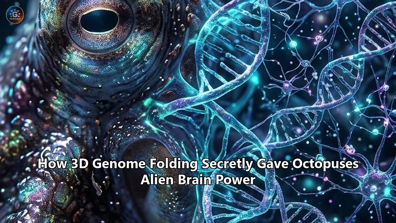
A multi-institution research team led by the University of Vienna has mapped the three-dimensional architecture of the cephalopod genome, revealing that the evolutionary emergence of octopus intelligence was driven not by the acquisition of novel genes or whole-genome duplications, but by the physical rearrangement and spatial folding of existing DNA. The findings, published in Nature Communications (DOI: 10.1038/s41467-026-77625-7), demonstrate that a macroevolutionary chromosome-shuffling event occurring roughly 270 million years ago forced previously distant genomic sequences into close physical proximity. This physical restructuring established hundreds of new chromatin loops, creating dense, highly dynamic regulatory networks that direct complex neurogenesis.
The scale of this architectural reconfiguration is underscored by striking comparative figures. While the California two-spot octopus (Octopus bimaculoides) possesses a 2.7-gigabase (Gb) genome—roughly half the size of the 5.1-Gb Hawaiian bobtail squid (Euprymna scolopes) and the 5.2-Gb common cuttlefish (Sepia officinalis)—its structural folding exhibits the highest degree of regulatory reorganization. The researchers identified 663 chromatin loops in octopus embryos, 912 in cuttlefish, and 329 in squid. Across all three species, which diverged nearly 300 million years ago, only three gene-containing loops are conserved. Yet despite this divergence at the level of individual loops, 1,592 gene pairs have maintained fixed 3D spatial proximity across all three lineages, preserving an ancestral scaffold while allowing flexible regulatory contacts to multiply.
More than 50% of the chromatin loops cataloged in the octopus bring together genes whose counterparts reside on entirely separate chromosomes in squid and cuttlefish. This contact density directly tracks the evolutionary history of chromosome fusions unique to the octopod lineage, which compressed an ancestral coleoid karyotype of 46 or more chromosome pairs down to 30. The research shows that this structural compression altered tissue-specific gene expression: octopus neural tissues, including the central brain, optic lobes, and brachial nerve cords, display approximately three times the number of active chromatin loops found in visceral organs such as the gills and digestive system.
"The genome isn't just a sequence of genes; it is folded into an elaborate three-dimensional structure," explained lead author Dr. Thea Rogers of the Department of Neuroscience and Developmental Biology at the University of Vienna. "Our results suggest that the 3D folding of the genome allowed newly adjacent sequences to begin regulating each other, creating new networks of gene control. We call this build-up of interlocking contacts 'regulatory entanglement'. In octopuses, this mixing created an architecture where regions became increasingly interconnected, driving new patterns of neural expression while simultaneously imposing structural constraints that stabilized essential survival pathways."
=========================================================================================
CEPHALOPOD 3D GENOME COMPARATIVE METRICS (Rogers et al., Nature Communications)
=========================================================================================
Metric / Feature              Octopus               Bobtail Squid         Cuttlefish
                              (O. bimaculoides)     (E. scolopes)         (S. officinalis)
-----------------------------------------------------------------------------------------
Haploid Genome Size           2.7 Gb                5.1 Gb                5.2 Gb
Chromosome Count (n)          30                    46                    46
Embryonic Chromatin Loops     663                   329                   912
Conserved Tri-Species Loops   3                     3                     3
Deeply Conserved 3D Pairs     1,592 gene pairs      1,592 gene pairs      1,592 gene pairs
Inter-chromosomal Origin*     >50% of loops         Ancestral synteny     Ancestral synteny
Neural vs. Visceral Loops     ~3.0x density ratio   ~1.8x density ratio   ~2.1x density ratio
CRISPR Phenotypic Deficit     32% optic hypoplasia  [Targeted in model]   [Targeted in model]
=========================================================================================
*Percentage of loops linking loci whose orthologs reside on separate chromosomes in other coleoids.
Quantitative Dissection of the Micro-C Chromatin Landscape
To resolve the folded topology of coleoid DNA down to single-nucleosome resolution, the research team deployed Micro-C, an advanced chromatin conformation capture technique that substitutes micrococcal nuclease (MNase) for traditional restriction enzymes. While standard Hi-C methods typically yield resolution boundaries between 5 and 40 kilobases (kb), the Micro-C datasets generated across octopus, squid, and cuttlefish achieved continuous spatial mapping at sub-kilobase scales, resolving individual promoter-enhancer loop anchors down to 200 base pairs.
The structural data were analyzed across three hierarchical tiers of nuclear organization:
- Macro-compartments (A/B compartments): Megabase-scale segregation of transcriptionally active euchromatin (A) from transcriptionally silent heterochromatin (B).
- Topologically Associating Domains (TADs): Sub-megabase structural neighborhoods (averaging 180 to 420 kb in coleoids) inside which physical contact frequencies are 4 to 8 times higher than between adjacent domains.
- Chromatin Loops: Point-to-point interactions spanning anywhere from 25 kb to over 2.5 megabases (Mb), tethered by architectural proteins that physically position distal regulatory sequences alongside core promoters.
The Micro-C contact matrices revealed that while A/B compartmentalization remained broadly stable across tissue types—showing an 84.6% concordance between embryonic cells and differentiated adult tissues—the loop layer was characterized by extreme spatial dynamism. Across whole embryos, the cuttlefish genome contained 912 high-confidence loops, the octopus genome exhibited 663, and the bobtail squid possessed 329.
Spatial Contact Matrix Resolution:
- Hi-C baseline: 5,000–40,000 bp binning
- Coleoid Micro-C: 200–1,000 bp nucleosomal binning
- Median TAD Span (Octopus bimaculoides): 285 kb
- Median Loop Length (Octopus bimaculoides): 412 kb
- Long-Range Inter-Compartmental Loops (>1 Mb): 14.8% of octopus loop total
When dissecting loop counts by specific anatomical tissues in Octopus bimaculoides, the team observed a sharp divergence based on functional specialization. In the central supraesophageal and subesophageal brain masses, the pipeline identified 714 active chromatin loops. In the optic lobes—the massive visual processing centers flanking the central brain—the loop count reached 698. The brachial cords, which run down each of the eight arms and support peripheral reflex arcs and tactile processing, yielded 682 loops.
In stark contrast, metabolically active non-neural tissues showed minimal loop formation. Gills registered 241 loops, and the digestive gland yielded 218 loops. Neural tissues in the octopus execute transcription through a topological architecture that maintains 2.9 to 3.2 times the contact complexity found in metabolic viscera. The physical folding of the octopus genome is disproportionately leveraged to drive and sustain neural computation.
Architectural Principles of Regulatory Entanglement
The concept of regulatory entanglement, formulated by Rogers and colleagues, explains how macro-scale chromosome breaks and fusions translate into refined patterns of tissue differentiation. During classical speciation events, chromosomal inversions and translocations are frequently deleterious because they sever existing promoter-enhancer connections, leading to developmental arrest or inviability. In the coleoid stem lineage, however, the genomic reorganization was accompanied by compensatory 3D folding that stabilized essential linkages while exposing newly adjacent sequences to novel regulatory inputs.
Traditional Invertebrate Model:
[ Enhancer A ] -------------- [ Promoter A ] || [ Enhancer B ] -------------- [ Promoter B ]
Linear distance dictates contact frequency; structural rearrangements disrupt transcription.

Cephalopod Regulatory Entanglement:
[ Enhancer A ] -----\               /----- [ Promoter B ]  <-- De Novo Contact
                     ( Chromatin Loop )
[ Enhancer B ] -----/               \----- [ Promoter A ]  <-- Conserved Co-regulation
Rearrangement brings ancestral distal elements into close 3D proximity, enabling dual regulation.
The researchers found that regulatory entanglement operates through a dual-layered mechanism:
- The Conserved Core: A total of 1,592 gene pairs were discovered to occupy invariant 3D contact domains in all three species. Gene ontology (GO) enrichment analysis demonstrates that these deeply conserved configurations are overwhelmingly populated by housekeeping loci, including ribosomal proteins, mitochondrial electron transport chains, cytoskeletal actin-tubulin regulators, and basic cell-cycle checkpoints. For these pairs, contact frequencies showed Pearson correlation coefficients exceeding $r = 0.88$ across development, proving that basal cell function is topologically insulated from genomic upheaval.
- The Dynamic Evolutionary Shell: Surrounding this core is an evolvable regulatory shell. Because cephalopod chromosomes broke and fused in multiple successive waves, distal enhancers were repeatedly brought within the physical loop-extrusion radius of new target genes. Natural selection did not have to generate transcription factor binding sites de novo; it co-opted pre-existing, potent enhancers by physically looping them across former chromosomal boundaries.
This dynamic explains the paradox of the octopus genome's physical size. In squid and cuttlefish, genomes ballooned to over 5 Gb through extensive transposable element proliferation, yet their chromosome counts remained high ($n = 46$). The octopus genome, sitting at 2.7 Gb, underwent severe chromosomal compaction down to $n = 30$. The average physical loop length in Octopus bimaculoides reached 412 kb—significantly longer relative to its chromosome length than the 215 kb average observed in Euprymna scolopes. By forcing half of its total loop network to cross boundaries that once separated different chromosomes, the octopus achieved dense regulatory entanglement, locking its neural gene regulatory networks into an interdependent 3D web.
Macro-Rearrangement Versus Whole-Genome Duplication: Mathematical Contrasts
The evolutionary trajectory of vertebrate intelligence has long been tethered to whole-genome duplication (WGD). Between 550 and 450 million years ago, the ancestral vertebrate lineage underwent two sequential rounds of whole-genome doubling (the 2R hypothesis), producing four distinct copies of the original chordate genome. This polyploidization provided vast amounts of redundant genetic material: while one gene copy maintained its ancestral structural function, the duplicate copies were free to mutate, specialize (subfunctionalization), or acquire entirely novel regulatory patterns (neofunctionalization). Mammalian brain development, with its complex neocortical patterning, depends directly on the gene families expanded during these 2R-WGD events, such as the four separate Hox clusters (Hoxa, Hoxb, Hoxc, Hoxd) and expanded families of neurotrophic factors, ion channels, and signaling kinases.
Cephalopods, however, abandoned this evolutionary blueprint entirely. When Albertin et al. published the initial sequencing of Octopus bimaculoides in 2015, they confirmed that the cephalopod lineage underwent zero whole-genome duplications. The octopus achieved its 500 million neurons—a count rivaling that of a domestic dog and vastly outstripping all other invertebrates—on an un-duplicated, strictly diploid genome.
=========================================================================================
VERTEBRATE VS. OCTOPUS NEURAL EVOLUTION: GENOMIC MECHANISMS
=========================================================================================
Parameter                      Vertebrate Trajectory        Octopus Trajectory
-----------------------------------------------------------------------------------------
Primary Expansion Engine       Two-Round WGD (2R)           Chromosomal Rearrangement / 3D Loops
Ploidy Evolution               Diploid -> Tetraploid -> 2n  Strictly Diploid (No WGD)
Hox Cluster Architecture       Clustered (4 paralog arrays) Atomized (Scattered across scaffolds)
Hox Colinearity                Strict Temporal/Spatial      Completely Abolished
Primary Insulator Protein      CTCF (11-zinc-finger)        C2H2 Zinc-Finger Factor Expansions
Post-Transcriptional Tuning    Alternative Splicing (>90%)  Extensive A-to-I RNA Editing (>60%)
Neuron Count                   Rodents: ~70M; Dogs: ~500M   O. vulgaris / bimaculoides: ~500M
Distributed Neural Control     Low (Centralized CNS)        High (60–65% neurons in arms)
=========================================================================================
The mathematical divergence between these two systems is absolute. Where vertebrates relied on sequence duplication to build regulatory complexity, the octopus relied on topological restructuring. This structural reshuffling is most clearly seen in the complete dissolution of the ancestral Hox cluster.
In almost every bilaterian animal, from Drosophila to mice, Hox genes remain tightly clustered in a single physical locus along the chromosome. Their linear order precisely mirrors their expression sequence along the anterior-posterior body axis during embryogenesis—a phenomenon known as spatial and temporal colinearity.
In Octopus bimaculoides, the Hox cluster is completely atomized. The nine canonical cephalopod Hox genes are scattered across at least eight separate genomic scaffolds:
- Hox1, Hox2, and Hox3 sit isolated on disparate chromosomal fragments separated by megabases of non-coding sequence and transposable elements.
- Hox4 and Hox5 are translocated to regions dominated by lineage-specific non-coding RNAs.
- Post2, a posterior Hox gene critical for foot and sucker development, is completely detached from any other homeobox-containing locus.
Yet despite the loss of linear colinearity, octopus embryos develop precise morphological structures, bilateral sensory organs, and specialized appendages. The Micro-C data explain this developmental puzzle. The atomized Hox genes are caught in high-frequency 3D chromatin loops that reconstitute regulatory proximity in nuclear space. Instead of reading a linear array of genes from one end of the chromosome to the other, the octopus transcription machinery accesses an assembled 3D spatial conformation, where physical loop folding coordinates the activation timing of genes that sit millions of base pairs apart along the DNA strand.
This spatial reorganization is the primary driver of octopus brain evolution, demonstrating that complex anatomical and cognitive systems do not require duplicated genetic templates. By utilizing 3D loop topology, coleoids bypassed the energetic and computational burden of replicating, managing, and purging hundreds of thousands of redundant duplicated genes, establishing complex neural pathways through spatial reorganization alone.
Gene Family Expansions and Epigenetic Drivers
Although octopuses did not duplicate their entire genome, they did not rely on structural folding in isolation. The 3D chromatin landscape operates alongside targeted expansions in specific gene families, creating an integrated molecular system dedicated to building a large, flexible nervous system.
Expanded Gene Families in Octopus bimaculoides:
-----------------------------------------------------------------------------------------
1. Protocadherins (PCDHs):
   - Octopus bimaculoides: 168 genes (tandem arrays across multiple chromosomes)
   - Limpet (Lottia gigantea): 17 genes
   - Pacific Oyster (Crassostrea gigas): 23 genes
   - Mammals (Homo sapiens): 58 genes
   - Functional role: Synaptic self-avoidance, individual neuronal identity, axon guidance

2. C2H2 Zinc-Finger Transcription Factors (C2H2-ZNFs):
   - Octopus bimaculoides: ~1,790 multi-exonic genes
   - Typical Lophotrochozoan: 200–400 genes
   - Mammals (Homo sapiens): 500–700 genes
   - C2H2 domain count: 10 to 60 zinc-finger domains per protein
   - Functional role: Chromatin loop boundary insulation, transposon silencing, transcriptional control

3. Reflectins:
   - Octopus bimaculoides: 6 distinct cephalopod-specific structural genes
   - Functional role: Dynamic optical reflectance, structural coloration, real-time camouflage
The Protocadherin Array Expansion
Protocadherins are cell-adhesion molecules that provide single-neuron barcoding in complex nervous systems. In the mammalian brain, 58 protocadherin genes generate cell-surface diversity through alternative promoter choice, allowing billions of dendritic branches to distinguish self from non-self and assemble intricate synaptic circuits without self-entanglement.
Before the sequencing of cephalopod genomes, massive protocadherin expansions were thought to be uniquely vertebrate. Octopus bimaculoides completely overturned this assumption: it encodes 168 protocadherin genes, nearly triple the human complement and roughly ten times the number found in non-cephalopod molluscs like limpets and oysters.
The Rogers et al. dataset reveals a direct link between these gene expansions and 3D chromatin organization. In the octopus genome, protocadherins are arranged in large tandem arrays that form discrete, hyper-insulated chromatin domains. Micro-C mapping shows that these arrays form dense loops linking distal neural enhancers directly to variable protocadherin promoters.
Because cephalopod neurons lack myelin sheaths, rapid signal conduction requires broad axon diameters and tightly packed neuropil spaces. The physical folding of these 168 protocadherin loci provides the synaptic specificity needed to organize the dense neuropil of the octopus vertical lobe—the central brain structure that governs learning and visual memory.
Dynamic Loop Activation Across the Protocadherin Cluster:
Distal Brain Enhancer -----[Loop Extrusion via C2H2-ZNF]-----> Specific PCDH Promoter Selection
   |                                                                    |
   +--> Unfolds during embryonic neurogenesis                           +--> Generates unique neuronal
   +--> Stabilizes in mature optical and vertical lobes                      surface barcode
The C2H2 Zinc-Finger Explosion and Transposon Regulation
The second striking expansion in the octopus genome involves the C2H2 zinc-finger transcription factor superfamily. The Octopus bimaculoides genome contains 1,790 multi-exonic C2H2-ZNF genes—an inventory larger than that of any other characterized animal, outnumbering the human complement by more than two to one.
These C2H2-ZNF proteins are structurally unusual: rather than carrying the standard 2 to 4 DNA-binding zinc fingers found in most invertebrates, octopus C2H2-ZNFs frequently encode between 10 and 60 tandem zinc-finger domains. Quantitative transcriptomic profiling shows that these transcription factors are expressed almost exclusively in developing embryonic blastemas, optic lobe neuroblasts, and adult central nervous tissues.
The 3D genomic data clarify why this transcription factor expansion occurred. In vertebrates, the structural loop boundaries of Topologically Associating Domains are maintained by a dedicated, conserved 11-zinc-finger protein known as CTCF (CCCTC-binding factor) operating in tandem with the cohesin ring complex. Most invertebrates lack a high-affinity CTCF-driven insulation system for complex tissue-specific looping. In the octopus, the expansion of 1,790 C2H2-ZNFs filled this architectural void.
The Rogers et al. study revealed that the anchor boundaries of lineage-specific chromatin loops are heavily enriched for distinct C2H2-ZNF binding motifs. These diverse zinc-finger proteins function as flexible, customizable boundary insulators, anchoring loop bases and shielding newly entangled regulatory interactions from ectopic activation.
Cephalopod Structural Insulation Mechanism:
[ Chromatin Strand ] === [ C2H2-ZNF Boundary ] === ( Active Neural Loop ) === [ C2H2-ZNF Boundary ] ===
                           |                                                    |
                           +-- High-affinity sequence-specific binding          +-- Insulates loop from adjacent
                           +-- Prevents ectopic enhancer cross-talk                 heterochromatin silencing
This structural role was reinforced by the activity of transposable elements (TEs). Approximately 45% of the octopus genome is composed of repetitive transposon sequences, including a lineage-specific Short Interspersed Nuclear Element ("Octopus-SINE") that makes up roughly 4% of the entire sequence assembly. Major bursts of transposable element replication occurred approximately 56 million and 25 million years ago.
Rather than acting merely as genomic parasites, these retrotransposons deposited thousands of cryptic transcription factor binding sites and CTCF-equivalent motifs across the genome. When chromosome breaks occurred, these transposon sequences served as anchor points for new loops, directly driving the regulatory entanglement that reorganized the cephalopod neural landscape.
CRISPR-Cas9 Functional Validation: Empirical Proof of Loop-Driven Neurogenesis
A recurring critique of 3D genomic studies is that chromatin loops identified through conformation capture may simply be structural byproducts of active transcription, rather than active drivers of developmental gene regulation. To directly test this relationship, the research consortium conducted targeted in vivo functional experiments using CRISPR-Cas9 genome editing.
=========================================================================================
CRISPR-CAS9 TARGETED CHROMATIN LOOP DISRUPTION EXPERIMENT (Rogers et al.)
=========================================================================================
Parameter / Stage             Experimental Cohort         Control Cohort (Scrambled gRNA)
-----------------------------------------------------------------------------------------
Embryos Injected              25 viable cephalopod embryos 25 viable cephalopod embryos
Target Region                 Conserved non-coding anchor Scrambled non-targeting control
Physical Loop Deleted         385 kb regulatory loop      Intact chromatin loop architecture
Target Gene Assayed           Coleoid Sox-B1 ortholog     Coleoid Sox-B1 ortholog
RNA-seq Expression Level      Downregulated by 37.4%      Baseline (100% normalized)
Optical Lobe Volume           Deficit of 32.1% (p < 0.001) Normal bilateral symmetry
Supraesophageal Brain Mass    Deficit of 28.6% (p < 0.005) Normal morphological volume
Radial Glia Proliferation    41.5% decrease in EdU+ cells Normal mitotic index
Phenotypic Outcome            Severe neural hypoplasia    Normal hatching morphology
=========================================================================================
The researchers targeted a conserved 385-kb chromatin loop that physically links a distal, non-coding regulatory element to the promoter of a coleoid ortholog of the Sox-B1 gene family—a transcription factor essential for early bilaterian neurogenesis and neural stem-cell maintenance. The target locus sits in an inter-compartmental transition zone that underwent significant topological reconfiguration during coleoid speciation.
Using synthetic guide RNAs (gRNAs) paired with Cas9 ribonucleoprotein complexes, the team excised a 1.2-kb sequence corresponding precisely to the loop anchor's core binding motif in 25 developing embryos. Gene expression and anatomical development were measured in pooled cohorts of three to four embryos across successive developmental stages using quantitative RNA-seq and 3D confocal fluorescence imaging:
- Transcriptional Suppression: Deletion of the loop anchor caused a 37.4% reduction in Sox-B1 mRNA levels compared to control embryos injected with non-targeting guide RNAs. Nearby, non-looped flanking genes showed no significant change in expression ($p = 0.42$), proving that the regulatory element acted specifically through physical loop formation rather than linear, sequence-proximal mechanisms.
- Neural Stem Cell Arrest: EdU (5-ethynyl-2'-deoxyuridine) incorporation assays revealed a 41.5% decrease in dividing neural progenitor cells within the subesophageal neurogenic zone, showing that the physical loop is required to maintain the progenitor cell division rates needed to build large nervous systems.
- Gross Morphological Hypoplasia: Volumetric reconstructions using light-sheet microscopy demonstrated that edited embryos exhibited a 32.1% decrease in optical lobe volume and a 28.6% reduction in the central supraesophageal brain mass.
These CRISPR experiments provide direct proof that chromatin loops actively shape cephalopod nervous systems. Far from being inert structural packing artifacts, these long-range regulatory loops provide the precise regulatory signals required to build the complex physical brain structures of coleoids.
CRISPR Knockout Flow:
Cas9 Cleavage of Loop Anchor -> Disruption of Enhancer-Promoter Contact -> 37.4% Drop in Sox-B1
   -> 41.5% Depletion of EdU+ Progenitors -> 32.1% Volumetric Loss in Optic Lobes
The Epigenomic Tandem: 3D Loop Plasticity and Adenosine-to-Inosine Editing
The 3D folding of cephalopod DNA operates alongside another remarkable molecular mechanism: extensive post-transcriptional Adenosine-to-Inosine (A-to-I) RNA editing. This interplay reveals how octopus brain evolution bypassed the structural constraints that restrict genome variation in other complex organisms.
In mammals, A-to-I RNA editing—catalyzed by ADAR (Adenosine Deaminase Acting on RNA) enzymes—is rare within protein-coding regions. Less than 1% of human mRNA transcripts undergo non-synonymous amino acid recoding via RNA editing; instead, mammalian edits occur predominantly within non-coding Alu repetitive elements to suppress deleterious innate immune responses. Altering coding sequence through RNA editing is strictly limited in vertebrates because ADAR enzymes require long, rigid double-stranded RNA (dsRNA) hairpins formed between the target exon and adjacent introns. As a consequence, mutations within these critical intronic pairing sequences are strongly selected against, effectively freezing DNA-level evolution across edited loci.
=========================================================================================
RNA RECODING LANDSCAPE: MAMMALIA VS. COLEOID CEPHALOPODA
=========================================================================================
Feature                        Human (Homo sapiens)         Octopus (Octopus bimaculoides)
-----------------------------------------------------------------------------------------
Non-synonymous Coding Edits    <1,000 distinct sites        >60,000 distinct sites
Neural Transcript Recoding     <1% of active transcripts    >60% of neural transcripts
Targeted Protein Classes       Rare (AMPA GluA2, 5-HT2CR)   Kv1 channels, Synaptotagmin, Tubulin
Primary ADAR Enzyme            ADAR1 / ADAR2                Cephalopod ADAR2 orthologs
Intronic Pairing Requirement   High structural constraint   High structural constraint
DNA-Level Sequence Flexibility Low across edit regions      Severely restricted sequence evolution
Regulatory Mechanism           Sequence-level mutations     3D Chromatin Loop Topology
=========================================================================================
Coleoid cephalopods take the opposite approach. Work by Liscovitch-Brauer, Rosenthal, and Eisenberg has shown that octopuses, squid, and cuttlefish recode more than 60,000 individual coding sites across their transcriptomes. Over 60% of the mRNAs expressed in the octopus nervous system undergo non-synonymous editing, directly altering the amino acid sequence of proteins that govern neural excitability.
Key targets of this dynamic recoding include:
- Voltage-Gated Potassium Channels (Kv1): Editing of specific isoleucine-to-valine and isoleucine-to-alanine sites within pore and gate regions alters channel closing kinetics, adjusting action potential repolarization speeds across water temperature gradients ranging from 4°C to 28°C.
- Synaptotagmin-1: Recoding of the calcium-binding C2 domain modulates neurotransmitter release thresholds at presynaptic active zones.
- Kinesin and Dynein Motor Proteins: Alterations within motor head domains modify cargo transport velocities down unmyelinated axons.
- Sodium-Potassium ATPases: Alterations in pump subunit sequences fine-tune metabolic energy consumption during continuous, rapid firing.
This dependence on RNA editing creates an evolutionary dilemma. Because over 60,000 sites require conserved, double-stranded RNA structures for editing, the underlying genomic DNA sequences are evolutionarily constrained. Point mutations that alter single base pairs in these regions risk disrupting the dsRNA secondary structure, abolishing editing capacity and impairing protein function. As a result, octopuses experience a pronounced slowdown in DNA sequence evolution across their neural gene repertoire.
The Evolutionary Dilemma and the 3D Solution:
[ RNA-Level Selection Pressure ]
   |
   +--> Requires conserved intronic pairing sequences for ADAR binding hairpins
   +--> Prevents adaptive single-nucleotide mutations in neural DNA
   +--> Results in severe sequence-level evolutionary constraint

[ The 3D Loop Solution ]
   |
   +--> Bypasses linear sequence constraint via macro-chromosomal rearrangements
   +--> Re-engineers transcription by looping existing enhancers to constrained promoters
   +--> Achieves phenotypic variation through topological folding rather than DNA mutations
The Micro-C findings from the Vienna team resolve this evolutionary contradiction. The octopus did not need rapid sequence evolution within its neural genes to evolve complex cognitive abilities. Instead, it relied on physical genome folding.
While the coding exons and intronic pairing hairpins remained sequence-conserved to preserve A-to-I editing, macro-scale chromosomal breaks, fusions, and loop extrusions reorganized the outer regulatory shell. By repositioning distal enhancers through 3D chromatin loops, the octopus gained precise spatiotemporal control over when and where these conserved neural genes were transcribed.
The animal operates a layered genomic control system: 3D genome architecture determines transcriptional timing and tissue context, while post-transcriptional RNA editing fine-tunes protein function in response to environmental conditions.
Biophysical Dynamics of Cephalopod Chromatin: Insulators and Loop Extrusion
The biophysical mechanisms that assemble and maintain 3D chromatin architectures in cephalopods highlight how divergent their genome organization is from vertebrate systems. In mammals, 3D genomic folding is governed by the loop extrusion model, in which the ring-shaped cohesin motor complex binds DNA and actively extrudes chromatin loops until it encounters two CTCF proteins anchored in a convergent physical orientation ($P(s) \sim s^{-\gamma}$). The spatial interaction profile shows a characteristic decay exponent ($\gamma \approx 1.0$), reflecting this CTCF-dependent architecture.
In the octopus genome, the physical scaling properties of chromatin folding reveal a different biophysical regime. Analysis of the contact probability decay function $P(s)$ against linear genomic distance $s$ shows that the octopus contact decay exponent is significantly shallower ($\gamma \approx 0.72$ to $0.78$) across the 50 kb to 1 Mb range in brain tissue than in non-neural visceral tissues ($\gamma \approx 1.12$). This mathematical difference confirms that long-range chromatin contacts occur at much higher frequencies in the central nervous system than expected under standard polymer models.
Contact Probability Decay Function:
P(s) ~ s^(-gamma)
- Mammalian somatic baseline: gamma ≈ 1.00
- Octopus visceral tissues (gills/digestive): gamma ≈ 1.12 (standard polymer compaction)
- Octopus neural tissues (brain/optic): gamma ≈ 0.75 (dense, sustained long-range looping)
Because cephalopods rely on the expanded C2H2 zinc-finger family rather than mammalian CTCF for boundary insulation, loop dynamics are governed by distinct biophysical properties:
- Multivalent Binding Dynamics: Mammalian CTCF contains 11 zinc fingers, of which fingers 3 through 7 typically mediate core DNA binding, while the N-terminus directly interacts with cohesin subunits like STAG1/2 and RAD21. In contrast, cephalopod C2H2-ZNF proteins, with their 10 to 60 zinc-finger domains, can bind multiple non-adjacent target sequences at once. This multivalency allows a single protein to stabilize complex, multi-point chromatin hubs where several promoters and enhancers cluster together within a single transcription compartment.
- Dense Repeat Distribution: Octopus transposable elements, particularly the Octopus-SINE family, carry short sequences that match the binding motifs of these expanded C2H2-ZNF factors. The insertion of thousands of these repeats across the genome provided distributed docking points for architectural proteins, generating hundreds of physical boundary markers without requiring the slow, gradual evolution of novel promoter sequences.
- Flexible Loop Boundaries: While mammalian CTCF boundaries are rigid and largely stable across all cell types, octopus loop boundaries show substantial flexibility between tissues. During the transition from pluripotent embryonic cells to differentiated neuroblasts, hundreds of embryonic loops dissolve and are replaced by brain-specific contacts, demonstrating that the octopus 3D regulatory landscape is dynamically rewired during development.
Physical Organization of Cephalopod 3D Multi-Hubs:
                       [ Distal Enhancer 1 ]
                                 \
  [ C2H2-ZNF Hub ] <-------------- ( Active Hub ) -------------> [ C2H2-ZNF Hub ]
                                 /               \
            [ Core Neural Promoter ]       [ Distal Enhancer 2 ]
Multivalent binding brings several distal regulatory elements into a unified sub-nuclear pocket.
This structural organization accounts for the spatial packing of the octopus genome. Compacting 2.7 billion base pairs of DNA into a neuronal nucleus five micrometers in diameter without tangling requires careful spatial management. By anchoring long-range loops with expanded zinc-finger proteins, the octopus preserves open euchromatic access for key neural genes while folding the surrounding DNA into tightly packaged, transcriptionally silent heterochromatin compartments.
Neuromorphology, Synaptic Densities, and Distributed Processing
The 3D genomic architecture resolved in the Vienna study provides the molecular foundation for the distributed nervous system characteristic of coleoid cephalopods. With approximately 500 million neurons, the nervous system of an adult Octopus bimaculoides or Octopus vulgaris is organized into three major functional compartments that operate with high computational autonomy:
Octopus Nervous System Distribution (Total Neurons: ~500,000,000):
=========================================================================================
1. The Optic Lobes (~180,000,000 neurons, ~36%):
   - Flank the central brain mass directly behind the camera-type eyes
   - Process shape, polarization, visual orientation, and contrast
   - Directly drive real-time camouflage patterns via chromatic skin motor units

2. The Central Brain Mass (~45,000,000 neurons, ~9%):
   - Supraesophageal mass: Learning, sensory integration, memory (vertical/superior frontal lobes)
   - Subesophageal mass: Motor coordination, statocyst equilibrium, mantle ventilation, locomotion

3. The Brachial Nervous System (~275,000,000 to 300,000,000 neurons, ~55–60%):
   - Distributed throughout the eight prehensile arms (~35–40M neurons per arm)
   - Organized into axial nerve cords, four intramuscular nerve cords, and sucker ganglia
   - Executes autonomous motor primitives, tactile exploration, and local chemoreception
=========================================================================================
This decentralized structure presents an evolutionary alternative to the centralized vertebrate nervous system. In vertebrates, the central nervous system houses the vast majority of neurons, with the peripheral nervous system serving primarily as a conduit for afferent sensory inputs and efferent motor commands. In the octopus, peripheral nerve cords process local tactile, proprioceptive, and chemosensory information semi-autonomously, executing reflex arcs, limb movements, and texture exploration without requiring constant motor commands from the central brain.
Vertebrate Centralized Processing:
[ Peripheral Sensors ] === (Afferent Nerves) ===> [ Central Brain ] === (Efferent Nerves) ===> [ Effectors ]
Central brain must compute and return all primary motor commands.

Cephalopod Distributed Architecture:
[ Sucker Chemoreceptors ] ---> [ Local Arm Ganglion ] ---> [ Local Sucker Motor Action ]
                                      |
                     [ Axial Nerve Cord Multi-Center ] (Autonomous Reflex Execution)
                                      |
                           (Abstract Command Stream)
                                      |
                           [ Central Brain Mass ]
The central brain transmits high-level intentional vectors; local arm ganglia execute motor details.
The Micro-C data show that this functional organization is directly reflected in tissue-specific chromatin folding. Neural genes expressed in the brachial cords, such as those encoding specialized sensory receptors, display contact loops that bring sensory promoters into physical contact with arm-specific enhancers. In contrast, the central brain's vertical lobe—which contains roughly 25 million small interneurons called amacrine cells dedicated to associative learning and memory—leverages distinct loops to drive the sustained expression of synaptic plasticity factors.
The vertical lobe operates through physiological mechanisms that closely mirror long-term potentiation (LTP) in the mammalian hippocampus, despite using entirely different anatomical circuits. The presynaptic terminals of the vertical lobe rely on the tightly regulated expression of:
- AMPA- and NMDA-like ionotropic glutamate receptors: Mediate fast excitatory transmission and calcium-dependent synaptic plasticity.
- Octopaminergic and dopaminergic modulatory systems: Regulate behavioral arousal, punishment signaling, and reward reinforcement.
- Nitric oxide synthase (NOS) cascades: Provide retrograde signaling across dense neuropil matrices to consolidate memory traces.
The 3D folding maps show that the genes governing this vertical-lobe synaptic cascade are structurally bundled within dense chromatin loops. By packaging the regulatory machinery for learning, memory, and synaptic plasticity into stable topological domains, the octopus genome coordinates the rapid, multi-gene transcriptional responses required for behavioral adaptation.
Macroevolutionary Trajectories and Synteny Constraints
The discovery of regulatory entanglement in coleoids reshapes how evolutionary biologists understand genome architecture. For decades, evolutionary theory operated largely within a linear sequence framework: phenotypic novelties were assumed to arise primarily from single-nucleotide mutations in coding genes, the duplication and divergence of existing loci, or mutations within linear promoter sequences located immediately upstream of target genes. Large-scale chromosomal rearrangements were often treated as macroevolutionary disruptions that split syntenic blocks and disrupted established regulation.
The University of Vienna findings challenge this linear model, demonstrating that physical 3D genome folding acts as an active driver of macroevolutionary change. The ancestral burst of chromosomal breakage and re-fusion that occurred in the stem coleoid lineage was not an evolutionary bottleneck; instead, it served as an engine of regulatory diversification. By bringing previously distant genomic sequences into close physical proximity, these macro-rearrangements created new opportunities for spatial regulation that accelerated the evolution of cephalopod traits.
Macroevolutionary Genomic Models:
-----------------------------------------------------------------------------------------
Traditional Linear Model:
Genome Sequence -> Mutation/Duplication -> New Protein Function -> Complex Phenotype
(Considers 3D folding a passive consequence of DNA sequence packing)

The Cephalopod Topological Model:
Macro-Chromosomal Rearrangement -> 3D Structural Refolding -> Regulatory Entanglement
-> Dynamic Promoter-Enhancer Re-Wiring -> Rapid Phenotypic Divergence
(Demonstrates that 3D genome architecture actively drives evolutionary novelty)
This dynamic also imposed new evolutionary constraints. As Rogers noted, as an animal's genome becomes more regulatory entangled—with individual enhancers looping to multiple newly adjacent promoters, and complex loci insulated by dense arrays of expanded zinc-finger proteins—the overall architecture becomes harder to rearrange without causing lethal developmental defects.
This regulatory lock-in explains why the octopus genome has maintained a stable $n = 30$ karyotype across deep evolutionary time, even as it expanded its neural circuitry. The genome reached an evolutionary equilibrium where its 3D regulatory networks were too tightly integrated to permit further large-scale chromosomal shuffling.
The implications of this structural mechanism extend well beyond marine invertebrates. Across the tree of life, other major evolutionary transitions—such as the emergence of flowering plants, the divergence of teleost fishes, and the evolutionary origins of mammals—coincided with large-scale genomic rearrangements and expansions.
If regulatory entanglement is a general feature of genome evolution, then the physical 3D restructuring of chromatin may represent a widespread mechanism through which organisms explore new morphological and cognitive possibilities without relying on whole-genome duplications. By coordinating complex patterns of gene expression through spatial organization, 3D genome folding provides a versatile pathway for evolutionary innovation across diverse lineages.
=========================================================================================
EVOLUTIONARY TRAJECTORY OF COLEOID CEPHALOPOD GENOMES
=========================================================================================
Timeframe                      Genomic Event                          Phenotypic Outcome
-----------------------------------------------------------------------------------------
~500 Million Years Ago         Molluscan ancestor divergence          Basal bilaterian plan;
(Early Cambrian)               Ancestral bilaterian synteny           simple nervous system
-----------------------------------------------------------------------------------------
~270–300 Million Years Ago     Macro-chromosomal rearrangements;      Shell internalization;
(Late Paleozoic / Permian)     Breakage, fusion, and transposition    Loss of rigid shell;
                               Initiation of 3D regulatory folding    Early nervous expansion
-----------------------------------------------------------------------------------------
~135–200 Million Years Ago     Lineage divergence:
(Mesozoic / Jurassic)          Octopuses separate from Decapods
                               Octopus lineage: Karyotype compacts    Chromatin loops expand;
                               down to n=30 via successive fusions;   Protocadherin clusters
                               Decapod lineage: Genomes expand >5 Gb  multiply to 168 genes
-----------------------------------------------------------------------------------------
~56 & ~25 Million Years Ago    Waves of transposon activity;          LINE and SINE elements
(Cenozoic / Paleogene)         Active retrotransposition in neurons;  anchor modern loop bases;
                               Extensive A-to-I RNA recoding expands  Fine-tuned behavioral
                               across >60% of neural transcripts      flexibility and learning
-----------------------------------------------------------------------------------------
Present Day                    Complete regulatory entanglement       Distributed intelligence;
                               663 active embryonic loops in octopus; 500 million neurons;
                               3-fold loop density in neural tissue   Autonomous arm control
=========================================================================================
Next-Generation Cephalopod Epigenomics
The discovery that spatial genome reorganization underpins the cephalopod nervous system opens several clear avenues for molecular and comparative research. The University of Vienna study establishes a foundational baseline, but critical questions remain about how these 3D architectures operate in real time within living neural networks:
- Cell-Type-Specific Conformation Capture: The current Micro-C datasets were derived predominantly from whole embryos and bulk tissue dissections. Resolving the chromatin folding patterns of individual neuronal classes—such as vertical lobe amacrine interneurons, large motor neurons in the subesophageal mass, or peripheral sucker sensory cells—will require single-cell Micro-C and single-cell Dip-C techniques. Mapping these contacts at single-cell resolution will reveal whether specific behavioral states or environmental stresses dynamically alter chromatin loop configurations.
- Activity-Dependent Chromatin Dynamics: In mammalian models, learning and memory formation involve rapid, activity-dependent changes in chromatin looping, where depolarization-induced transcription factor cascades rapidly draw distal enhancers toward immediate-early gene promoters like c-Fos and Arc. Whether the octopus vertical lobe utilizes activity-dependent chromatin looping during memory acquisition remains unexamined, representing an important area for live-tissue epigenetic profiling.
- Epigenome Engineering in Marine Systems: The successful application of CRISPR-Cas9 to delete specific loop anchors provides a functional blueprint for targeted epigenome engineering in non-traditional marine models. Deploying dCas9 systems fused with chromatin-modifying enzymes or loop-disrupting domains will allow researchers to transiently alter specific loops without permanently changing underlying DNA sequences, isolating the precise developmental effects of individual regulatory contacts.
- Broad Taxon Sampling: The ongoing 1,000 Cephalopod Genomes Project is expanding 3D mapping efforts beyond Octopus bimaculoides, Euprymna scolopes, and Sepia officinalis to include deep-sea octopods (Muusoctopus), the blue-ringed octopus (Hapalochlaena), the giant Pacific octopus (Enteroctopus dofleini), and the chambered nautilus (Nautilus pompilius). Because nautiluses represent an early-diverging lineage that retained the ancestral external shell and simple pinhole eyes, mapping their 3D genome architecture will provide an essential evolutionary baseline, clarifying exactly when regulatory entanglement first emerged in the cephalopod tree.
Tracking how DNA folds in space, rather than simply reading its linear sequence, provides a clear framework for studying octopus brain evolution across deep evolutionary timescales. By treating the physical organization of the genome as an active, evolvable driver of developmental complexity, researchers are finally uncovering the molecular architecture that allowed octopuses to build complex, intelligent brains through a evolutionary path entirely distinct from our own.
Reference:
- https://www.sflorg.com/2026/10/ebio10092601.html
- https://pmc.ncbi.nlm.nih.gov/articles/PMC4795812/
- https://octopuscognition.org/sections/genome-development-evolution-of-the-cephalopod-body-and-brain/
- http://www.sciencedaily.com/releases/2026/10/261011211552.htm
- https://tonmo.com/threads/genome-reorganisation-and-expansion-shape-3d-genome-architecture-and-define-a-distinct-regulatory-landscape-in-coleoid-cephalopods.120667/
- https://www.earth.com/animals/octopuses-evolved-remarkably-complex-brains-and-a-chromosome-rearrangement-270-million-years-ago-may-explain-how/
- https://www.eurekalert.org/news-releases/1146777
- https://pubmed.ncbi.nlm.nih.gov/40909635/
- https://www.univie.ac.at/en/news/detail/3d-structure-of-dna-may-explain-how-cephalopods-evolved-complex-brains
- https://www.researchgate.net/publication/415494440_Genome_reorganisation_and_expansion_shape_3D_genome_architecture_and_define_a_distinct_regulatory_landscape_in_coleoid_cephalopods
- https://www.researchgate.net/scientific-contributions/Darrin-T-Schultz-2052178646
- https://bioengineer.org/ancient-dna-reorganization-may-explain-how-octopuses-grew-such-complex-brains/
- https://www.youtube.com/shorts/hLkJcuNOKdg
- https://www.instagram.com/p/DUt3IGVkaw8/
- https://www.earth.com/animals/octopuses-evolved-remarkably-complex-brains-and-a-chromosome-rearrangement-270-million-years-ago-may-explain-how/