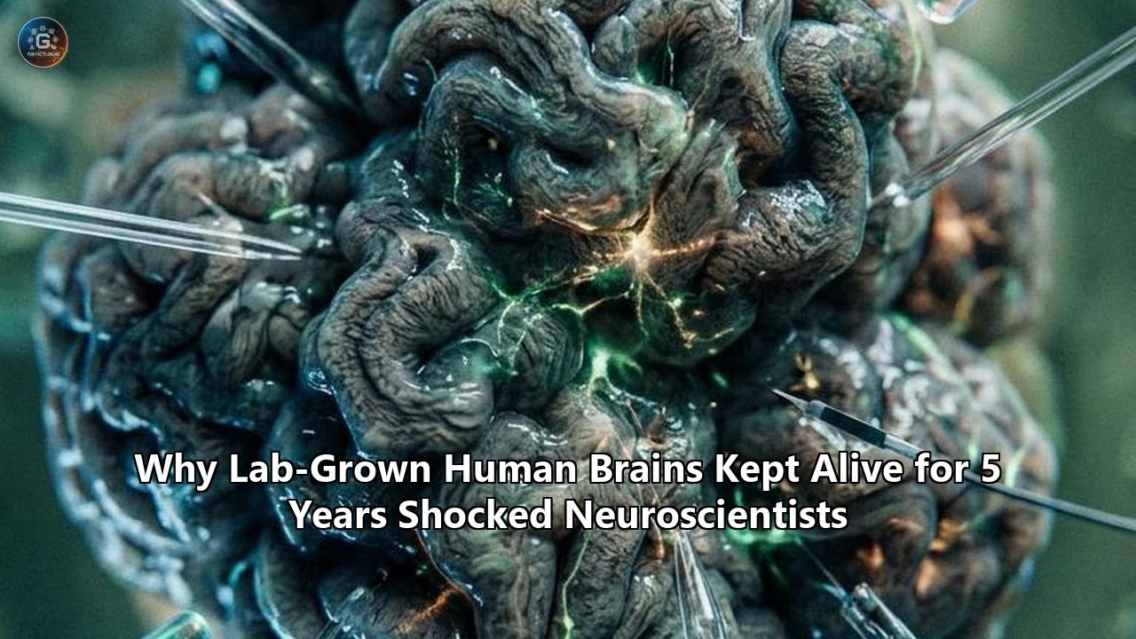
In an incubator tucked inside a secure Harvard University laboratory, several clusters of living human cerebral tissue recently reached a milestone once widely dismissed as biological science fiction: they crossed their fifth consecutive year of continuous life, maturation, and electrical signaling outside a living human body.
The findings, published in the journal Nature, mark the first time that disembodied human cortical tissue has been sustained in culture for more than half a decade. But what has caught the international neuroscience community off guard is not simply that the tissue survived. The real shock is what happened inside those miniature cerebral spheres across those five years: entirely cut off from blood vessels, sensory organs, hormonal systems, and bodily inputs, the cells continued to develop on an intrinsic, human-specific developmental calendar, methodically switching on genetic programs, maturing synaptic networks, and accumulating epigenetic marks that mirror the transitions seen in human infants and young children.
"We didn't know how far the development and maturation of human brain tissue could occur outside the context of the normal brain inside the head," said Paola Arlotta, the Golub Family Professor of Stem Cell and Regenerative Biology at Harvard University and senior author of the study. "This work showed that it's actually possible to not just have these organoids survive in culture, but also continue to change, develop, and mature over stretches of time that had never been reached before".
THE FIVE-YEAR EXPERIMENTAL TIMELINE
┌───────────────────────────────────────────────────────────────────────────────┐
│ Month 0–6       │ Progenitor proliferation; early radial glia emerge         │
│ Month 6–12      │ Deep-layer cortical neurons form; early spontaneous bursts   │
│ Year 1–2        │ Shift to postnatal gene signatures; upper-layer maturation   │
│ Year 2–3        │ Astrocytic and oligodendrocytic lineages stabilize          │
│ Year 4–5+       │ Epigenetic clocks match early childhood; temporal memory retained│
└───────────────────────────────────────────────────────────────────────────────┘
The experiment shatters decades of neurodevelopmental assumptions. For generations, biologists maintained that after initial embryonic morphogenesis, human brain tissue required a torrent of systemic signals—circulating endocrine hormones, sensory inputs from eyes and ears, metabolic cues from the gut and liver, and vascular flow—to advance past fetal stages. The discovery that lab grown human brains harbor an autonomous, self-contained internal clock rewires the scientific understanding of human neurobiology, offering new pathways to study neuropsychiatric disorders, while forcing bioethicists to reckon with long-term disembodied neural culture.
The Biological Clock That Needs No Body
The human brain takes roughly two decades to fully mature, a developmental trajectory that is uniquely drawn out compared to any other species on Earth. While a mouse brain completes its structural wiring in weeks and a macaque monkey reaches neural maturity in a few years, human cerebral cortex development unfolds across years of infancy, childhood, and adolescence.
DEVELOPMENTAL TIMELINE COMPARISON ACROSS SPECIES
Human:   [==================================================] ~20 Years
Macaque: [============] ~3–4 Years
Rodent:  [===] ~6–8 Weeks
Historically, scientists attempting to model this protracted process in laboratory dishes hit a wall. Brain organoids—three-dimensional cell aggregates derived from human induced pluripotent stem cells (iPSCs)—typically degenerated or stalled within several months. The previous longevity record, established in 2021 by teams at UCLA and Stanford University, capped out at 694 days, just under two years. Even in those long-running cultures, the cells frequently suffered from metabolic distress, core necrosis, and developmental stagnation, leaving researchers uncertain whether true postnatal maturation was biologically possible in a dish.
The Harvard-led team, which included lead authors Noelia Antón-Bolaños (now an assistant professor at University Medical Center Utrecht) and Irene Faravelli (now an assistant professor at the University of Milan), set out to answer whether human cerebral tissue possessed an inherent timeline or if it inevitably decayed without systemic organismal cues.
To evaluate the biological age of the tissue over time, the team deployed high-resolution single-cell RNA sequencing and surveyed DNA methylation patterns—the chemical modifications on DNA that function as epigenetic clocks. In total, the researchers profiled 110 organoids comprising nearly 425,000 individual cells sampled at eight distinct timepoints between six months and five years of age.
+-----------------------------------------------------------------------------+
|                     DATA PROFILE OF THE FIVE-YEAR COHORT                     |
+------------------------------------+----------------------------------------+
| Total Organoids Analyzed           | 110 individual cortical structures     |
| Total Single Cells Sequenced       | ~425,000 individual neural cells       |
| Timepoints Profiled                | 6 mo, 9 mo, 1 yr, 1.5 yr, 2 yr, 3 yr,  |
|                                    | 4 yr, 5+ years                         |
| Primary Assays                     | Single-cell RNA-seq, DNA methylation   |
|                                    | arrays, microelectrode electrophysiology|
+------------------------------------+----------------------------------------+
The molecular readouts matched post-mortem human brain benchmarks with startling precision. Between months six and twelve, the organoids exhibited signatures characteristic of mid-to-late fetal cortex. But as the cultures crossed the 365-day mark, their transcriptomic profiles transitioned: fetal gene programs shut down, and postnatal markers associated with early infancy switched on.
"In the human brain, these epigenetic changes accumulate according to a characteristic developmental pattern," Antón-Bolaños explained. "We observed the same pattern in the brain organoids. The cells are outside the body, yet they still follow approximately the same developmental timeline as we do—and even more closely than we had anticipated".
The cells followed the exact canonical sequence seen in human neurodevelopment:
- Early Progenitor Expansion: Neural stem cells organized into ventricular-like zones, multiplying to generate foundational pools.
- Sequential Cortical Layering: Deep-layer projection neurons formed first, followed systematically by upper-layer callosal neurons.
- Glial Diversification: Astrocytes, the metabolic support cells of the brain, shifted from immature states to functionally mature phenotypes capable of regulating neurotransmitters.
- Synaptic Maturation: Neurons formed dense, organized synaptic networks, accompanied by the molecular machinery required for receptor subunit switching and synaptic refinement.
How Researchers Kept Delicate Neural Cultures Alive Across Half a Decade
Sustaining disembodied human brain tissue for 1,800 days required solving fundamental biological and biochemical challenges that had plagued the field for more than a decade.
Brain organoids lack a vascular circulatory system. Without capillaries to deliver oxygen and glucose directly into the interior, organoids larger than a few millimeters invariably develop a "necrotic core"—a central pocket of suffocated, dying cells that leaches inflammatory factors and toxic metabolites into the surrounding tissue. Furthermore, as neurons mature, their energy requirements surge exponentially to fuel the ion pumps required for action potentials, making older cultures notoriously fragile.
THE NUTRIENT DIFFUSION BARRIER IN AVASCULAR CULTURES
[ Standard Culture Media ]
       │  Oxygen / Glucose Inflow (Diffusion limit ~300–400 μm)
       ▼
 ┌───────────────┐
 │ Outer Layer   │  ◄── Healthy, metabolically active cortical neurons
 ├───────────────┤
 │ Intermediate  │  ◄── Stressed progenitor cells & astrocytes
 ├───────────────┤
 │ Core Center   │  ◄── Risk of hypoxia, apoptosis, and necrotic decay
 └───────────────┘
The Harvard researchers discovered that under standard culture conditions, neural cells began to show significant signs of functional exhaustion around the one-year mark. The electrical signals that indicate healthy neural communication began to stutter and fade.
"Yes, the organoid was maturing, everything was great, but the neurons were suffering," Arlotta said.
To solve the breakdown, the research team engineered a customized biochemical culture medium designed specifically to support long-term physiological activity. During natural brain development, spontaneous synchronized electrical firing between neighboring neurons is not just an output—it is a vital survival signal that instructs cells to maintain metabolic pathways, extend dendrites, and build sturdy synaptic junctions.
By systematically adjusting the concentrations of key neurotrophic factors, trace minerals, and electrolyte balances, the researchers stabilized the membrane potentials of the neurons, encouraging spontaneous rhythmic bursting. This electrical pacing kept the cell populations healthy.
KEY METHODOLOGICAL ADVANCEMENTS
┌─────────────────────────────────────────────────────────────────────────────┐
│ 1. Tailored Media Formulations                                              │
│    • Optimized neurotrophic balances (BDNF, GDNF, cyclic AMP)               │
│    • Electrolyte titration to foster spontaneous, non-excitotoxic bursting  │
├─────────────────────────────────────────────────────────────────────────────┤
│ 2. Isolated Environmental Incubation                                        │
│    • Dedicated cleanroom incubators isolated from atmospheric drift         │
│    • Strict feeding cadences maintained across >1,800 consecutive days      │
├─────────────────────────────────────────────────────────────────────────────┤
│ 3. Spatial Morphology Regulation                                            │
│    • Managed physical cluster size to optimize oxygen diffusion thresholds  │
│    • Prevented cell-layer compaction to minimize inner core necrosis        │
└─────────────────────────────────────────────────────────────────────────────┘
The result was an unprecedented level of tissue preservation. Sections of five-year-old organoids revealed intricate arborizations of dendritic trees, clear laminar distribution of distinct neuron subtypes, and an abundance of glial cells that closely mirrored post-mortem tissue taken from early childhood brains.
The "Time-Travel" Discovery: Cells Carry an Unbreakable Memory of Their Age
Beyond showing that cortical tissue could survive and mature over multi-year intervals, the study produced an even more unexpected finding: individual human brain cells possess a resilient "temporal memory" that cannot be easily erased by their local environment.
To test whether the developmental age of a cell was dictated by internal programming or external chemical signals from neighboring cells, the research team conducted what Arlotta termed a "crazy experiment". They built "chimeric organoids"—composite structures created by blending neural progenitor cells of radically different ages into a single culture.
THE CHIMERIC ORGANOID (CHIMEROID) EXPERIMENT
  [ 5-Year-Old Progenitor Cells ]    +    [ 2-Week-Old Naive Progenitors ]
          (Aged Epigenetic State)                 (Early Embryonic State)
                                   │
                                   ▼
                ┌─────────────────────────────────────┐
                │        CHIMERIC CO-CULTURE          │
                └─────────────────────────────────────┘
                                   │
                                   ▼
 ┌────────────────────────────────────┬────────────────────────────────────┐
 │  Behavior of Young Cells:          │  Behavior of 5-Year-Old Cells:      │
 │  • Generated early deep-layer      │  • Skipped early developmental      │
 │    cortical neurons                │    checkpoints                     │
 │  • Followed standard slow embryonic│  • Immediately produced mature,     │
 │    trajectory                      │    upper-layer neurons in 2 weeks   │
 └────────────────────────────────────┴────────────────────────────────────┘
The scientists took progenitor cells from organoids that had been cultured for years and dissociated them into single cells. They then mixed these aged progenitors with fresh, two-week-old naive neural stem cells that were just beginning their developmental journey.
Conventional biological models suggested two possible outcomes:
- The older cells might be reset by the potent embryonic growth factors secreted by the young cells, reverting to an early developmental state.
- The older cells might fail to divide altogether, having reached the end of their replicative capacity.
Neither happened. When exposed to the neurogenic signals of the young environment, the aged progenitor cells woke up and began dividing to create new neurons. However, they did not produce early embryonic cell types. Instead, they completely bypassed the initial stages of neurogenesis, generating mature, upper-layer cortical neurons in just two weeks—a process that normally takes months of sequential division.
"It was as if they knew that they had already undergone development," Arlotta said. "They could do different things because they were older".
NEUROGENESIS BYPASS MECHANISM
Standard Pathway (Months of differentiation):
[Radial Glia] ──► [Deep Layer VI/V Neurons] ──► [Intermediate Neurons] ──► [Upper Layer II/III Neurons]

Aged Progenitor in Chimeric Co-Culture (2-Week Shortcut):
[5-Year Progenitor] ───────────────────────────────────────────────────► [Upper Layer II/III Neurons]
This temporal memory proves that neural progenitors undergo irreversible, progressive chromatin and epigenetic remodeling as they age. The cells maintain a biological ledger of every developmental step they have completed, allowing them to resume their program precisely where they left off.
For the broader biomedical community, this finding is practical as well as theoretical. Researchers will not need to wait five chronological years every time they want to study mature brain tissue. By harvesting and banking aged progenitor cells or engineering the molecular pathways that encode this temporal memory, scientists can rapidly generate mature human cortical models on demand.
Why Animal Models and Short-Lived Dishes Failed Neuroscience
To understand why this five-year breakthrough sent shockwaves through the biomedical field, one must examine the long-standing crisis in neurological drug development and disease modeling.
For more than half a century, neuroscience relied primarily on two experimental systems: animal models (chiefly mice and rats) and post-mortem human brain tissue. Both carry severe structural limitations that have contributed to a staggering failure rate—consistently exceeding 90%—in clinical trials for psychiatric and neurodegenerative therapies.
THE THREE EXPERIMENTAL PARADIGMS IN NEUROSCIENCE
┌─────────────────────┬────────────────────────────┬────────────────────────────┐
│ Experimental Model  │ Core Advantages            │ Critical Limitations       │
├─────────────────────┼────────────────────────────┼────────────────────────────┤
│ Rodent Models       │ Full organismal physiology;│ Radically different cortex │
│ (Mice / Rats)       │ intact vascular, immune,   │ architecture; lacks human  │
│                     │ and behavioral systems     │ cell diversity & timeline  │
├─────────────────────┼────────────────────────────┼────────────────────────────┤
│ Post-Mortem Human   │ Genuine human genetics     │ Static snapshot; endpoint  │
│ Brain Samples       │ and complete physiological │ tissue; impossible to run  │
│                     │ context                    │ longitudinal interventions │
├─────────────────────┼────────────────────────────┼────────────────────────────┤
│ Long-Term Cortical  │ Dynamic human developmental│ Lacks sensory input and    │
│ Organoids (5+ Years)│ biology; longitudinal data;│ full vascular architecture;│
│                     │ cell-type fidelity         │ high technical maintenance │
└─────────────────────┴────────────────────────────┴────────────────────────────┘
The human cerebral cortex is fundamentally distinct from that of a rodent. It possesses unique cell populations—such as outer radial glia (oRGs)—that drive the massive evolutionary expansion and folding of the human cerebral surface. Rodents possess virtually no outer radial glia and have a smooth, lissencephalic brain surface. Furthermore, genes that are critical in human brain diseases often have completely different expression profiles or functions in rodents.
Post-mortem human tissue, while biologically authentic, offers only an end-stage snapshot. By the time a pathologist examines brain tissue from an individual who died with schizophrenia, Alzheimer's disease, or autism spectrum disorder, decades of secondary damage, medication effects, and terminal cell death have obscured the primary molecular events that initiated the condition.
Early lab grown human brains partially bridged this gap, but their brief lifespans restricted researchers to studying conditions that originate in the first trimester of pregnancy, such as microcephaly caused by the Zika virus.
They were essentially useless for modeling conditions that emerge in later stages of human life:
- Autism Spectrum Disorder (ASD): While some synaptic anomalies occur prenatally, major circuit disruptions and behavioral onset occur between 12 and 36 months of age.
- Schizophrenia: Typically remains completely silent through early childhood, only manifesting symptoms during late adolescence and early adulthood when synaptic pruning peaks.
- Bipolar Disorder: Characterized by progressive alterations in neuronal excitability and circuit stability that develop over decades.
- Neurodegenerative Disorders: Conditions like Alzheimer's disease, Parkinson's disease, and Amyotrophic Lateral Sclerosis (ALS) take 50 to 70 years to manifest, requiring cells with mature mitochondrial, epigenetic, and proteostatic profiles.
NEUROLOGICAL CONDITIONS MAPPED TO DEVELOPMENTAL TIME
Timeline: [ Prenatal ] ──► [ Infancy (0–2 yr) ] ──► [ Childhood / Adolescence ] ──► [ Adulthood / Aging ]
                │                   │                           │                            │
                ▼                   ▼                           ▼                            ▼
         Early Organoids      5-Year Organoid Cohort      5-Year Organoids + Chimeras    Chimeric Aging Models
         (Zika, Microcephaly) (Autism, Early Epilepsy)    (Schizophrenia, Bipolar)       (Alzheimer's, ALS, FTD)
By proving that human cortical organoids can survive for five years and autonomously advance into postnatal epigenetic and transcriptomic states, the Harvard team has opened the door to modeling postnatal human brain diseases directly in living human tissue.
Unlocking New Horizons for Drug Discovery and Precision Medicine
The immediate beneficiary of this technical leap is pharmaceutical research and therapeutic development.
In typical drug screening pipelines, pharmaceutical companies expose young, fetal-like organoids to potential drug candidates. These tests often misfire because fetal cells express entirely different receptor subunits, metabolic enzymes, and ion channels than mature adult neurons. A compound designed to stabilize an NMDA receptor or modulate a GABAergic circuit might show zero efficacy—or unexpected toxicity—simply because the target receptor has not yet transitioned to its mature configuration.
ION RECEPTOR SUBUNIT TRANSITIONS DURING MATURATION
Fetal / Immature State:               Postnatal / Mature State (Seen in 5-Yr Organoids):
┌────────────────────────────┐        ┌────────────────────────────┐
│ NMDA Receptor: GluN2B      │ ─────► │ NMDA Receptor: GluN2A      │
│ (Slow decay, high calcium) │        │ (Fast kinetics, plasticity)│
├────────────────────────────┤        ├────────────────────────────┤
│ GABA Receptor: Depolarizing│ ─────► │ GABA Receptor: Hyperpolar- │
│ (Excitatory action)        │        │ izing (Inhibitory action)  │
└────────────────────────────┘        └────────────────────────────┘
During human development, NMDA glutamate receptors undergo a well-documented shift, swapping out embryonic GluN2B subunits for mature GluN2A subunits, fundamentally changing how synapses process signals and adapt to learning. Simultaneously, GABA neurotransmission flips from being primarily excitatory in early embryonic stages to inhibitory in postnatal tissue.
The five-year organoid cultures demonstrated these precise molecular transitions. This means that biotechnology and pharmaceutical laboratories now have access to a human testing platform that accurately mirrors the pharmacological targets found in mature human brains.
"For biomedicine, we might see a renaissance of new therapies that will emerge, where for decades and decades, nothing worked," Arlotta said in an interview discussing the translational impact. "This system is an approach and a pipeline that must be used in the context of biotech, startups and industry. It has demonstrated to be able to do some powerful things that no other model in the past could do. It is a link to patients".
Patient-Specific "Brain Avatars"
Because induced pluripotent stem cells can be generated from a simple blood draw or skin biopsy taken from any individual, this multi-year cultivation framework enables the creation of patient-specific "brain avatars".
PRECISION MEDICINE PIPELINE WITH LONG-TERM ORGANOIDS
Patient Blood Draw
       │
       ▼
Reprogram to iPSCs (Induced Pluripotent Stem Cells)
       │
       ▼
Generate Cortical Organoids (Retain Patient's Exact Genetic Architecture)
       │
       ▼
Long-Term Maturation or Rapid Chimeric Acceleration
       │
       ▼
Direct Drug Screening / Gene Therapy Testing on Patient-Specific Mature Brain Cells
Consider a child diagnosed with a rare, severe form of pediatric epilepsy or an individual carrying a complex genetic predisposition to schizophrenia. Clinicians can now generate lab grown human brains using that patient's exact genome, mature the tissue past postnatal thresholds, and observe how the genetic mutations disrupt network synchronization, synaptic pruning, or astrocytic support over years of developmental time.
Candidate therapeutics, antisense oligonucleotides (ASOs), and CRISPR gene-editing therapies can then be tested directly on the patient's living neural tissue before ever administering them into a human subject, drastically reducing safety risks and improving clinical trial success rates.
Ethical Boundaries: Consciousness, Sentience, and Disembodied Neural Networks
Whenever scientists announce that clusters of human brain cells have lived for five years, developed complex network architectures, and produced continuous electrical bursting, a fundamental question immediately arises: Can these lab-grown tissues feel, think, or experience their existence?
ORGANOID COMPLEXITY VS. CONSCIOUS EXPERIENCE
┌──────────────────────────────────────┬──────────────────────────────────────┐
│ What 5-Year Organoids CAN Do:        │ What 5-Year Organoids CANNOT Do:     │
├──────────────────────────────────────┼──────────────────────────────────────┤
│ • Exhibit spontaneous bursting       │ • Process sensory input (no eyes,    │
│ • Fire coordinated action potentials │   ears, or tactile nerve endings)    │
│ • Form micro-scale synaptic circuits │ • Form macro-scale brain regions     │
│ • Transition gene expression states  │   (no thalamus, brainstem, or limbic)│
│ • Self-organize into layered laminar │ • Experience pain, emotion, intent,  │
│   cortical architectures             │   or conscious self-awareness        │
└──────────────────────────────────────┴──────────────────────────────────────┘
The scientific consensus among neuroscientists and bioethicists remains unequivocal: five-year-old brain organoids are not conscious, cannot feel pain, and do not possess subjective awareness.
Consciousness in the human brain is not merely the product of individual neurons firing action potentials. It is an emergent property that relies upon immense, macro-scale anatomical systems. It requires reciprocal thalamocortical loops (continuous feedback loops between the cerebral cortex and the sensory-routing thalamus), an intact ascending reticular activating system in the brainstem to modulate arousal and wakefulness, and sensory afferents providing a continuous stream of embodied data from the physical world.
The five-year organoids described in Nature are peppercorn-sized spheres containing roughly 1 to 2 million cells. For comparison, an adult human brain contains approximately 86 billion neurons and another 85 billion non-neuronal cells, wired together across hundreds of distinct, specialized anatomical regions.
SCALE COMPARISON: CELLULAR COMPLEXITY
Organoid (5-Year Cohort):  [█] ~1–2 Million Neurons
Honeybee Brain:            [█] ~1 Million Neurons
Mouse Brain:               [██████████] ~70 Million Neurons
Adult Human Brain:         [████████████████████████████████████████] ~86 Billion Neurons
However, the fact that current organoids lack consciousness does not mean the field is free of complex ethical considerations. As culture techniques advance and scientists integrate microfluidic vascularization, sensory organ inputs, and multi-region "assembloids"—fusing cortical organoids with thalamic, striatal, and cerebellar spheres—the line between simple cellular models and complex neural processing will inevitably narrow.
The International Society for Stem Cell Research (ISSCR) has established rigorous ethical guidelines governing brain organoid research, mandating:
- Continuous monitoring of electrophysiological complexity.
- Strict restrictions on chimera studies involving non-human primates.
- Prohibitions against maintaining models that could plausibly generate unmitigated nociception (pain) or distress.
- Rigorous informed consent protocols for cell donors whose biological material is used to generate long-lived neural lines.
Bioethicists emphasize that transparent oversight is essential. Society must balance the clear, life-saving medical potential of modeling intractable human brain diseases against the need for clear moral boundaries as disembodied neural systems become increasingly complex.
Organoid Intelligence: The Intersection of Living Tissue and Biological Computing
While medical researchers focus on curing disease, a parallel branch of science is watching long-lived brain organoid experiments through a very different lens: biocomputing and Organoid Intelligence (OI).
Over the past three years, computer scientists and neural engineers have increasingly experimented with interfacing living neural cultures with silicon microelectrode arrays (MEAs) to perform computational tasks. Biological neural networks operate with remarkable energy efficiency, processing complex, non-linear pattern recognition tasks using a fraction of the power demanded by modern silicon artificial intelligence clusters.
ENERGY EFFICIENCY: BIOLOGICAL VS. SILICON PROCESSING
Human Brain:            ~20 Watts   (Complete multimodal intelligence)
Advanced AI Data Center: Megawatts  (LLM training & real-time inference)
Until now, the fundamental limiting factor for biological computing was longevity. Neural cultures mounted on microchips typically degraded after a few weeks or months, rendering them impractical for ongoing computational tasks or long-term algorithmic training.
The demonstration that human cortical networks can be maintained in a stable, electrically active state for five years fundamentally alters that equation. By providing a blueprint for long-term neuronal health and synchronized bursting, this research provides the bioengineering foundation required to build durable, living biocomputers capable of adaptive plasticity and long-term computational stability.
THE BIOCOMPUTING INTEGRATION ARCHITECTURE
┌─────────────────────────────────────────────────────────────────────────────┐
│ High-Density Microelectrode Array (MEA)                                     │
│   ▲                                                                     │   │
│   │ Bidirectional Electrical Interface (Spike Detection & Stimulation)  │   │
│   ▼                                                                     │   │
│ Long-Lived Cortical Organoid (Maintained via 5-Year Culture Protocols)      │
│   ▲                                                                     │   │
│   │ Neurochemical Perfusion & Metabolic Regulation                      │   │
│   ▼                                                                     │   │
│ Computational Closed-Loop System (Pattern Recognition / Adaptive Learning)  │
└─────────────────────────────────────────────────────────────────────────────┘
Engineers are already exploring whether these long-lived cultures can be trained to recognize sensory patterns, control robotic limbs in closed-loop simulations, or serve as biological accelerators for hybrid AI systems. The ability of aged progenitor cells to retain a developmental memory could even be leveraged to pre-program specific circuit architectures before interfacing them with silicon hardware.
Technical Hurdles Still Ahead
Despite the historic achievement of sustaining neural cultures for half a decade, significant biological hurdles remain before these systems can fully replicate human brain architecture.
CURRENT CAPABILITIES VS. FUTURE ROADMAP MILESTONES
┌──────────────────────────────────────────┬──────────────────────────────────────────┐
│ Achieved in 5-Year Nature Study:         │ Critical Unresolved Technical Hurdles:   │
├──────────────────────────────────────────┼──────────────────────────────────────────┤
│ • 5+ years of continuous tissue survival │ • True perfusable capillary networks     │
│ • Postnatal epigenetic clock maturation  │ • Full microglia & systemic immune cells │
│ • Autonomous laminar cell specification  │ • Long-range tract wiring & white matter │
│ • Retention of progenitor memory         │ • Sensory sensory input transduction     │
│ • Sustained spontaneous firing bursts    │ • Cortical folding (gyrification)        │
└──────────────────────────────────────────┴──────────────────────────────────────────┘
The most pressing challenge remains true vascular perfusion. While adjusting culture media and managing cluster size allowed the Harvard team to prevent catastrophic necrosis, diffusion limits still restrict the overall volume of avascular organoids to a few millimeters. To grow tissue that matches the thickness and anatomical complexity of an intact human cortex, bioengineers must incorporate endothelial cells that spontaneously assemble into functional, perfusable capillary beds that can be hooked up to microfluidic pumps.
The second major missing element is the brain's resident immune system: microglia. Microglia do not arise from the neural ectoderm that produces neurons and astrocytes; they migrate into the brain from the embryonic yolk sac during early development. Without microglia, organoids lack the primary cellular agents responsible for synaptic pruning—the process of eliminating weak synapses that shapes mature neural circuits during late childhood and adolescence.
Teams around the world are currently working to create "immunocompetent" and vascularized organoids by co-culturing neural stem cells with yolk-sac-derived primitive macrophages and endothelial progenitors, aiming to create a complete cellular ecosystem in vitro.
What to Watch Next: Milestones on the Horizon
The successful cultivation and profiling of five-year-old human brain tissue represents a clear turning point in experimental biology. As research laboratories and pharmaceutical pipelines adopt these methodologies, several critical developments and upcoming milestones will signal where the science goes from here:
- First Clinical Drug Candidates from Mature Organoids: Watch for the initiation of clinical trials for neuropsychiatric drugs specifically discovered or validated using mature, postnatal organoid platforms, particularly for schizophrenia and treatment-resistant bipolar disorder.
- Industrialization of the "Chimera Shortcut": Expect biotechnology startups to commercialize banked libraries of aged neural progenitor cells, allowing academic and commercial labs to generate 2- to 3-year-equivalent human cortical tissue in just a few weeks.
- Integration of Perfusable Microfluidic Vasculature: Research groups are racing to publish protocols demonstrating long-term survival of vascularized organoids that exceed one centimeter in diameter, breaking through the physical diffusion ceiling.
- Refined Bioethical Frameworks: Regulatory agencies—including the U.S. National Academy of Sciences, the European Research Council, and international bioethics panels—are expected to issue updated guidance specifically addressing long-lived, multi-region assembloids and biological computing interfaces.
- Multi-Regional Assembloid Longevity: The next structural frontier involves connecting distinct brain regions—fusing five-year-old cortical organoids with long-term striatal, hippocampal, and spinal cord organoids to map how human brain tracts develop and degenerate across multi-year lifespans.
The revelation that lab grown human brains carry their own autonomous clock, ticking away on an authentic human timeline inside an incubator, proves that the blueprint for human brain maturation is far more resilient, self-contained, and programmable than anyone had previously dared to imagine. Science has stepped into an era where disembodied human brain tissue can grow, change, and age outside the human skull—opening a direct window into the development of our most complex and mysterious organ.
Reference:
- https://current.fas.harvard.edu/stories/lab-grown-brain-organoids-set-longevity-record
- https://www.thetransmitter.org/organoids/five-year-old-human-brain-organoids-aged-on-schedule/
- https://www.eurekalert.org/news-releases/1140771
- https://www.malaymail.com/news/life/2026/08/20/lab-grown-human-brain-cells-survive-seven-years-and-appear-to-record-passage-of-time-scientists-say/231983
- https://www.scimex.org/newsfeed/five-years-of-lab-minibrains-could-show-how-our-brain-cells-develop-after-birth
- https://nautil.us/the-remarkable-lives-of-5-year-old-brain-organoids-1284120
- https://www.alzforum.org/news/research-news/neurons-born-and-raised-organoids-act-their-age
- https://www.fiercebiotech.com/research/renaissance-biomedicine-human-brain-organoids-model-development-5-years
- https://bbsphd.hms.harvard.edu/news/2026/08/lab-grown-brain-organoids-set-longevity-record