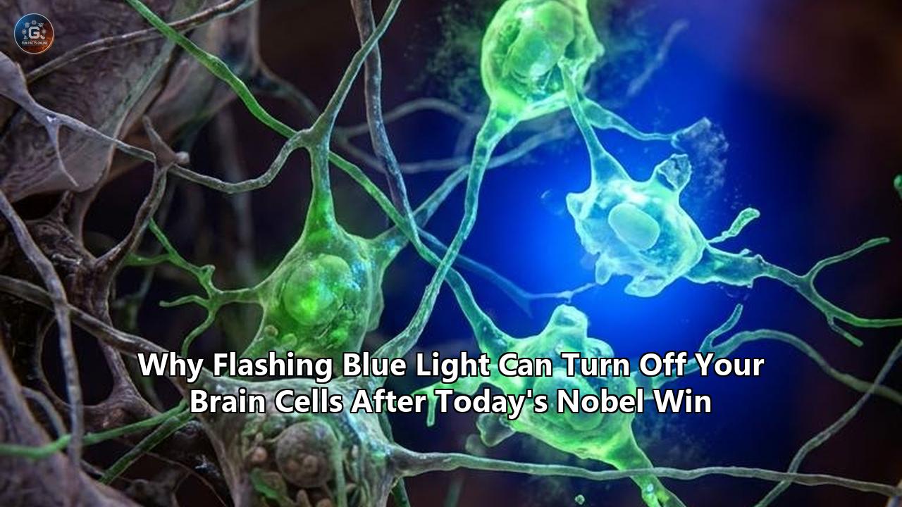
The Nobel Assembly at Karolinska Institutet in Stockholm awarded the 2026 Nobel Prize in Physiology or Medicine to Karl Deisseroth, Peter Hegemann, and Georg Nagel for discoveries that made it possible to control nerve cells using targeted pulses of light. The 12-million-Swedish-kronor ($1.2 million) prize honors the development of optogenetics, an approach that fuses genetic engineering with optical physics to switch individual neurons on and off inside living brain tissue.
While popular attention has centered on using flashes of blue light to ignite neural circuits, the central therapeutic frontier celebrated by the Nobel Committee hinges on the reverse process: turning hyperactive brain cells off. In laboratories worldwide, researchers have demonstrated that shining specific wavelengths of blue light onto genetically sensitized neurons can halt abnormal electrical storms in real time.
"Optogenetics provides opportunities for mapping the brain in a way that we could once only dream of," Per Svenningsson, Chair of the Nobel Committee for Physiology or Medicine, stated during the announcement in Stockholm. Deisseroth, a professor of bioengineering and psychiatry at Stanford University and an investigator at the Howard Hughes Medical Institute, underscored the clinical gravity of the mechanism shortly after receiving the call from Sweden: "People usually think about light as a way of collecting information to observe things. But this is the opposite of that. This is using light to control things, to make things happen, and to do it in a very precise way." That precision, Deisseroth explained, "gives you, inevitably, ideas for how to correct things that go wrong."
The ability to silence hyperactive circuits with optical millisecond precision addresses an urgent medical challenge. Pathological neural hyperactivity drives debilitating human disorders, ranging from pharmacoresistant focal epilepsy and severe treatment-resistant depression to chronic neuropathic pain and obsessive-compulsive disorders. For more than a century, clinical neurology has relied on blunt interventions—systemic pharmaceutical compounds that saturate the entire central nervous system, or implanted metal electrodes that discharge indiscriminate electrical currents into healthy and diseased tissue alike.
Attempting to extinguish pathological firing by shining blue light into living neural tissue uncovered an array of biophysical obstacles: paradoxical axon firing, ion gradient collapses, tissue overheating, and severe light scatter. Overcoming these dilemmas has driven a sweeping engineering overhaul across molecular biology and clinical neurotechnology, redefining how modern medicine plans to treat structural brain failure.
The Biophysical Switch: How Blue Photons Silence Firing Neurons
Understanding why a pulse of blue light can extinguish an action potential requires unpacking the molecular machinery Hegemann and Nagel first isolated from green algae. In the early 2000s, working at the Max Planck Institutes in Martinsried and Frankfurt, Hegemann and Nagel studied Chlamydomonas reinhardtii, a single-celled chlorophyte alga that navigates aquatic environments by swimming toward moderate light sources. They identified channelrhodopsin-1 and channelrhodopsin-2 (ChR2), seven-transmembrane retinal-binding proteins embedded within the organism's eyespot. Unlike mammalian visual pigments that require complex G-protein-coupled secondary messenger cascades to open adjacent channels, channelrhodopsin functions as a light-gated ion pore: a single protein that directly combines the photon sensor and the physical ion gate.
When blue photons with a wavelength of approximately 470 nanometers strike the all-trans-retinal chromophore inside ChR2, the chromophore undergoes ultrafast photoisomerization to 13-cis-retinal. This structural flip triggers a conformational rearrangement throughout the protein's helical bundle, opening a central pore within half a millisecond.
In native ChR2, this pore conducts positively charged cations—primarily sodium ($Na^+$) and calcium ($Ca^{2+}$)—down their electrochemical gradients into the cytoplasm. When Deisseroth, working alongside graduate students Edward Boyden and Feng Zhang at Stanford in 2005, packaged the ChR2 genetic sequence into an adeno-associated virus (AAV) and delivered it into mammalian nerve cells, shining blue light depolarized the cellular membrane. Once the transmembrane potential surged past the critical -55 millivolt threshold, voltage-gated sodium channels opened, driving the neuron to fire.
           470 nm Blue Light Pulse
                     │
                     ▼
       ┌───────────────────────────┐
       │     Channelrhodopsin      │ (Photoisomerization of Retinal)
       └─────────────┬─────────────┘
                     │
         [DIRECT PORE OPENING]
                     │
        ┌────────────┴────────────┐
        ▼                         ▼
   CATION INFLUX             ANION INFLUX
  (Na+, Ca2+ via ChR2)     (Cl- via GtACRs)
        │                         │
  Depolarization            Hyperpolarization
        │                         │
[ACTION POTENTIAL]        [NEURAL SILENCING]
Silencing a neuron, however, requires preventing that electrical spike. The optogenetic toolkit achieves this shutoff via two distinct mechanisms:
1. Direct Anion Shunting via Light-Gated Chloride Channels
Following the algal discoveries of Hegemann and Nagel, researchers isolated and engineered light-gated anion channelrhodopsins (ACRs), most notably GtACR1 and GtACR2 from the cryptophyte alga Guillardia theta. Like ChR2, GtACR2 responds rapidly to 470 nm blue light. Instead of passing cations, its pore lining is reconfigured with positively charged amino acids that selectively conduct negative chloride ions ($Cl^-$).
In a mature, healthy mammalian neuron, the intracellular concentration of chloride is kept low (roughly 4 to 10 millimoles per liter), maintained by the potassium-chloride cotransporter KCC2. As a result, the equilibrium potential for chloride ($E_{Cl}$) sits near -65 to -70 millivolts—at or below the resting membrane potential.
When blue light strikes GtACR2, negative chloride ions rush across the membrane into the cell. This hyperpolarizes the neuron, driving its membrane voltage away from the firing threshold. Simultaneously, the sudden drop in membrane electrical resistance creates an effect known as shunting inhibition: any incoming excitatory electrical currents leaking in from neighboring synapses are short-circuited across the open chloride channels before they can reach the axon initial segment. The neuron is effectively silenced within milliseconds.
2. Upstream Circuit Silencing via Inhibitory Interneuron Networks
The second silencing modality utilizes standard excitatory blue-light channelrhodopsins targeted exclusively to inhibitory brain cells. The mammalian cerebral cortex and hippocampus are populated by specialized local circuits of GABAergic (gamma-aminobutyric acid) interneurons, primarily parvalbumin-positive ($PV^+$) basket cells and somatostatin-positive ($SST^+$) interneurons.
By packaging ChR2 downstream of cell-specific genetic promoters, Deisseroth's group demonstrated that a flash of blue light can selectively activate these inhibitory regulators. The stimulated interneurons dump surges of GABA onto the bodies and dendrites of nearby excitatory pyramidal projection neurons. The GABA binds to endogenous $GABA_A$ receptors, flooding the surrounding pyramidal cells with chloride ions and arresting regional brain activity. Through this circuit architecture, shining blue light acts as an off-switch for entire computational modules in the brain.
In laboratory models engineered to evaluate how blue light brain cells interactions dictate circuit activity, researchers confirmed that this optical brake can terminate runaway seizure activity, suppress anxiety cascades, and disrupt compulsive behavioral loops within milliseconds.
The Neurological Crisis: Why Blunt Tools Fail Pathological Circuits
The therapeutic urgency of this optical off-switch stems from the limitations of modern clinical neurology. For decades, the medical field has attempted to manage brain disorders using treatments that lack spatial and temporal resolution.
┌───────────────────────────┬───────────────────────────┬───────────────────────────┐
│ Modality                  │ Spatial Resolution        │ Temporal Precision        │
├───────────────────────────┼───────────────────────────┼───────────────────────────┤
│ Systemic Pharmacology     │ Whole-body / Non-specific │ Hours to weeks            │
│ Electrical DBS            │ Millimeters (Spherical)   │ Milliseconds (Continuous) │
│ Optogenetic Inhibition    │ Single-cell / Micro-micron│ Milliseconds (Pulsed)     │
└───────────────────────────┴───────────────────────────┴───────────────────────────┘
Systemic pharmacology remains the standard of care for neuropsychiatric and neurological diseases, yet small-molecule drugs distribute across the entire brain and body. Antiepileptic compounds, such as sodium-channel blockers (carbamazepine, phenytoin) or GABA-enhancers (clonazepam, valproate), suppress overall cortical excitability. By altering ion kinetics globally, they cause chronic sedation, cognitive blunting, hepatotoxicity, and motor ataxia. More critically, approximately 30 percent of the world's 50 million epilepsy patients suffer from pharmacoresistant epilepsy, meaning their seizures continue to break through even under high doses of multi-drug regimens.
To overcome the broad wash of pharmaceuticals, neurosurgeons turned to Deep Brain Stimulation (DBS). Pioneered in the late 20th century, DBS uses surgically implanted, high-frequency metal electrodes to drive electrical current into specific anatomical nodes, such as the subthalamic nucleus for Parkinson's disease or the internal capsule for severe obsessive-compulsive disorder.
DBS remains hampered by an unavoidable biophysical reality: electrical currents obey Maxwell's equations in conductive saline media, dispersing spherically through brain tissue without distinguishing between cell types. The electrical field stimulates excitatory pyramidal neurons, inhibitory interneurons, glial cells, and myelin-insulated axon "fibers of passage" traveling to unrelated brain structures. Consequently, patients receiving DBS for motor disorders frequently suffer psychiatric side effects—including acute depression, hypomania, speech disturbances, and executive impairment—caused by the accidental activation of passing axonal highways.
"It has let us go from a 'Rand McNally' road atlas of the brain to something more akin to 'Google Earth,'" noted neuroscientist Patrick Forcelli of Georgetown University, evaluating the Nobel-winning technology's capacity to dissect these circuits. Electrical stimulation acts like an operational sledgehammer. Optogenetics provides the equivalent of a software compiler: the capacity to silence one specific genetic class of hyperactive neurons while leaving adjacent cells and intersecting wiring completely untouched.
The Biophysical Roadblock: What Went Wrong Inside the Silenced Cell
Despite the conceptual elegance of using light to halt neural firing, early attempts to implement blue-light optogenetic silencing revealed profound biological failure modes. Living brain tissue proved far more complex than isolated cell cultures, and researchers encountered major hurdles when trying to turn neurons off reliably.
                  THE AXONAL INVERSION TRAP

   SOMA                                      AXON TERMINAL
┌───────────────────────┐                  ┌───────────────────────┐
│ KCC2 Transporter High │                  │ NKCC1 Transporter High│
│ Intracellular Cl- Low │                  │ Intracellular Cl- High│
│ E_Cl = -70 mV         │                  │ E_Cl = -40 mV         │
└───────────┬───────────┘                  └───────────┬───────────┘
            │                                          │
            ▼                                          ▼
   470 nm Blue Light                          470 nm Blue Light
   Strikes GtACR2                             Strikes GtACR2
            │                                          │
            ▼                                          ▼
      Inward Cl- Flux                            Outward Cl- Flux
            │                                          │
            ▼                                          ▼
     HYPERPOLARIZATION                          DEPOLARIZATION
    (Neuron Silenced)                         (PARADOXICAL FIRING)
The Axonal Inversion Paradox
The most alarming discovery occurred when researchers attempted to silence projecting pathways using anion channelrhodopsins such as GtACR2. When blue light was directed at the neuronal cell body (soma), the neuron silenced as expected. However, when light was directed onto the downstream axons and presynaptic nerve terminals, the cells unexpectedly discharged bursts of neurotransmitters. The off-switch had turned into an on-switch.
The cause lies in developmental ion-transporter gradients:
- In the neuronal soma, the KCC2 transporter actively pumps chloride ions out of the cell, keeping internal chloride low and setting the chloride reversal potential ($E_{Cl}$) at an inhibitory -70 millivolts.
- In distal axons and presynaptic terminals, KCC2 is largely absent. Instead, the sodium-potassium-chloride cotransporter NKCC1 dominates, pumping chloride into the axon.
- This elevates axonal internal chloride, shifting its reversal potential to a depolarizing -40 millivolts.
When 470 nm blue light strikes GtACR2 channels located in the axon, chloride ions rush out of the terminal along their electrochemical gradient. This efflux leaves behind excess positive charge, depolarizing the axonal membrane, opening voltage-gated calcium channels, and triggering sudden neurotransmitter release. Instead of suppressing neural communication, shining blue light onto axonal arborizations induced antidromic action potentials that traveled backward into the brain, causing erratic circuit behavior.
Post-Inhibitory Rebound Spikes
A second challenge involves post-inhibitory rebound excitation. Mammalian neurons maintain hyperpolarization-activated cyclic nucleotide-gated (HCN) channels, which generate an inward, depolarizing current known as $I_h$. When a neuron is deeply hyperpolarized by a sustained pulse of blue light, these HCN channels open. Simultaneously, low-threshold T-type voltage-gated calcium channels de-inactivate.
The moment the blue light is switched off, the accumulated $I_h$ current and inward calcium flux produce a massive, coordinated rebound depolarization. Instead of returning smoothly to a baseline resting state, the silenced neuron discharges a volley of high-frequency action potentials. In animal models of epilepsy, abruptly terminating an optical silencing pulse occasionally triggered the exact seizure event the light had been deployed to prevent.
Membrane Potential (mV)
    │
-50 ┼─────────────────── Threshold ──────────────────
    │
-65 ┼─────┐             ┌──────────────────────────── Baseline
    │     │             │     ▲ (Rebound Spike Burst!)
    │     │             │    ╱ ╲
-80 ┼─────┴─────────────┴───╱───╲──────────────────── Hyperpolarized
          ◄── Light ON ──►  ◄─ Light OFF ─►
Severe Blue-Wavelength Tissue Attenuation and Thermal Deposition
The physics of light propagation through dense mammalian brain matter creates an engineering bottleneck. Blue light (470 nm) possesses high photon energy, but biological tissue acts as a turbid scattering medium packed with lipid bilayers, myelin sheaths, and absorbing chromophores like hemoglobin.
The scattering coefficient of mammalian gray matter at 470 nm exceeds $10\text{ mm}^{-1}$. Consequently, more than 90 percent of the optical power delivered by a flat-cleaved fiber optic cannula is scattered or absorbed within the first 500 to 800 micrometers of tissue.
To deliver the threshold power density (typically 1 to 5 milliwatts per square millimeter) required to activate microbial opsins distributed across several cubic millimeters of human cortex, operators must pump high optical power into the implant interface. This energy converts directly into localized thermal dissipation.
Brain tissue is vulnerable to heat: an elevation of just 1.0 degree Celsius alters endogenous synaptic kinetics and triggers heat-shock protein expression; an elevation of 2.0 to 3.0 degrees Celsius induces thermal necrosis and irreversible neurovascular damage. Pumping sustained blue light into deep subcortical structures to suppress seizures risked cooking the surrounding tissue.
Transgene Immunogenicity and Vector Capsid Limitations
Channelrhodopsins are foreign proteins derived from single-celled algae (Chlamydomonas, Guillardia) and archaeal prokaryotes (Halobacterium). When expressed in the mammalian central nervous system over months or years, these foreign peptides can trigger neuroinflammatory responses. Activated microglia and astrocytes migrate toward opsin-expressing regions, driving glial scar formation that insulates neurons from light and degrades long-term gene expression.
Furthermore, packaging opsin genes alongside complex cell-type-specific promoter sequences pushes the spatial carrying capacity of recombinant adeno-associated viruses (rAAVs), which hit a hard packaging ceiling of 4.7 kilobases. Early vector configurations often suffered from structural instability, poor membrane trafficking, and cytoplasmic aggregation, leaving opsin proteins trapped inside the endoplasmic reticulum where they caused cellular toxicity rather than functioning at the cell surface.
The Engineering Solutions: Modern Neurotechnology Countermeasures
To overcome these roadblocks, neuroengineers, biophysicists, and molecular biologists transformed optogenetic silencing into a clinically viable modality.
┌─────────────────────────────────┬─────────────────────────────────┬─────────────────────────────────┐
│ Biophysical Challenge           │ Molecular / Optical Solution    │ Engineering Mechanism           │
├─────────────────────────────────┼─────────────────────────────────┼─────────────────────────────────┤
│ Axonal Paradoxical Excitation   │ Soma-Targeted Opsin (st-GtACR2) │ Kv2.1 peptide targeting motif   │
│ Post-Inhibitory Rebound Spikes  │ Step-Function Inactivation      │ Tapered ramp-down photocycles   │
│ Blue Light Scattering & Heat    │ Near-Infrared Upconversion /    │ UCNP transducers & red-shifted  │
│                                 │ Red-Shifted Chloride Pumps      │ halo/arch-rhodopsins (Jaws)     │
│ Invasive Hardware & Tissue Tear │ Flexible Inorganic Micro-LEDs   │ Cellular-scale thin-film arrays │
└─────────────────────────────────┴─────────────────────────────────┴─────────────────────────────────┘
Subcellular Molecular Targeting: The Somatic Lock
To resolve the axonal inversion paradox—where blue light triggered accidental neurotransmitter release—biophysicists engineered subcellular compartmentalization tags. Drawing on work from the laboratories of Deisseroth, Hegemann, and their collaborators, teams fused GtACR2 to the intracellular targeting motif of the mammalian voltage-gated potassium channel Kv2.1.
The Kv2.1 motif acts as a molecular shipping label, directing the cell's transport machinery to insert the opsin exclusively into the plasma membrane of the soma and proximal dendrites, preventing the protein from entering the axon initial segment or distal terminals.
The resulting construct, designated soma-targeted GtACR2 (st-GtACR2), confines chloride-conducting pores to the cell body. When illuminated with blue light, st-GtACR2 produces high hyperpolarizing currents and shunts incoming signals without causing terminal depolarization. Axonal paradoxical spikes are eliminated, providing clean, predictable silencing across intact neural circuits.
Step-Function Inactivation and Tapered Photocyles
To prevent post-inhibitory rebound bursts, researchers re-engineered the channelrhodopsin binding pocket using structure-guided point mutations. By mutating the C128 and D156 residues within the retinal pocket of ChR2, Hegemann’s team created "Step-Function Opsins" (SFOs) and stabilized step-function chloride channels (SwiChR).
These engineered channels alter the photocycle kinetics: a single, sub-millisecond flash of 470 nm blue light switches the channel into an open, conducting state that remains active for minutes without further illumination. Because light does not need to shine continuously, the total optical power delivered to tissue drops by more than three orders of magnitude, eliminating the threat of photothermal heating.
Furthermore, to extinguish the inhibition without triggering rebound action potentials, researchers flash a pulse of red or amber light (590–630 nm) or slowly step down the blue light power in a tapered ramp. By closing the channels gradually over several hundred milliseconds, the membrane voltage drifts steadily back to resting potential, allowing HCN channels and T-type calcium channels to adjust without generating rebound bursts.
In experiments where targeted blue light brain cells switches were deployed to silence epileptic foci, this ramped closure protocol suppressed seizure activity without inducing secondary hyperexcitable cascades.
Standard Square Pulse Silencing (High Rebound Risk):
Light: ┌──────────────────────────────────────────┐
       │                                          │
───────┴──────────────────────────────────────────┴─────────────────
       ▼ SHUTOFF SHOCK -> Triggers HCN / T-Type Ca2+ Rebound Burst

Ramped Tapered Silencing (Zero Rebound):
Light: ┌────────────────────────────────────\
       │                                     \
───────┴──────────────────────────────────────\─────────────────────
       ▲ GRADUAL RECOVERY -> Membrane potential resets smoothly
Upconversion Nanoparticles and Deep-Tissue Optical Transduction
To bypass the severe scatter of blue light in deep brain structures, neuroengineers introduced lanthanide-doped upconversion nanoparticles (UCNPs). These microscopic ceramic transducers absorb deep-penetrating, low-energy near-infrared (NIR) light (typically 808 nm to 980 nm) and upconvert the photons through multiphoton mechanisms, emitting higher-energy blue light (470 nm) locally inside tissue.
Near-infrared wavelengths penetrate deep into lipid-rich mammalian brain matter with minimal scattering and low absorption by blood and water. By injecting biocompatible, silica-coated UCNPs into deep target areas—such as the subthalamic nucleus, amygdala, or hippocampus—clinicians can position an optical transducer directly next to opsin-expressing cells.
A near-infrared laser beam directed at the intact scalp penetrates the skull, travels through intervening cortex, and strikes the UCNPs. The nanoparticles absorb the invisible near-infrared light and emit localized 470 nm blue photons, activating the st-GtACR channels to silence target neurons without requiring invasive fiber-optic cannulas or causing thermal burns to surface tissue.
Cellular-Scale, Flexible Inorganic Micro-LEDs
For applications requiring permanent hardware implantation, mechanical engineers departed from rigid, brittle silica glass fibers. Collaborative efforts led by researchers such as John A. Rogers at Northwestern University and Philipp Gutruf at the University of Arizona produced flexible, microscale inorganic light-emitting diodes (micro-LEDs) thinner than a human hair (less than 10 micrometers thick).
Encapsulated in biocompatible polymers like polyimide and polydimethylsiloxane (PDMS), these micro-LED arrays integrate directly into neural tissue, flexing naturally alongside brain pulsations and vascular movement. By combining these arrays with microscale diamond heat sinks and integrated transparent platinum-iridium electrodes, engineers created closed-loop interfaces capable of recording local field potentials, detecting seizure spikes in real time, flashing a localized micro-burst of blue light to silence the offending cluster of cells, and confirming signal arrest within 10 milliseconds.
Clinical Translations: Where Light-Based Silencing Meets Patient Care
The Nobel Prize honors not just a laboratory discovery, but an approach actively transitioning into clinical trials to address historically intractable diseases.
                                  CLINICAL TRANSLATION PIPELINE

    PHASE I / II TRIALS                 PRECLINICAL VALIDATION                EARLY STAGE RESEARCH
┌──────────────────────────┐         ┌──────────────────────────┐         ┌──────────────────────────┐
│  Retinitis Pigmentosa    │         │  Focal Cortical Epilepsy │         │  Deep Brain Stimulation  │
│  - GenSight Biologics    │         │  - On-demand seizure     │         │  - Targeted silencing of │
│    GS030 trial           │         │    arrest within 50 ms   │         │    subthalamic pathways  │
│  - Bionic Sight system   │         │  - Closed-loop detection │         │  - Eliminates DBS        │
│  - Partial visual acuity │         │    via ECoG arrays       │         │    behavioral side-effects│
│    restoration in humans │         │  - Minimizes brain resec-│         │  - Next-gen Parkinson's  │
│                          │         │    tion surgery          │         │    and dystonia trials   │
└──────────────────────────┘         └──────────────────────────┘         └──────────────────────────┘
The First Human Milestone: Restoring Sight
The earliest clinical realization of optogenetics occurred in the human retina, an accessible extension of the central nervous system. In diseases like retinitis pigmentosa, genetic mutations destroy the eye's primary photoreceptors—rods and cones—leaving patients completely blind. Crucially, the downstream retinal ganglion cells (RGCs) that carry visual signals down the optic nerve to the visual cortex survive.
In clinical trials conducted by GenSight Biologics (the PIONEER study, utilizing GS030) and Bionic Sight, clinicians injected an AAV vector encoding a light-sensitive opsin into the vitreous cavity of blind patients' eyes. The vector selectively transduced surviving retinal ganglion cells, transforming them into de facto primary light sensors.
Patients wore specialized neuromorphic goggles equipped with an outward-facing camera that captured visual scenes and projected high-intensity optical patterns onto the retina. In published clinical outcomes, patients with complete vision loss regained the ability to detect, locate, and count objects on a table. The trial confirmed that microbial opsins can be expressed safely inside human neural tissue without triggering devastating neuroimmune responses.
Halting Intractable Focal Epilepsy
The next clinical milestone centers on drug-resistant focal epilepsy. In patients with focal cortical dysplasia or mesial temporal lobe epilepsy, unmanageable seizures originate from an identifiable, circumscribed cluster of hyperactive neurons before spreading across both hemispheres. Standard surgical treatment involves physically resecting or burning away the diseased brain tissue—an irreversible procedure that carries risks of cognitive, memory, or language deficits.
Preclinical work across academic medical centers has demonstrated closed-loop optogenetic seizure suppression in non-human primates and rodent models. A clinical-grade AAV vector delivering soma-targeted anion channelrhodopsins (st-GtACR) or inhibitory parvalbumin-targeted switches is delivered directly to the epileptic focus.
Flexible subdural electrocorticography (ECoG) grids constantly monitor the electrical rhythm of the cortex. The moment an automated algorithm detects the high-frequency oscillatory spikes signaling seizure emergence, an array of micro-LEDs flashes a calibrated burst of blue light. The hyperactive neurons are clamped at their resting chloride potential, terminating the seizure in under 50 milliseconds before it can propagate into motor pathways or clinical convulsion. The patient experiences no convulsion, no loss of consciousness, and none of the cognitive fog associated with high-dose antiepileptic medications.
Silencing the Neural Substrates of Pain and Psychiatric Despair
Optogenetic silencing is also transforming the study of intractable neuropathic pain. Chronic neuropathic pain—arising from diabetic neuropathy, spinal cord trauma, or post-herpetic neuralgia—is maintained by hyperactive, spontaneous firing inside primary nociceptive neurons located within the dorsal root ganglion (DRG). Standard interventions rely heavily on opioid agonists, which carry high addiction liability, respiratory depression risks, and tolerance build-up.
By delivering light-sensitive inhibitory opsins into the peripheral nerves of the DRG, researchers can direct transdermal light through flexible wireless patches, turning off nociceptive transmission at the primary spinal gateway. Pain signals are extinguished before they can ascend the spinothalamic tract into the thalamus and somatosensory cortex.
In neuropsychiatry, Deisseroth’s clinical practice as a psychiatrist informed his laboratory’s mapping of psychiatric symptom circuits. In deep, treatment-resistant depression, functional neuroimaging reveals severe, unyielding metabolic hyperactivity within the subgenual anterior cingulate cortex (Brodmann Area 25).
Similarly, severe obsessive-compulsive disorder is driven by uncontrolled, hyperactive reverberating loops traversing the orbitofrontal cortex and striatum. In animal models, optogenetically silencing these hyperactive corticostriatal projection circuits resolves depressive-like social withdrawal and stops compulsive grooming behaviors instantly.
By mapping these pathways with optogenetics, clinicians are now developing targeted, non-viral neuromodulation protocols—such as accelerated transcranial magnetic stimulation (TMS) and low-intensity focused ultrasound (FUS)—that mimic the cell-type silencing discovered through optical science.
In neurodegenerative disorders, demonstrating that in these specialized blue light brain cells models, selective inhibition of overactive striatal pathways could rebalance the direct and indirect basal ganglia loops has guided cleaner, lower-voltage deep brain stimulation programming protocols in clinical neurology suites.
Resolving Public Misconceptions: Screen Wavelengths vs. Neural Switches
The news that flashing blue light can turn off brain cells has triggered understandable public confusion, fueled by widespread warnings about the dangers of blue-emitting smartphones, tablets, and LED computer screens. The public dilemma requires drawing a sharp line between environmental light striking the human eye and targeted optogenetic silencing delivered inside brain tissue.
┌─────────────────────────────────┬─────────────────────────────────┬─────────────────────────────────┐
│ Feature                         │ Consumer Device Blue Light      │ Optogenetic Blue Light (ACR)    │
├─────────────────────────────────┼─────────────────────────────────┼─────────────────────────────────┤
│ Target Sensor                   │ Melanopsin in retinal ipRGCs    │ Transgenic Algal Opsins         │
│ Mechanism of Action             │ GPCR-mediated G_q signaling     │ Direct ion channel pore gating  │
│ Biological Effect               │ Suppresses Pineal Melatonin     │ Direct Cl- influx / Hyperpolar- │
│                                 │ Shifts Circadian Master Clock   │ ization of Action Potentials    │
│ Target Tissue Location          │ Unmodified Human Retina         │ Genetically Transduced Neurons  │
│ Light Power Density             │ Nanowatts to Microwatts / cm²   │ Milliwatts / mm² (Localized)    │
│ Action Potential Arrest?        │ NO (Increases alertness)        │ YES (Arrests firing in 10 ms)   │
└─────────────────────────────────┴─────────────────────────────────┴─────────────────────────────────┘
The human retina contains approximately 120 million rod cells, 6 million cone cells, and a tiny subset (roughly 1 to 2 percent) of specialized, non-image-forming sensors known as intrinsically photosensitive retinal ganglion cells (ipRGCs). These ipRGCs express an endogenous, mammalian photopigment called melanopsin. Melanopsin responds to blue wavelengths between 460 and 480 nm.
When blue light from an evening smartphone display or an office monitor strikes the eye:
- Melanopsin absorbs the photon and triggers an intracellular G-protein cascade ($G_{q/11}$), activating phospholipase C.
- This opens transient receptor potential (TRP) channels, depolarizing the ipRGC.
- The resulting electrical spikes travel along the retinohypothalamic tract to the suprachiasmatic nucleus (SCN), the brain’s central circadian clock.
- The SCN transmits inhibitory signals to the pineal gland, suppressing the secretion of melatonin, the hormone that promotes physiological sleep.
Consumer screen blue light does not turn off brain cells; in intact, non-genetically modified human brains, environmental blue light is activating. It signals morning sunlight, increases autonomic arousal, raises cortisol, and keeps executive neural networks awake.
The Nobel-winning technology functions through an entirely different paradigm. A normal, unmodified human neuron does not express algal channelrhodopsin and is biologically indifferent to light. If a neurosurgeon shines a blue laser onto an unmodified human brain, the light will scatter and produce minor warmth, but the neurons will not change their electrical firing rates.
The brain cells turn off if and only if they have undergone genetic transduction via viral vectors to manufacture algal anion channels like GtACR2, and are illuminated with focused, high-density light delivered directly to the cell membrane. Far from a hazard of looking at an iPhone, optogenetic blue-light silencing is a synthetic, engineered intervention designed to arrest targeted electrical dysfunction.
Ethical and Regulatory Frontiers: The Path to Clinical Safety
As optogenetics transitions from academic laboratories into translational medical centers, regulatory bodies and bioethicists are building governance frameworks to shepherd these therapies safely into human medicine.
                     REGULATORY PIPELINE FOR OPTOGENETIC SYSTEMS

         GENE THERAPY                       ACTIVE IMPLANTABLE DEVICE
         (FDA CBER)                                (FDA CDRH)
              │                                         │
              ▼                                         ▼
   AAV Capsid & Opsin Vector                Flexible Micro-LED Hardware
   - Biodistribution & Shedding             - ISO 10993 Biocompatibility
   - Off-target Expression Testing          - Thermal Dissipation (<1.0°C)
   - Transgene Immunogenicity               - Chronic Mechanical Strain
              │                                         │
              └───────────────────┬─────────────────────┘
                                  ▼
                     COMBINATION PRODUCT REVIEW
                      (FDA Office of Combination
                              Products)
                                  │
                                  ▼
                 FIRST-IN-HUMAN CLINICAL TRIALS
                 - Dose-Escalation & Safety
                 - Objective Electrophysiology
                 - Long-term Neural Integration
The Combination Product Regulatory Gauntlet
The U.S. Food and Drug Administration (FDA) and the European Medicines Agency (EMA) classify optogenetic silencing platforms as "combination products." They involve both a biologic (a genetically engineered viral vector delivering foreign algal DNA) and a Class III active implantable medical device (an optoelectronic pulse generator, micro-LED array, or near-infrared laser system).
This dual identity requires concurrent review by the FDA’s Center for Biologics Evaluation and Research (CBER) and the Center for Devices and Radiological Health (CDRH). Approval hinges on proving:
- Long-Term Genetic Stability: Demonstrating that the AAV vector does not integrate into host genomic DNA, causing insertional mutagenesis or oncogene activation.
- Immune Tolerability: Verifying that sustained expression of foreign algal proteins like GtACR does not provoke chronic, cytotoxic CD8+ T-cell infiltration, blood-brain-barrier breakdown, or neurodegenerative auto-reactivity.
- Hardware Durability and Thermal Margins: Demonstrating that flexible implanted micro-LEDs will not mechanically degrade, leach toxic packaging materials, or heat neural tissue beyond the strict 1.0 degree Celsius safety limit over decades of continuous operation.
To address these hurdles, neurotechnology consortia are developing non-viral gene delivery platforms, such as lipid nanoparticles (LNPs) functionalized with brain-targeting peptides, and engineered "humanized" opsins. By mutating the amino acid surface residues of algal rhodopsins to match endogenous human channel proteins, synthetic biologists aim to "camouflage" the light switches from circulating surveillance lymphocytes, eliminating long-term immune rejection risks.
Neuroethics and Circuit Governance
The prospect of using light to extinguish activity in targeted brain circuits raises profound neuroethical questions. If an implant can turn off fear circuits in the amygdala, rage circuits in the hypothalamus, or reward circuits in the ventral tegmental area on command, where does medical treatment end and behavioral engineering begin?
During the Nobel Prize proceedings, ethicists and clinical leaders emphasized that optogenetics must adhere to clear boundaries of patient autonomy. The primary near-term indications—such as halting epileptic seizures, treating pharmacoresistant psychiatric distress, or arresting intractable physical pain—are restorative, aimed at returning a pathologically locked system to healthy homeostatic baselines.
Nevertheless, the development of closed-loop optogenetics—where machine-learning algorithms make autonomous, real-time decisions to silence neural circuits without conscious patient intervention—demands strict regulatory standards. The scientific community, through bodies such as the International Brain Initiative and the NIH BRAIN Initiative, has established governance standards requiring patient access to transparent audit trails of every optical silencing event, explicit therapeutic parameter locking, and ironclad fail-safe cutoffs to prevent unintentional circuit suppression.
By standardizing how blue light brain cells connections are mapped and manipulated in primate cortex, international teams are building the precise physiological models required to satisfy these rigorous regulatory benchmarks before widespread surgical adoption.
What Comes Next: The Horizon of Light-Controlled Brains
The awarding of the 2026 Nobel Prize in Physiology or Medicine to Deisseroth, Hegemann, and Nagel honors a scientific journey that began with basic curiosity about single-celled pond algae and matured into one of the most sophisticated platforms in modern medicine. The capacity to turn brain cells off with light has resolved basic limitations that frustrated neuroscience for over a century.
           THE DECADE AHEAD: ANTICIPATED MILESTONES (2026–2035)

  2026                 2028                 2031                 2035
   │                    │                    │                    │
   ▼                    ▼                    ▼                    ▼
Nobel Prize       First-in-Human       Phase II Multi-Center     Fully Integrated
Awarded for       Clinical Trials      Clinical Trials for       Non-Invasive
Optogenetic       for Cortical         Intractable Depres-       Opto-Nanomedicine
Foundations       Focal Epilepsy       sion & Neuropathic Pain   Commercialization
Over the next two to four years, the biomedical landscape will witness the launch of Phase I and Phase II clinical trials applying soma-targeted anion channelrhodopsins directly to the cerebral cortex of patients with pharmacoresistant focal epilepsy. These trials will mark the transition of optogenetic silencing from an invaluable discovery tool in academic laboratories into a lifesaving clinical reality in neurological operating rooms.
Concurrently, developments in near-infrared upconversion nanomedicine and non-invasive two-photon focal illumination will reduce the need for permanent, penetrating brain hardware. Patients suffering from intractable central nervous system disorders may soon receive targeted viral or non-viral vector infusions, followed by therapeutic sessions under non-invasive optical helmets that focus invisible, harmless near-infrared beams through the scalp and skull to extinguish malfunctioning circuits deep within the brain without a single incision.
Beyond clinical applications, the optogenetic off-switch continues to illuminate the fundamental logic of human consciousness, cognition, and emotion. By selectively silencing individual nodes within the brain's computational networks, scientists are systematically identifying the exact neural signatures that generate memory recall, mediate voluntary decision-making, and sustain subjective conscious awareness.
As the mechanisms of optogenetics progress from foundational pond-scum biophysics to next-generation human therapeutics, the ability to control neural electrical storms with flashes of light stands as a profound testament to biological science—transforming what was once an intractable mystery into a solvable engineering problem.
Timeline: The Evolution of Optogenetics
- 1979: Francis Crick suggests that the ultimate challenge in neuroscience is to control one specific type of cell in the brain while leaving others untouched, speculating that light might serve as the ideal control tool.
- 2002–2003: Peter Hegemann and Georg Nagel isolate and characterize channelrhodopsin-1 and channelrhodopsin-2 from Chlamydomonas reinhardtii, demonstrating that a single algal protein functions directly as a light-gated ion channel.
- 2005: Karl Deisseroth, along with Edward Boyden and Feng Zhang at Stanford University, successfully expresses channelrhodopsin-2 in mammalian neurons, proving that pulses of blue light can fire brain cells on command with millisecond temporal precision.
- 2007: Deisseroth’s laboratory achieves optical neural control in awake, freely moving mice using thin, flexible optical fibers, proving causal links between specific neural circuits and complex behaviors.
- 2010: Identification and engineering of inhibitory optogenetic tools, including halorhodopsins (chloride pumps) and archaerhodopsins (proton pumps), enabling optical neural silencing.
- 2015: Discovery and characterization of natural anion channelrhodopsins (GtACR1 and GtACR2) from Guillardia theta, providing light-gated chloride channels that silence neurons with orders-of-magnitude higher light sensitivity.
- 2018: Engineering of soma-targeted GtACR variants (st-GtACR), resolving the axonal inversion paradox and eliminating accidental antidromic terminal excitation during neural silencing experiments.
- 2021: First clinical milestone published: A blind retinitis pigmentosa patient partially recovers visual object detection following optogenetic AAV delivery into retinal ganglion cells combined with light-projecting goggles.
- October 5, 2026: The Nobel Assembly at Karolinska Institutet awards the 2026 Nobel Prize in Physiology or Medicine to Karl Deisseroth, Peter Hegemann, and Georg Nagel for their foundational discoveries in light-gated ion channels and optogenetics.
Reference:
- https://www.nobelprize.org/prizes/medicine/2026/press-release/
- https://www.dailywire.com/news/scientists-win-nobel-prize-for-turning-brain-cells-on-and-off-it-began-with-pond-scum
- https://news.inbox.eu/150tym6-the-nobel-prize-in-medicine-awarded-for-technology-that-allows-to-turn-on-and-turn-off-brain-cells?language=en
- https://www.hhmi.org/news/karl-deisseroth-2026-nobel-prize-physiology-medicine
- https://www.caledonianrecord.com/news/national/nobel-medicine-prize-goes-to-3-scientists-for-shining-light-on-brain-activity/article_5cedcd02-90c3-5c39-b74c-2f4386d354e9.html
- https://neurosciencenews.com/optogenetics-nobel-prize-2026-31290/
- https://stanforddaily.com/2026/10/06/karl-deisseroth-2026-nobel-prize/
- https://thenextweb.com/news/nobel-prize-medicine-2026-optogenetics
- https://opg.optica.org/boe/viewmedia.cfm?URI=cleo_at-2013-AF1L.1&seq=0&origin=search
- https://www.mightexbio.com/optogenetic-stimulation/
- https://scoop.upworthy.com/scientists-created-an-on-and-off-switch-for-operating-human-brain
- https://en.wikipedia.org/wiki/Anion-conducting_channelrhodopsin
- https://www.pnas.org/doi/10.1073/pnas.2519786123
- https://pmc.ncbi.nlm.nih.gov/articles/PMC6175909/
- https://www.researchgate.net/post/Has_anyone_used_anion_channel_rhodopsins_ACR-GtACR1_or_GtACR2_in_vivo
- https://www.thetransmitter.org/spectrum/flash-light-shifts-social-behavior-autism-mice/
- https://en.ara.cat/society/nobel-of-medicine-to-the-technology-that-can-help-to-understand-why-we-remember-or-fall-in-love_1_5869982.html
- https://www.facebook.com/fromquarktoquasars/posts/nobel-prize-has-been-awarded-for-controlling-brain-cells-with-light-karl-deisser/1671884041216188/
- https://www.facebook.com/cnbctv18india/videos/the-2026-nobel-prize-in-physiology-or-medicine-goes-to-karl-deisseroth-peter-heg/1644632934116027/
- https://news.wttw.com/2026/10/05/nobel-medicine-prize-goes-3-scientists-shining-light-brain-activity
- https://pmc.ncbi.nlm.nih.gov/articles/PMC5670698/
- https://www.reddit.com/r/science/comments/1wyb2z9/the_nobel_prize_in_physiology_or_medicine_2026/
- https://www.facebook.com/vajiramandraviofficial/videos/nobel-prize-2026-winners-in-physiology-or-medicine-what-if-light-could-control-b/1582864739711967/
- https://www.facebook.com/NBCNews/posts/three-scientists-win-the-2026-nobel-prize-in-physiology-or-medicine-for-discover/1492240356101158/
- https://pmc.ncbi.nlm.nih.gov/articles/PMC3225502/
- https://www.nobelprize.org/prizes/medicine/2026/popular-information/