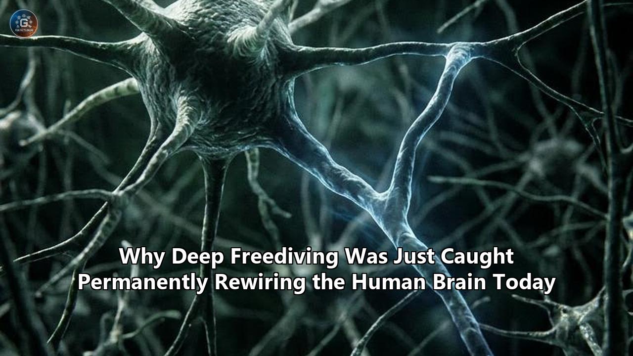
A neuroimaging study conducted by researchers at Université Paris-Saclay, the French Alternative Energies and Atomic Energy Commission (CEA) NeuroSpin center, and Inserm has established that deep freediving permanently alters the human brain's functional architecture.
The research, led by cognitive neuroscientist Julia Micaux and senior investigator Marion Noulhiane, tracked athletes over seven months of intensive breath-hold training. Using high-field functional magnetic resonance imaging (fMRI) paired with episodic memory testing, the research team discovered that the brains of freedivers undergo an extensive reorganization of large-scale neural networks.
Crucially, the hippocampus—the seahorse-shaped structure essential for navigation and episodic memory, long considered the human central nervous system’s most hypoxia-sensitive structure—did not wither or sustain cell death from repeated oxygen deprivation. Instead, it systematically disconnected from sensory networks and forged new functional pathways to the cerebellum and visual cortex.
These findings challenge a cornerstone of modern emergency medicine and clinical neurology: the doctrine that severe, repetitive hypoxia invariably damages mammalian brain tissue. When systemic arterial oxygen saturation falls below 60%, or when arterial partial pressures of oxygen ($PaO_2$) plummet to levels typically seen in acute respiratory distress or near-fatal strangulation, neurons in the hippocampus typically experience metabolic collapse, calcium excitotoxicity, and apoptosis.
Yet elite freedivers routinely push their arterial oxygen saturation down into the 50% to 25% range during static and dynamic apnea—depths of hypoxia where non-divers slip into comas. Instead of manifesting cognitive decline, the Paris-Saclay cohort demonstrated preserved memory performance directly correlated with their newly wired brain networks.
The discovery has ignited an intense physiological debate. For decades, hyperbaric physicians, neuroscientists, and pulmonologists have held conflicting views over what happens to the central nervous system beneath hundreds of feet of water.
One camp has long maintained that deep breath-hold diving constitutes uncontrolled, cumulative brain injury disguised as sport, pointing to circulating biomarkers of central nervous system damage and microvascular lesions. The competing camp has asserted that the human body possesses dormant, deep-seated adaptive mechanisms—dormant evolutionary legacies shared with marine mammals—that can be awakened to shield brain metabolism.
The Paris-Saclay findings provide the first longitudinal functional imaging evidence showing that the brain adapts to extreme environmental hypoxia not through passive survival, but through active, structural, and network-level reconfiguration.
Clinical Damage or Hormetic Shield: Competing Paradigms in Deep Freediving Brain Effects
The medical debate over how the central nervous system withstands apnea centers on two conflicting frameworks: the Pathological Degradation model and the Hormetic Neuroplasticity model. Each approach interprets identical physiological stress markers through entirely different clinical lenses, arriving at opposing conclusions about the safety and consequences of extreme breath holding.
       PATHOLOGICAL DEGRADATION MODEL                HORMETIC NEUROPLASTICITY MODEL
 (Clinical Neurology / Emergency Medicine)       (Applied Physiology / Paris-Saclay Discovery)
                     │                                                 │
  Repetitive Voluntary Hypoxia & Hypercapnia        Repetitive Voluntary Hypoxia & Hypercapnia
                     │                                                 │
         ┌───────────┴───────────┐                         ┌───────────┴───────────┐
         ▼                       ▼                         ▼                       ▼
Blood-Brain Barrier     Microvascular Shearing     Massive Cerebral         Astrocytic Glycolysis
Disruption (S100B Rise)  (Tau & Amyloid Release)    Vasodilation (+107% CBF) & Lactate Shuttling
         │                       │                         │                       │
         ▼                       ▼                         ▼                       ▼
Cumulative Ischemic     Subclinical White Matter    Preservation of Core   Network Rewiring
   Neural Atrophy              Lesions                Tissue Oxygenation     (Hippocampus-Cerebellum)
         │                       │                         │                       │
         └───────────┬───────────┘                         └───────────┬───────────┘
                     ▼                                                 ▼
        Long-Term Cognitive Decline                     Enhanced Resilience & Cognitive
       & Irreversible Encephalopathy                              Preservation
The Pathological Degradation Model
Rooted in emergency medicine, intensive care, and sleep medicine, the pathological model interprets severe hypoxia as inherently cytotoxic. Clinicians operating under this view draw direct parallels between freediving and clinical disorders such as obstructive sleep apnea (OSA), ischemic stroke, and carbon monoxide poisoning.
In patients suffering from obstructive sleep apnea, recurrent drops in oxygen saturation produce systemic inflammation, elevated oxidative stress, endothelial dysfunction, and measurable gray matter loss in the hippocampus and prefrontal cortex. The neurological outcome is well documented: progressive deficits in executive function, reduced working memory, and heightened vulnerability to neurodegenerative conditions.
When hyperbaric researchers applied this perspective to competitive freediving, they uncovered alarming biochemical indicators:
- S100B Protein Elevations: In pioneering work conducted at Lund University by Dr. Johan Andersson and colleagues, serum levels of S100B—a classic peripheral biomarker for astrocyte damage and blood-brain barrier permeation—were found to spike by an average of 37% within ten minutes of divers concluding a maximal static breath hold. In several participants, S100B reached levels seen following acute concussions.
- Axonal and Amyloid Biomarkers: Subsequent investigations identified post-apnea surges in plasma concentrations of total tau and amyloid-beta 42. These proteins are classically tied to axonal shearing and the degenerative cascades of Alzheimer's disease.
- Hypercapnic Acidosis: Arterial blood gas analyses have shown that as oxygen drops to critical thresholds, arterial carbon dioxide ($PaCO_2$) climbs past 45 to 55 mmHg. This hypercapnia induces profound respiratory acidosis, leading some clinicians to warn of cumulative, subclinical micro-infarctions and disrupted vascular autoregulation.
- Arterial Gas Embolism and White Matter Spots: Neuroimaging surveys have revealed asymptomatic white-matter hyperintensities in the brains of veteran divers. Proponents of the damage model argue these spots result from microscopic venous gas emboli passing through a patent foramen ovale (PFO) or unprevented capillary bubbling during decompression.
For advocates of this model, extreme breath holding is a game of biological roulette. In their view, any sport that results in arterial oxygen desaturations under 50% must inevitably cause microstructural brain damage, irrespective of how conditioned an athlete appears.
The Hormetic Neuroplasticity Model
The opposing camp—dominated by extreme environmental physiologists, evolutionary biologists, and the team behind the latest Paris-Saclay study—approaches the phenomenon through the principle of hormesis. Hormesis dictates that biological systems exposed to intermittent, non-lethal doses of an otherwise toxic stressor mount compensatory adaptations that leave the organism more resilient than before.
Researchers within this camp emphasize fundamental differences between clinical hypoxia and elite freediving:
| Physiological Parameter | Obstructive Sleep Apnea (OSA) | Clinical Ischemic Stroke | Elite Deep Freediving |
|---|---|---|---|
| Exposure Nature | Involuntary, fragmented, chronic nightly cycles | Acute, involuntary, uncompensated focal blockage | Voluntary, controlled, intermittent training cycles |
| Systemic Autonomic State | Sympathetic hyperactivation, hypertension, panic | Sympathetic storm, localized metabolic starvation | Strong parasympathetic tone, diving bradycardia |
| Cerebrovascular Response | Impaired vasodilation, endothelial sclerosis | Blocked regional perfusion, rapid ischemic core | Explosive vasodilation (+100–150% cerebral blood flow) |
| Carbon Dioxide Balance | Erratic hypercapnia without preconditioning | Variable, uncoupled from tissue demand | Planned hypercapnic buffering with pre-dive ventilation |
| Neurological Outcome | Apoptosis, hippocampal atrophy, cognitive loss | Necrosis, penumbral damage, functional loss | Functional network rewiring, preserved memory |
Damian Bailey, a professor of physiology and biochemistry at the University of South Wales, has demonstrated that the human brain can unlock compensatory mechanisms when hypoxia is paired with controlled hypercapnia.
Rather than starving the brain, the concurrent buildup of carbon dioxide triggers an immediate, profound relaxation of the cerebrovascular tree. Global cerebral blood flow (CBF) surges by up to 107% or more, allowing the body to sustain overall cerebral oxygen delivery ($CDO_2$) despite falling arterial blood oxygen levels.
Furthermore, magnetic resonance spectroscopy (MRS) examinations have revealed that freedivers do not suffer catastrophic neuronal bioenergetic failure. Instead, astrocytic glycolysis steps in to supplement oxidative phosphorylation, safely shuttling lactate to neighboring neurons as a supplemental metabolic fuel.
The Paris-Saclay fMRI study provides the structural validation this hormetic model previously lacked. When analyzing deep freediving brain effects, Micaux and her colleagues established that the brain does not simply survive the metabolic storm; it systematically reorganizes its connectivity to insulate its computational machinery from hypoxia-induced memory loss.
Diagnostic Showdown: Telemetric NIRS vs. High-Field fMRI in Tracking Deep Freediving Brain Effects
Understanding how the brain reconfigures itself under severe hypoxia has historically been limited by instrumentation. Scientists cannot easily wheel an 8-ton MRI scanner onto a dive boat, nor can delicate laboratory instruments survive the crushing pressures of the open ocean.
The breakthrough findings from Paris-Saclay, alongside earlier field studies, highlight a contrast between the technologies used to analyze deep freediving brain effects: laboratory-based High-Field Functional MRI (fMRI) and field-deployable Marinized Continuous-Wave Near-Infrared Spectroscopy (CW-NIRS).
┌─────────────────────────────────────────────────────────────────────────────────────────┐
│                                 DIAGNOSTIC TRADE-OFF SPACE                              │
└─────────────────────────────────────────────────────────────────────────────────────────┘

     High Spatial Resolution /
     Subcortical Circuitry
               ▲
               │                                      ★ HIGH-FIELD fMRI / BOLD
               │                                        (NeuroSpin 3T/7T Platforms)
               │                                        • Maps subcortical structures (Hippocampus)
               │                                        • Longitudinal whole-brain functional connectivity
               │                                        • Cannot operate in deep water environments
               │
               │
               │
               │                  ★ ARTERIAL LINE CATHETERIZATION
               │                    (Mitchell et al., Dominica Trench)
               │                    • Direct in situ blood gas profiling ($PaO_2$, $PaCO_2$)
               │                    • Invasive, high physical risk, zero spatial imaging
               │
               │
               │   ★ MARINIZED CW-NIRS
               │     (Telemetry at 107m Depth)
               │     • Real-time hemodynamics in open ocean
               │     • High temporal data under hydrostatic load
               │     • Restricted to outer cerebral cortex
               │
               └─────────────────────────────────────────────────────────────────────────►
               Low Ecological Validity                             High Ecological Validity
               (Dry Lab / Simulated Apnea)                         (Open Sea / True Hydrostatic Depth)
High-Field Functional Magnetic Resonance Imaging (fMRI)
The methodology deployed by Julia Micaux’s group at the NeuroSpin facility utilized a 3-Tesla MRI platform running Blood Oxygenation Level Dependent (BOLD) functional sequences, supported by high-resolution T1-weighted structural segmentation.
                 fMRI / BOLD PIPELINE (NEUROSPIN PLATFORM)

┌───────────────────────┐      ┌─────────────────────────┐      ┌─────────────────────────┐
│ 17 Freedivers         │      │ 4 Scan Rounds per Diver │      │ Whole-Brain Seed-Based  │
│ 7 Months Standardized │ ───► │ • 2-Min Static Apnea    │ ───► │ Connectivity Matrices & │
│ Training Regimen      │      │ • 90-Sec Recovery Norm  │      │ Hippocampal Covariance  │
└───────────────────────┘      └─────────────────────────┘      └─────────────────────────┘
The fMRI approach offers unmatched spatial resolution. It can probe deep subcortical structures, segment hippocampal subfields (such as the dentate gyrus, CA1, CA2, CA3, and subiculum), and map functional connectivity across distinct neural networks.
By capturing resting-state scans and real-time voluntary apneas before and after a seven-month training cycle, the Paris-Saclay researchers mapped alterations in seed-to-voxel connectivity across the entire human cerebrum.
However, fMRI has clear ecological limitations:
- Absence of Hydrostatic Pressure: Divers in an MRI scanner lie supine in an air-conditioned room at one atmosphere of absolute pressure (1 ATA). They experience no thoracic compression, lung squeeze, or water temperature drop.
- Absence of the Full Diving Reflex: Without facial submersion in cold water, stimulation of the ophthalmic branch of the trigeminal nerve is attenuated, blunting the full vagal activation that characterizes deep descents.
- Apnea Duration Restrictions: For participant safety and imaging stability inside the bore, scan apneas are restricted to short durations (two minutes), falling well short of the five- to nine-minute limits seen in elite competition.
Marinized Continuous-Wave Near-Infrared Spectroscopy (CW-NIRS)
To observe real-time hemodynamics in open water, an international research consortium engineered marinized CW-NIRS systems. These miniaturized, pressure-proofed optoelectronic sensors mount directly to the diver’s forehead beneath a neoprene hood, transmitting near-infrared light through the scalp and skull into the cerebral cortex down to depths of 107 meters.
             MARINIZED CW-NIRS DEPLOYMENT (OPEN WATER TO 107m)

┌───────────────────────┐      ┌─────────────────────────┐      ┌─────────────────────────┐
│ Pressure-Proofed Dual │      │ Real-Time Light Optical │      │ Continuous Tracking:    │
│ Wavelength Sensors    │ ───► │ Attenuation Under       │ ───► │ Oxyhemoglobin, Deoxy-   │
│ Sealed to Forehead    │      │ 11 Atmospheres Pressure │      │ hemoglobin, Heart Rate  │
└───────────────────────┘      └─────────────────────────┘      └─────────────────────────┘
By measuring differential absorption spectra, CW-NIRS measures real-time changes in oxyhemoglobin ($HbO_2$), deoxyhemoglobin ($HHb$), and total blood volume. The technology proved that cerebral oxygenation drops to levels below those seen in clinical trauma, showing brain tissue saturations dropping toward 25% near the end of deep ocean dives.
Yet CW-NIRS presents its own set of trade-offs:
- Shallow Penetration Depth: Photons migrate through tissue in a banana-shaped trajectory, reaching only 1.5 to 2.5 centimeters beneath the scalp. This restricts data capture to the superficial cerebral cortex.
- Inability to Image Deep Structures: CW-NIRS cannot measure the hippocampus, amygdala, basal ganglia, or cerebellum—the specific structures undergoing adaptation in the Paris-Saclay study.
- Motion Artifacts and Hemodynamic Confounders: Extreme shifts in cranial venous pooling, changes in ambient sunlight near the surface, and facial muscle contractions can distort optical signals.
Invasive Arterial Line Catheterization at Depth
A third technological approach bypassed non-invasive imaging entirely in favor of direct vascular access. Led by Professor Simon Mitchell and Dr. Tom Scott from the University of Auckland, an expedition to Dominica backed by Inkfish Expeditions successfully placed indwelling radial arterial catheters in elite freedivers before they dove to open-water depths of 80 meters.
Mixed-gas support divers met the freedivers at depth intervals (20, 40, 60, and 80 meters) to draw pressurized arterial blood directly from the wrist. This provided precise, in situ laboratory blood gas profiles during open-sea descents.
Mitchell's team established that while hypercapnia mounts rapidly, blood gas dynamics change during descent and ascent due to ambient hydrostatic pressure shifts. This mechanism helps explain why divers often black out not at the bottom of a dive, but within the final ten meters of the surface, where ambient pressure drops and partial pressures of oxygen plummet.
While Mitchell's direct catheterization yields definitive metabolic metrics, the procedure carries distinct risks of arterial thrombosis, hemorrhage, and gas embolism under pressure. Furthermore, it provides systemic arterial metrics without imaging the brain tissue itself.
The following matrix compares these diagnostic modalities:
| Diagnostic Feature | High-Field fMRI (Paris-Saclay) | Marinized CW-NIRS (In Situ) | Arterial Line Profiling (Dominica) |
|---|---|---|---|
| Primary Metric | BOLD contrast & functional connectivity | Optical tissue hemoglobin saturation | Direct arterial blood gases ($PaO_2, PaCO_2, pH$) |
| Spatial Scope | Whole brain, subcortical & cerebellar | Superficial prefrontal cortex (top 2 cm) | Systemic radial artery (no spatial data) |
| Temporal Resolution | Moderate (~1–2 seconds per volume) | High (continuous millisecond sampling) | Discrete points (samples at fixed depths) |
| Environmental Realism | Very Low (dry, 1 ATA, supine, no cold) | Very High (ocean, to 107m, cold water) | Maximum (ocean, to 80m, true dive profile) |
| Clinical Risk | Minimal (standard non-invasive scan) | Minimal (non-invasive surface sensor) | High (arterial puncture under pressure) |
Network Architecture: How the Hippocampus Rewires Its Circuitry
The discovery reported by Julia Micaux, Marion Noulhiane, and colleagues centers on how the freediver’s brain functionally alters its communication architecture.
The study examined 17 recreational male freedivers across seven months of standardized training and evaluated them alongside 20 matched control athletes who completed an identical volume of weekly aerobic conditioning without breath holding.
       BASELINE BRAIN CONNECTIVITY              POST-TRAINING FUNCTIONAL REORGANIZATION
           (Standard Control)                                (Freediver Brain)

       ┌────────────────────────┐                        ┌────────────────────────┐
       │   Sensory & Motor      │                        │   Sensory & Motor      │
       │       Cortices         │                        │       Cortices         │
       └───────────┬────────────┘                        └───────────┬────────────┘
                   │                                                 ┆ (Decoupled /
                   │ (Direct, High Coupling)                         ┆  Downregulated)
                   ▼                                                 ▼
       ┌────────────────────────┐                        ┌────────────────────────┐
       │      HIPPOCAMPUS       │                        │      HIPPOCAMPUS       │
       └───────────┬────────────┘                        └───────────┬────────────┘
                   │                                                 │
                   │ (Baseline Links)                                │ (Hyper-Synchronized
                   ▼                                                 ▼  Functional Highway)
       ┌────────────────────────┐                        ┌────────────────────────┐
       │   Default Mode Network │                        │       CEREBELLUM       │
       │        (DMN)           │                        │   & Visual Processing  │
       └────────────────────────┘                        └────────────────────────┘
Before training, the divers exhibited neural connectivity patterns typical of healthy adults. The hippocampus maintained strong functional coupling with the Default Mode Network (DMN)—involved in mind-wandering and self-referential thought—as well as the primary sensorimotor cortices, which process bodily sensations, respiratory urges, and physical movement.
Following seven months of voluntary hypoxic conditioning, this circuitry changed dramatically:
1. Hippocampal-Sensorimotor Decoupling
The researchers identified a drop in functional connectivity between the hippocampus and the sensorimotor cortex during both rest and breath holding.
Under normal physiological conditions, the brain continually monitors internal sensations: the stretch of the intercostal muscles, the rise of thoracic pressure, and the burning sensation caused by carbon dioxide accumulation. This visceral sensory feedback travels through the brainstem and thalamus to the sensorimotor regions and hippocampus, registering as distress and triggering air hunger.
In trained freedivers, the brain uncouples this communication line. By muting the connection between memory centers and sensory inputs, the diver's brain reduces its reaction to severe somatic distress.
This uncoupling prevents panic-induced spikes in metabolic rate, helping conserve remaining oxygen stores. It also allows the central nervous system to remain stable despite extreme hypercapnia and acidosis.
2. The Cerebellar-Hippocampal Highway
While somatic connections were damped down, connectivity between both the left and right hippocampi and the cerebellum increased significantly.
Long regarded simply as an engine for motor coordination and balance, the cerebellum is now recognized as a key player in non-motor cognitive operations, pattern processing, and working memory. The Paris-Saclay team observed that after seven months of training, the cerebellum became functionally integrated with the hippocampus.
Cognitive testing corroborated this neuroimaging shift:
- Freedivers with the highest functional connectivity between the left hippocampus and the cerebellum scored highest on pattern separation tasks.
- Pattern separation requires the brain to distinguish between two highly similar visual or contextual memories without conflating them.
- Rather than developing the memory deficits typically caused by hypoxia, divers with enhanced cerebellar-hippocampal coupling showed an improved capacity to distinguish between subtle memory cues.
3. Frontoparietal and Salience Network Reconfiguration
Beyond the hippocampus, whole-brain analyses revealed functional reorganization spanning the frontoparietal control network (FPN) and the salience network.
The frontoparietal network governs executive control, goal-directed focus, and the deliberate suppression of automatic responses. Connectivity between frontoparietal hubs and visual areas strengthened considerably over the seven months of training.
This pattern reflects an enhanced capacity for focused, inward attention. By shifting neural activity away from external sensory distractions and toward a controlled, meditative state, the brain minimizes electrical and metabolic activity across unnecessary cortical regions, channeling available oxygen to critical homeostatic centers.
This network configuration shares similarities with brain states observed during advanced meditation practices, such as Vipassana or Tummo, where practitioners report sensory detachment and lowered metabolic consumption.
However, freediving introduces a severe physiological element absent from dry-land meditation: progressive, profound cellular asphyxia. The freediver's brain establishes these communication routes out of metabolic necessity, developing a functional architecture that protects cognitive processing in low-oxygen environments.
Physiological Tradeoffs: Human Adaptations vs. Marine Mammal Evolution
To understand how distinctive these functional brain changes are, human physiological responses must be compared against the evolutionary specializations of marine mammals.
Over millions of years, obligate divers—such as Weddell seals, sperm whales, and bottlenose dolphins—evolved anatomical and biochemical adaptations designed to withstand deep water and prolonged breath holds. Humans possess no such genetic modifications; instead, our bodies rely on phenotypic flexibility, activating ancestral mammalian reflexes supported by newly identified neural remodeling.
┌─────────────────────────────────────────────────────────────────────────────────────────┐
│                    COMPARATIVE PHYLOGENETIC ADAPTATION SPECTRUM                         │
└─────────────────────────────────────────────────────────────────────────────────────────┘

        CETACEANS & PINNIPEDS                                  HUMAN FREEDIVERS
 (Evolutionary / Genetic Specialization)              (Phenotypic / Neuroplastic Adaptation)
                     │                                                 │
  • Massive Endogenous Neuroglobin                    • Minimal Neuroglobin; Relies on
    Concentrations in Brain Tissue                      Massive Cerebrovascular Vasodilation
                     │                                                 │
  • Retia Mirabilia (Vascular Damping                 • Rigid Cerebral Arteries Subject to
    Plexuses) Absorb Pressure Spikes                    High Transmural Shear Stress
                     │                                                 │
  • Splenic Contraction Releases Massive               • Modest Splenic Contraction (~15–20%
    Erythrocyte Stores (Up to 60% Hct)                  Boost in Circulating Hematocrit)
                     │                                                 │
  • Complete Passive Lung Collapse at                 • Mechanical Thoracic Blood Shift;
    Depth; Gas Exchange Halts Safely                    Risk of Hydrostatic Alveolar Edema
                     │                                                 │
  • Inherited Evolutionary Wiring;                    • Rapid Structural Network Rewiring;
    Fixed Biochemical Substrates                        Selective Functional Uncoupling
The Oxygen Storage and Buffering Deficit
The primary difference between humans and marine mammals lies in endogenous tissue oxygen storage:
TISSUE MYOGLOBIN & NEUROGLOBIN DENSITY
Whale/Seal Muscle & Brain:  ██████████████████████████████ (Up to 10x Human Levels)
Human Elite Freediver:      ████ (Unaltered by Training)
Marine mammals maintain large concentrations of myoglobin in their skeletal muscles and neuroglobin in their brain parenchyma. These specialized globin proteins store substantial amounts of oxygen locally, buffering nervous tissue against hypoxic drops during deep dives.
Humans possess minimal cerebral neuroglobin. When a human dives, the brain relies almost entirely on oxygen carried by circulating hemoglobin in the bloodstream. Consequently, while a Weddell seal comfortably relies on internal tissue reserves, a human freediver depends entirely on active circulatory adjustments:
- Peripheral Vasoconstriction: Arterioles in the extremities, skin, viscera, and kidneys constrict, shunting warm arterial blood toward the heart and brain.
- Profound Bradycardia: Heart rate decelerates rapidly upon facial contact with cold water, occasionally falling below 15 beats per minute during deep descents.
- Splenic Contraction: The human spleen contracts, expressing concentrated red blood cells into systemic circulation and providing a temporary 10% to 20% boost in circulating hematocrit.
Vascular Architecture and the "Blood Shift"
Under water, hydrostatic pressure increases by one atmosphere (760 mmHg) for every 10 meters of depth. At a depth of 100 meters (11 atmospheres of pressure), an initial six-liter lung volume is compressed to less than 550 milliliters, shrinking below residual volume.
Marine mammals handle this compression through anatomically flexible rib cages and collapsible alveoli, safely pushing air into non-absorptive upper airways to prevent both thoracic trauma and decompression sickness.
Humans lack collapsible rib cages. To prevent the chest wall from fracturing under hydrostatic load, the body triggers a dramatic vascular response known as the "blood shift".
Vascular beds in the thoracic cavity engorge with venous blood drawn from the periphery. Capillaries line the alveolar walls, stiffening the lungs to resist the ambient ocean pressure.
                 HYDROSTATIC LOAD & THE BLOOD SHIFT (100 METERS DEPTH)

┌─────────────────────────┐      ┌─────────────────────────┐      ┌─────────────────────────┐
│ Ocean Pressure (11 ATA) │      │ Peripheral Vessels      │      │ Pulmonary Microvessels  │
│ Compresses Lungs Below  │ ───► │ Constrict; Venous Blood │ ───► │ Engorge to Prevent      │
│ Residual Volume (<0.5L) │      │ Shunted Into Thorax     │      │ Thoracic Cavity Crushing│
└─────────────────────────┘      └─────────────────────────┘      └─────────────────────────┘
However, this compensatory blood shift introduces severe physiological tradeoffs:
- Pulmonary Barotrauma: Extreme thoracic engorgement can elevate capillary pressures until delicate membranes rupture, leading to alveolar hemorrhage—known in diving as "lung squeeze."
- Cranial Venous Hypertension: Blood pooling centrally impedes venous drainage from the brain through the internal jugular veins, elevating intracranial pressure.
- Vascular Shear Stress: Unlike cetaceans, which possess retia mirabilia (dense vascular networks that cushion arterial pressure waves), human cerebral vessels bear the full force of sudden blood pressure shifts and hypercapnic dilation.
The Divergence in Neuroplasticity
These anatomical constraints highlight what makes the Paris-Saclay discovery so significant.
Unable to evolve higher neuroglobin concentrations or a compliant chest wall over the course of a human lifetime, the human central nervous system adapts through neuroplasticity.
The brain compensates for its evolutionary vulnerabilities by restructuring its communication pathways—downregulating high-energy sensory monitoring and strengthening links between memory hubs and subcortical control centers. The human brain adapts to the deep ocean not by changing its anatomy, but by altering its computational software.
Therapeutic Frontiers vs. Extreme Sport Realities
The confirmation that repeated voluntary hypoxia reorganizes central nervous system connectivity has implications extending beyond extreme sports.
A central question emerges: Can the mechanisms that protect freedivers be translated into clinical therapies for patients suffering from hypoxia-related brain disorders? Or does the physical reality of deep apnea carry long-term risks that make it unsafe outside elite athletic contexts?
Translational Promise: Controlled Hypoxic Conditioning
Neurologists and rehabilitation specialists are examining whether controlled intermittent hypoxic-hypercapnic conditioning (IHHC) could treat clinical conditions where hippocampal function is compromised:
                                  POTENTIAL CLINICAL PATHWAYS

┌───────────────────────────────┐               ┌─────────────────────────────────────────┐
│ Pathologies Involving         │               │ Adaptive Mechanisms Identified          │
│ Hippocampal Vulnerability     │               │ in Freedivers                           │
├───────────────────────────────┤               ├─────────────────────────────────────────┤
│ • Early-Stage Alzheimer's     │               │ • Cerebellar-Hippocampal Rewiring       │
│ • Vascular Dementia           │ ◄───────────► │ • Astrocytic Glycolysis / Lactate Use   │
│ • Post-Stroke Ischemia        │               │ • Attenuation of Sensory Distraction    │
│ • Traumatic Brain Injury      │               │ • Enhanced Cerebrovascular Reactivity   │
└───────────────────────────────┘ └─────────────────────────────────────────┘- Combating Neurodegeneration: Hippocampal atrophy and pattern separation breakdown are early hallmarks of Alzheimer's disease and vascular dementia. If non-damaging, controlled hypoxic conditioning can stimulate cerebellar-hippocampal connectivity in clinical cohorts, it could help maintain functional episodic memory even as structural damage develops.
- Stroke Preconditioning and Neuroprotection: Repeated, controlled hypoxic cycles stimulate the expression of Hypoxia-Inducible Factor 1-alpha (HIF-1α) and Vascular Endothelial Growth Factor (VEGF). These transcription factors promote angiogenesis, increase capillary density, and elevate baseline brain-derived neurotrophic factor (BDNF). Delivering this stimulus under clinical supervision could build ischemic tolerance in patients at high risk of recurrent stroke.
- Rehabilitation Protocols: Controlled intermittent hypoxia is being evaluated in motor rehabilitation for incomplete spinal cord injury and Parkinson's disease, where brief hypoxic exposures stimulate motor neuron plasticity and synaptic output.
The Realities of Extreme Sport
Despite these therapeutic possibilities, translating extreme diving adaptations into medical treatments presents significant hurdles. Competitive freediving carries acute physiological risks that run counter to clinical safety:
┌─────────────────────────────────────────────────────────────────────────────────────────┐
│                              THE SPECTRUM OF DIVING HAZARDS                             │
└─────────────────────────────────────────────────────────────────────────────────────────┘

     SHALLOW-WATER BLACKOUT                  PULMONARY SQUEEZE                 GAS EMBOLISM (AGE)
               │                                     │                                  │
  Rapid drop in ambient pressure        Hydrostatic compression leads        Microbubbles cross from
  near surface causes cerebral          to alveolar capillary rupture        venous to arterial system;
  $PO_2$ to collapse; immediate         and hemorrhage into lungs;           causes focal cerebral
hypoxic syncope and drowning risk. induces profound gas exchange drop. infarctions and paralysis.- Loss of Motor Control (LMC) and Blackout: Divers pushing their limits frequently experience LMC (known colloquially as "samba"), characterized by uncontrolled tremors upon surfacing. If hypoxia progresses further, the brain initiates a protective shutdown: instantaneous loss of consciousness. Without immediate rescue, a shallow-water blackout can lead to fatal drowning within seconds.
- Repetitive Subclinical Micro-Trauma: While the Paris-Saclay cohort showed preserved memory over a seven-month period, questions remain regarding long-term, multi-decade careers. Does the recurrent opening of the blood-brain barrier—evidenced by S100B leaks—eventually lead to chronic microvascular scarring, perivascular inflammation, or early-onset cognitive decline in a diver's sixties and seventies?
- Individual Variability and Fatal Hypoxic Collapse: The line between adaptive hormesis and irreversible cellular death is narrow. Variations in an individual's vascular anatomy, such as incomplete Circles of Willis, undiscovered cardiac shunts (PFO), or sub-optimal carotid chemoreceptor sensitivity, can cause one athlete to adapt successfully while another suffers an ischemic event or severe pulmonary edema.
The challenge for researchers is to isolate the beneficial neuroplastic triggers—the specific timing, duration, and depth of hypoxic exposure—from the dangerous extremes of the sport.
What the Rewired Brain Reveals About Human Limits
The discovery that deep freediving restructures the human brain provides a clear answer to a longstanding medical question.
The brain does not endure severe hypoxia as a rigid, static organ that simply breaks down when oxygen levels fall. When pushed through disciplined training, the brain activates neuroplastic mechanisms, altering its communication networks to shield vital cognitive operations from metabolic stress.
                                  SUMMARY OF RESEARCH TIMELINE

   2009–2018             2021–2025              AUG 2026               OCT 2026
┌──────────────┐      ┌──────────────┐      ┌──────────────┐      ┌─────────────────────────┐
│ Lund Studies │ ───► │ CW-NIRS &    │ ───► │ Auckland 80m │ ───► │ Paris-Saclay Study      │
│ Reveal S100B │      │ fMRI Link    │      │ Arterial Gas │      │ Proves Longitudinal     │
│ BBB Leaks    │      │ Apnea to DMN │      │ Profiling    │      │ Network Reorganization  │
└──────────────┘      └──────────────┘      └──────────────┘      └─────────────────────────┘
This structural adaptation reframes our understanding of human endurance. For over a century, traditional neurology treated brain tissue as uniformly fragile, assuming that episodic memory and the hippocampus were among the first casualties of oxygen deprivation.
The longitudinal fMRI data from Paris-Saclay challenges that view: under repeated, controlled exposure, the human brain reorganizes its functional connections to maintain performance.
Significant scientific questions remain. Researchers must still determine whether these functional shifts persist if an athlete ceases training, or whether the brain returns to its baseline configuration once the hypoxic stimulus disappears.
Follow-up studies will need to track divers over decades to ensure that adaptations protecting memory in early adulthood do not accelerate vascular stiffness later in life.
Upcoming investigations at facilities like NeuroSpin plan to deploy 7-Tesla and 11.7-Tesla MRI scanners to evaluate microstructural gray matter integrity and axonal myelin packing, seeking to confirm whether this functional reorganization is matched by microscopic physical remodeling.
The ocean has long served as a testing ground for human endurance. As modern imaging technologies reveal what happens beneath the surface, they demonstrate that the greatest transformation during a deep descent is not occurring in the lungs, spleen, or heart.
The most significant change occurs between the ears: a human brain rewiring its own circuits to keep thinking in an environment without breath.
Reference:
- https://www.sciencealert.com/freediving-seems-to-reorganize-the-human-brain-on-a-large-scale
- https://www.researchgate.net/publication/415203007_Hippocampal_and_large-scale_functional_connectivity_reorganization_following_freediving_training_and_relationships_with_episodic_memory
- https://www.biorxiv.org/content/10.64898/2026.09.24.753809v1.full.pdf
- https://www.biorxiv.org/content/10.64898/2026.09.24.753809v1
- https://www.biorxiv.org/content/10.64898/2026.09.24.753809v1.full.pdf
- https://pmc.ncbi.nlm.nih.gov/articles/PMC6498754/
- https://royalsocietypublishing.org/rstb/article/376/1831/20200349/31734/When-the-human-brain-goes-diving-using-near
- https://www.sciencedaily.com/releases/2009/08/090804071410.htm
- https://pmc.ncbi.nlm.nih.gov/articles/PMC8144079/
- https://pubmed.ncbi.nlm.nih.gov/19574501/
- https://dan.org/alert-diver/article/effects-of-diving-on-the-brain/
- https://www.sciencenews.org/article/freediving-physical-mental-limits-risks
- https://pmc.ncbi.nlm.nih.gov/articles/PMC7410726/
- https://scholarship.claremont.edu/cgi/viewcontent.cgi?article=3010&context=scripps_theses
- https://www.auckland.ac.nz/en/news/2026/08/17/freediving-research-reaches-new-depths.html
- https://gigazine.net/gsc_news/en/20261006-freediving-reorganize-human-brain/
- https://pubmed.ncbi.nlm.nih.gov/34420066/
- https://bengreenfieldlife.com/article/biohacking-articles/what-is-freediving/
- https://alchemyfreedivingcenter.com/blogs/news/recovery-is-part-of-the-training
- https://www.sciencealert.com/freediving-seems-to-reorganize-the-human-brain-on-a-large-scale