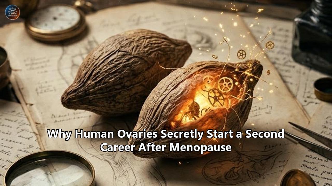
On October 14, 2026, researchers from Northwestern University’s Feinberg School of Medicine and the Buck Institute for Research on Aging released a study in Aging Cell that dismantles a foundational tenet of modern gynecology: the assumption that human ovaries become dormant, non-functional relics once a woman finishes her final menstrual period.
Analyzing 5,812 distinct protein groups extracted from cancer-free, non-pathological ovarian tissue collected from women aged 50 to 75, the scientific team established that ovaries do not retire at menopause. Instead, the organs undergo progressive, continuous molecular remodeling that persists for at least 25 years after reproductive function ceases. Far from being inert fibrous husks, the postmenopausal organs initiate an active, biologically demanding "second career" characterized by specialized androgen synthesis, metabolic restructuring, matrisome remodeling, and a profound shift toward immune surveillance.
These findings—co-authored by Francesca Duncan, an associate professor of reproductive science in medicine at Northwestern, and Birgit Schilling, director of the Mass Spectrometry Center at the Buck Institute—were reinforced by a companion Perspective published days earlier in Science, which detailed how post-reproductive ovaries actively transform into immune-regulatory and steroid-secreting centers.
The data reveal that while the follicles responsible for egg maturation vanish, the surrounding ovarian stroma remains an endocrine engine. Under intense stimulation from pituitary gonadotropins, it produces critical androgens—specifically testosterone and androstenedione—that circulate systemically and are converted by peripheral tissues into vital estrogens, sustaining the heart, brain, and skeleton. Concurrently, the organ alters its protein expression profile, trading structural-maintenance proteins for immune-signaling molecules, complement factors, and extracellular matrix remodelers.
           HYPOTHALAMIC-PITUITARY-OVARIAN AXIS POST-MENOPAUSE

                     Hypothalamus (GnRH Release)
                                 │
                                 ▼
                     Anterior Pituitary Gland
                                 │
                   Elevated LH (3x to 10x surge)
                                 │
                                 ▼
                    POSTMENOPAUSAL OVARIAN STROMA
         ┌───────────────────────┴───────────────────────┐
         ▼                                               ▼
   Endocrine Career                                Immune & Structural Career
   • Interstitial Cell Steroidogenesis            • BuckPOMS Expression (WNT4, FMOD)
   • Testosterone & Androstenedione               • Collagen Stiffening & Fibrosis
   • Peripheral Aromatization to Estrone (E1)     • SASP & Infiltration (Macrophage/T-Cell)
         │                                               │
         ▼                                               ▼
Sustains Bone, Heart, & Brain                   Alters Tissue Stiffness & Inflammatory Milieu
The medical and surgical implications are profound. For decades, routine prophylactic oophorectomy—the elective removal of both ovaries during hysterectomies for non-cancerous conditions—has been widely performed under the clinical rationalization that the organs are biological dead-weight carrying a dormant threat of malignancy. By demonstrating that the biological activity of ovaries after menopause directly shapes female health decades into late life, this research turns that legacy practice into a critical public health miscalculation, forcing physicians, surgeons, and patients to rethink the biological value of retaining native ovarian tissue.
Who Is Affected: Patients, Surgeons, and Health Systems
The reclassification of postmenopausal ovarian biology directly touches a vast demographic:
- Postmenopausal and Perimenopausal Women: Globally, more than 1 billion women are postmenopausal. In North America and Europe alone, tens of millions of women spend more than a third of their lives beyond the reproductive transition. For these women, medical management has historically focused almost entirely on what the ovaries have lost (cyclical estradiol and progesterone), while completely overlooking the hormones and active cellular processes the organs retain.
- Surgical Patients Facing Benign Gynecological Procedures: Every year, approximately 600,000 women in the United States undergo hysterectomies, the overwhelming majority for benign indications such as symptomatic uterine fibroids, severe adenomyosis, pelvic organ prolapse, and abnormal uterine bleeding. For over fifty years, surgeons routinely paired these operations with bilateral salpingo-oophorectomy (BSO) in women near or past age 50. Hundreds of thousands of patients who have already surrendered their ovaries under the presumption of organ uselessness—and countless future candidates evaluating surgical risks—are directly affected by this evidence.
- Surgical Specialists and Gynecologic Oncologists: Pelvic surgeons are confronting a clinical contradiction. Removing healthy ovaries effectively eliminates the roughly 1.4% lifetime population risk of ovarian cancer, but population-level registry data confirm that this preventive excision comes at the cost of accelerated multi-system aging and elevated all-cause mortality.
- Endocrinologists and Longevity Researchers: Medical specialists treating osteoporosis, cardiovascular decline, neurocognitive impairment, and metabolic syndrome must fundamentally recalculate hormonal baselines. Hormone replacement therapies (HRT) have long treated women as an endocrine blank slate once menses stop; understanding that the stroma continues to secrete endogenous androgens necessitates an integrated, multi-hormone diagnostic approach.
What Changes: The Molecular Mechanics of the Ovary’s Second Career
To comprehend the transformation of the ovary, one must examine what actually happens to its tissue architecture when follicles run out.
Menopause is classically defined by the exhaustion of the primordial follicle reserve. At gestational week 20, a female fetus possesses approximately 7 million oocytes. By birth, that figure drops to roughly 1 to 2 million; at menarche, fewer than 400,000 remain. Through steady monthly recruitment and relentless atresia, the follicular bank falls below the critical threshold of approximately 1,000 follicles around age 51. At this point, cyclic ovulation halts, cyclic surges of estradiol plummet, and menstruation stops.
For roughly a century, clinical textbooks assumed that when the follicular granulosa and theca layers degenerated, the organ essentially died. However, the human ovary is not composed exclusively of follicles. Up to 70% of the organ’s mass consists of the ovarian stroma: a dense connective tissue mesh populated by specialized spindle-shaped interstitial cells, fibroblasts, pericytes, endothelial cells, and immune populations.
               POSTMENOPAUSAL OVARIAN STROMAL STEROIDOGENESIS

                     Pituitary Luteinizing Hormone (LH)
                                    │
                                    ▼
                         Stromal LHCGR Activation
                                    │
                         Cholesterol Transport
                                    │ (StAR)
                                    ▼
                               Cholesterol
                                    │ (CYP11A1 / P450scc)
                                    ▼
                              Pregnenolone
                                    │ (CYP17A1 - 17α-hydroxylase)
                                    ▼
                       17α-Hydroxypregnenolone
                                    │ (CYP17A1 - 17,20-lyase)
                                    ▼
                             Dehydroepiandrosterone
                                    │ (HSD3B2 / 17β-HSD)
                                    ▼
                             Androstenedione
                                    │ (17β-HSD)
                                    ▼
                          Active Testosterone
                                    │
                Secreted into Ovarian Vein / Circulation
                                    │
                                    ▼
                  Peripheral Aromatization (CYP19A1)
                     in Adipose, Muscle, & Bone
                                    │
                                    ▼
                           Estrone (E1) / Estradiol (E2)
The Northwestern and Buck Institute team, along with preceding structural work by investigators like Yousin Suh at Columbia University and Ariella Shikanov at the University of Michigan, demonstrated that this stromal compartment undergoes an organized phenotypic pivot.
1. Stromal Androgen Synthesis Driven by Gonadotropins
When follicles disappear, the primary source of negative feedback on the hypothalamic-pituitary-ovarian axis—specifically estradiol and follicular inhibin B—collapses. In response, the anterior pituitary gland dramatically increases production of luteinizing hormone (LH) and follicle-stimulating hormone (FSH). Circulating LH concentrations spike three- to tenfold, where they remain elevated for decades.
While FSH finds few responsive targets once granulosa cells vanish, the postmenopausal ovarian stroma expresses high concentrations of functional LH/choriogonadotropin receptors (LHCGR). In response to continuous LH bombardment, the stromal interstitial cells differentiate into luteinized steroid-secreting factories.
Biochemical assays confirm that stromal cells preserve active expressions of:
- Steroidogenic acute regulatory protein (StAR): The rate-limiting transporter shuttling cholesterol across the inner mitochondrial membrane.
- CYP11A1 (cholesterol side-chain cleavage enzyme): Converts cholesterol into pregnenolone.
- CYP17A1 (17α-hydroxylase / 17,20-lyase): Catalyzes the conversion of 17-hydroxypregnenolone into dehydroepiandrosterone (DHEA).
- 17β-hydroxysteroid dehydrogenase (17β-HSD): Reduces androstenedione into biologically active testosterone.
Because postmenopausal stroma contains virtually no aromatase (CYP19A1)—which was historically restricted to follicular granulosa cells—it cannot convert these androgens into estrogens on-site. Instead, the stroma routes its output through the delta-5 steroidogenic pathway, synthesizing large amounts of androstenedione, DHEA, and testosterone.
Catheter-based venous sampling studies have verified this mechanism in real-time. Blood extracted directly from the ovarian veins of postmenopausal women reveals testosterone concentrations up to 15 times higher than matching samples taken from their peripheral arm veins, and substantially higher than adrenal venous effluents. Up to 50% of total circulating testosterone in an aging woman originates straight from the stroma of her postmenopausal ovaries.
This ovarian testosterone does not act solely as an androgen. Through systemic blood flow, these ovarian androgens travel to peripheral tissues: subcutaneous and visceral adipocytes, skeletal myocytes, and osteoblasts. These target tissues express high levels of aromatase, converting stromal testosterone into estradiol and androstenedione into estrone (E1).
Through this distributed biochemical circuit, ovaries after menopause serve as an endocrine engine, providing the raw hormonal substrate required for baseline peripheral estrogen production without the cyclical, pro-inflammatory peaks of the menstrual cycle.
2. The BuckPOMS Discovery: Proteomic Proof of Lifelong Remodeling
The study led by Mark Watson, Francesca Duncan, and Birgit Schilling in Aging Cell evaluated whether postmenopausal ovaries remain biologically static over time.
By stratifying cancer-free ovarian tissue across three specific postmenopausal cohorts—women aged 50–59, 60–69, and 70 and older—the investigators quantified 5,812 individual protein groups using high-resolution data-independent acquisition mass spectrometry.
Their findings revealed a continuous molecular drift:
- 117 proteins demonstrated statistically significant, divergent alteration when comparing women in their 50s directly against women aged 70 and older.
- Multivariate analysis showed completely segregated protein profiles between younger and older postmenopausal groups. In women in their 50s, ovarian tissue proteomes were overwhelmingly populated by proteins regulating RNA processing, DNA repair, chromatin architecture, and core cellular structural maintenance.
- In women aged 70 and above, the proteome transitioned to an entirely different biochemical identity: downregulating structural proteins while massively upregulating pathways involved in vesicle trafficking, metabolic regulation, innate immunity, and complement cascade activation.
From this analysis, the investigators distilled the Buck Postmenopausal Ovary Molecular Signature (BuckPOMS): a consensus 26-protein network that tracks post-reproductive biological aging. Among these proteins were secreted matrisome modulators like WNT4 (a critical regulator of sex determination and cellular fate) and Fibromodulin (FMOD) (a small leucine-rich proteoglycan essential for collagen fibrillogenesis and matrix remodeling), both of which were validated in the tissue using immunohistochemistry.
               BUCKPOMS MOLECULAR TRANSITION TRAJECTORY

Age Cohort       Primary Molecular Focus               Core Biological Output
──────────────────────────────────────────────────────────────────────────────────
Ages 50–59       RNA Processing & Cellular Identity    Structural retention; high
                 Chromatin organization; Keratins      collagen flexibility; low
                                                       senescence load
──────────────────────────────────────────────────────────────────────────────────
Ages 60–69       Intermediate Stromal Drift            Progressive loss of elastic
                 SASP onset; matrisome remodeling      fibers; initial collagen
                                                       crosslinking; low immune surge
──────────────────────────────────────────────────────────────────────────────────
Ages ≥ 70        Innate Immunity & Complement Drive    High FMOD and WNT4 expression;
                 Metabolic and vesicle trafficking;    dense collagenous fibrosis;
                 High DAMP and SASP release            macrophage/T-cell activation
3. Transition to an Immune-Regulatory Organ
Beyond steroid synthesis, the aging postmenopausal ovary undergoes a cellular metamorphosis. As detailed by Duncan and colleagues in Molecular Human Reproduction, the post-reproductive organ systematically shifts from a gametogenic structure to an immune-like organ.
Histological and spatial-transcriptomic profiling demonstrates that as human and mammalian ovaries age past natural menopause, the stromal compartment fills with organized immune populations:
- Infiltration of CD68+ and CD163+ Macrophages: These phagocytic cells scavenge the scarred remnants of collapsed atretic follicles (corpora albicantia) and clear cellular debris.
- Accumulation of CD3+, CD4+, and CD8+ T Lymphocytes: The ovarian cortex gradually takes on structural characteristics resembling tertiary lymphoid structures, complete with local cytokine production and antigen-presenting machinery.
- Presence of Multinucleated Giant Cells: Formed by the fusion of persistent macrophages attempting to process insoluble lipid-protein aggregates (lipofuscin) and dense, crosslinked collagen scars.
This progressive immune accumulation means the postmenopausal ovary is a dynamic immunological microenvironment, continually producing signals that interact with the systemic immune apparatus.
The Surgical Fallout: Reassessing Prophylactic Castration
The demonstration that ovaries retain crucial physiological roles long after menopause exposes decades of aggressive surgical overreach.
Throughout the late 20th and early 21st centuries, surgical gynecology maintained a clear protocol: if a woman undergoing a hysterectomy for a benign condition (such as heavy menstrual bleeding or leiomyomas) was older than 45, surgeons routinely recommended the concurrent excision of both healthy fallopian tubes and ovaries—a bilateral salpingo-oophorectomy (BSO).
The rationale appeared self-evident:
- Epithelial ovarian cancer is notoriously lethal, clinically silent, and lacks effective early screening tools.
- The population-level baseline risk of developing ovarian cancer over a woman’s lifetime is roughly 1.3% to 1.4%.
- If the ovaries are "useless" post-reproductive glands that no longer release eggs or produce premenopausal levels of cyclical estrogen, removing them appeared to provide cancer prophylaxis with minimal physiological cost.
                EPIDEMIOLOGICAL SURGICAL COMPARISON

Metric / Outcome               Conserved Ovaries          Prophylactic BSO
──────────────────────────────────────────────────────────────────────────────────
28-Year All-Cause Mortality    13.3% Baseline             16.8% (HR 1.13, 95% CI 1.06–1.21)
Fatal Coronary Heart Disease   Reference                  23% to 135% Elevated Risk
Dementia & Cognitive Decline   Reference                  45% to 82% Elevated Hazard
Parkinsonism                   Reference                  68% Elevated Hazard
Colorectal Cancer Death        Reference                  49% Higher Mortality
Bone Fracture Incidence        Baseline                   Substantially Elevated
Ovarian Cancer Incidence       0.1% to 0.75%              Near 0.0%
──────────────────────────────────────────────────────────────────────────────────
Surgeon's Calculation:         300 BSO Surgeries to prevent 1 ovarian cancer death,
                               while causing excess cardiovascular, neurologic,
                               and all-cause deaths across the treated cohort.
By the late 1990s, more than 300,000 American women were undergoing elective, non-cancerous bilateral oophorectomies each year. Prophylactic BSO accompanied roughly 55% to 80% of all benign hysterectomies performed in the United States.
This standard began to fracture when large-scale, prospective epidemiology linked bilateral oophorectomy to significant long-term systemic damage:
1. The Nurses’ Health Study
In a benchmark prospective study tracking 30,117 women who underwent hysterectomy for benign disease over a 28-year follow-up, William Parker and colleagues at the John Wayne Cancer Institute and Harvard Medical School uncovered unexpected mortality statistics:
- Among women who had their ovaries preserved, 13.3% died from all causes during follow-up.
- Among women who underwent concurrent bilateral oophorectomy, all-cause mortality climbed to 16.8%—a statistically significant 13% increase in the multivariable-adjusted hazard ratio (HR = 1.13; 95% CI, 1.06–1.21).
- While oophorectomy virtually eradicated ovarian cancer deaths (4 deaths in the oophorectomy cohort vs. 44 in the ovarian conservation cohort), this benefit was overshadowed by mortality in other organ systems.
- Oophorectomized women suffered a 23% higher risk of fatal coronary heart disease (HR = 1.23), a 29% higher risk of death from lung cancer (HR = 1.29), and a 49% higher risk of death from colorectal cancer (HR = 1.49).
- Crucially, these survival penalties persisted regardless of the age at which the ovaries were excised: at no age, from 40 up through 65, was prophylactic bilateral oophorectomy associated with an overall survival benefit.
The authors calculated the Number Needed to Treat (NNT): A surgeon must perform approximately 300 bilateral oophorectomies in women at low genetic risk to prevent a single death from ovarian cancer. Over that same surgical cohort, the resulting endocrine deprivation accelerates multiple premature deaths from fatal myocardial infarction, stroke, and systemic illness.
2. The Mayo Clinic Cohort Study of Oophorectomy and Aging
Directed by neurologist and epidemiologist Walter A. Rocca, the Mayo Clinic Cohort Study tracked thousands of women in Olmsted County, Minnesota, who underwent unilateral or bilateral oophorectomy for non-malignant indications between 1950 and 1987.
The neuro-cognitive and somatic consequences were severe:
- Women undergoing bilateral oophorectomy before natural menopause faced a 45% increase in the risk of developing cognitive impairment or overt dementia.
- For women under age 46 at the time of ovarian removal, the risk of developing dementia surged by 82%.
- The hazard ratio for developing parkinsonism—including classical Parkinson's disease and parkinsonian tremors—surged by 68% in oophorectomized women.
- Post-surgical evaluation revealed substantial increases in diagnosed depressive disorders, chronic anxiety states, and accelerated bone mineralization loss predisposing to hip fractures.
- Critically, while exogenous estrogen replacement therapy through age 50 partially mitigated cognitive and cardiovascular decay, it did not eliminate the increased incidence of parkinsonism, mood disorders, or all-cause mortality.
This shortfall strongly indicated that the ovary provided active neuroprotective and metabolic agents beyond simple estradiol—principally stromal androgens and local neurosteroid precursors that conventional hormone pills failed to supply.
3. The Discovery of Fallopian Origins
Compounding the tragedy of unnecessary oophorectomies was a major discovery in gynecological oncology: the realization that the vast majority of lethal, high-grade serous ovarian carcinomas do not originate in the ovary at all.
Pathological studies tracing early neoplastic lesions demonstrated that these malignancies typically arise in the fimbriated ends of the fallopian tubes as Serous Tubal Intraepithelial Carcinomas (STIC lesions), subsequently detaching and seeding the ovarian surface.
This finding gave rise to opportunistic salpingectomy: the practice of completely excising the fallopian tubes while carefully sparing the ovaries during benign pelvic surgeries.
Opportunistic salpingectomy strips away the primary anatomical source of ovarian carcinomas while preserving the hormone-producing ovarian stroma intact.
Yet, despite guidelines from the American College of Obstetricians and Gynecologists (ACOG) endorsing opportunistic salpingectomy, thousands of postmenopausal women continue to have functional ovaries removed due to outdated clinical habits.
Short-Term Consequences: Clinical Realities and Patient Management
The confirmation that human ovaries remain biologically dynamic long after menopause triggers immediate, near-term adjustments across everyday clinical practice.
                    SHORT-TERM CLINICAL REALIGNMENTS

Legacy Gynecology Protocol              Updated Impact-Driven Protocol
──────────────────────────────────────────────────────────────────────────────────
"Ovaries after 50 are dormant."         "Ovaries after 50 are dynamic endocrine hubs."

Routine prophylactic BSO with           Selective bilateral salpingectomy with
benign hysterectomy.                    deliberate ovarian conservation.

Monitoring estrogens alone (E2) in      Comprehensive steroid profiling: Total & Free
menopausal hormone management.          Testosterone, DHEA-S, Androstenedione, and LH.

Treating surgical menopause like        Recognizing surgical castration as an acute
natural menopause.                      neurovascular and endocrine emergency.
1. Overhauling Surgical Consent and Operative Indications
The most immediate operational change occurs in surgical pre-op clinics. Surgeons can no longer tell a 53-year-old or 62-year-old patient that her ovaries are non-functional tissue that can be safely discarded during a hysterectomy.
Unless a patient carries a verified high-penetrance genetic mutation—such as BRCA1, BRCA2, or Lynch syndrome-associated DNA mismatch repair mutations—ovarian conservation must now be the default clinical baseline. When women at average genetic risk are fully informed that removing their postmenopausal ovaries carries an elevated hazard for coronary artery disease, cognitive decline, structural bone loss, and shortened overall lifespan, rates of elective oophorectomy decline sharply.
Surgical training programs are actively revising pelvic dissection techniques to ensure that blood supply to the conserved ovarian stroma—via the infundibulopelvic ligament and ovarian arcade—remains undamaged when the uterus and fallopian tubes are excised.
2. Recalibrating Postmenopausal Endocrine Assessments
Clinical endocrinologists and menopause practitioners must overhaul their standard diagnostic blood panels.
Historically, when evaluating a postmenopausal woman suffering from debilitating brain fog, profound chronic fatigue, rapid loss of lean muscle mass, or suppressed libido, blood draws looked primarily at estradiol, progesterone, and thyroid-stimulating hormone (TSH).
The recognition of the ovary’s secondary career requires measuring the complete steroidogenic pathway:
- Total and free testosterone (via high-precision liquid chromatography-tandem mass spectrometry, LC-MS/MS).
- Dehydroepiandrosterone sulfate (DHEA-S).
- Androstenedione.
- Luteinizing Hormone (LH), which reflects how vigorously the pituitary is driving the ovarian stroma.
When these androgen markers drop precipitously—whether following bilateral oophorectomy or primary ovarian stromal failure—clinicians can identify why a patient remains symptomatic despite using standard estradiol skin patches.
The systemic effects of androgen deficiency can then be addressed directly, leading to more appropriate, targeted therapeutic choices.
3. Managing Acute Castration vs. Gradual Menopausal Transition
Understanding the stromal endocrine role highlights the biological divergence between natural menopause and surgical menopause.
Natural menopause is a gradual, buffered systemic adjustment. The follicular engine powers down over several years, but the ovarian stroma steps up androgen production to soften the endocrine drop.
In contrast, bilateral oophorectomy causes an abrupt, unnatural crash: testosterone, androstenedione, and peripheral estrogens fall by more than 50% in a single day.
This sudden hormonal drop explains why women subjected to surgical menopause suffer far more severe vasomotor hot flashes, faster drops in bone mineral density, accelerated microvascular endothelial dysfunction, and elevated psychiatric vulnerability compared to women who undergo natural menopause with their ovaries intact.
Surgeons performing indicated oophorectomies must treat surgical castration as an acute endocrinopathy requiring immediate, multi-pathway hormone optimization, rather than simply offering a short-course estrogen prescription.
Long-Term Consequences: The Longevity Trade-Off and the Oncogenic Paradox
While the ovarian stroma’s ongoing androgen production is a major metabolic asset, the Aging Cell study demonstrates that the aging ovary is also a double-edged sword.
The lifelong activity of the organ comes at a measurable molecular cost, creating an intricate biological trade-off between endocrine vitality, chronic inflammation, and oncogenic risk.
                 THE POSTMENOPAUSAL OVARIAN DILEMMA

               CONTINUED RETENTION OF POSTMENOPAUSAL OVARIES
                                     │
           ┌─────────────────────────┴─────────────────────────┐
           ▼                                                   ▼
PHYSIOLOGICAL ASSETS                                PATHOLOGICAL RISKS
• Constant Stromal Androgens                        • Progressive Matrisome Stiffening
• Peripheral Estrone Sourcing                       • BuckPOMS Secretory Transition
• Preserved Bone Mineral Density                    • Ovarian Senescence-Associated Secretory
• Neurovascular Endothelial Protection                Phenotype (SASP) Release
• Reduced Long-Term All-Cause Mortality             • Creation of an Immunologically Hospitable
                                                      Niche for Migrating Carcinomas
1. Cellular Senescence and the Matrisome Shift
As postmenopausal ovaries age, their cellular architecture undergoes marked alterations. The mass spectrometry analysis by Watson, Duncan, and Schilling showed that by age 70, the ovary expresses elevated concentrations of proteins associated with cellular senescence.
Senescent cells are damaged or aging cells that permanently arrest their cell division cycle while remaining metabolically hyperactive.
In the aged ovary, these senescent stromal cells acquire a robust Senescence-Associated Secretory Phenotype (SASP).
This phenotype pumps a cocktail of inflammatory mediators into the surrounding tissue:
- Pro-inflammatory cytokines (Interleukin-6, Interleukin-8, TNF-α).
- Matrix Metalloproteinases (MMPs), which break down extracellular matrices.
- Transforming Growth Factor-beta (TGF-β), a master driver of tissue fibrosis.
This process explains why the Buck Postmenopausal Ovary Molecular Signature (BuckPOMS) captures extensive extracellular matrix remodeling.
Duncan’s previous pioneering biomechanical studies demonstrated that aging mammalian ovaries become progressively fibrotic, hyper-stiff, and collagen-dense, mimicking cirrhotic liver or scarred myocardial tissue.
As the elastic fibers of younger stromal tissue are replaced by rigid, crosslinked collagen bundles driven by enzymes like lysyl oxidase (LOX), the physical compliance of the postmenopausal ovary alters completely.
2. The Oncogenic Seedbed Paradox
This structural stiffening sheds light on why ovarian cancer incidence peaks late in life—typically in a woman’s late 60s and 70s—decades after ovulation has ceased.
Cancer cell biologists recognize that high-grade serous carcinoma cells migrating from the fallopian tube thrive in stiff, collagen-dense environments rich in inflammatory cytokines.
The dense extracellular matrix of the aged ovary, combined with the presence of SASP factors and complement proteins, forms an ideal microenvironmental niche for circulating tumor cells to anchor, survive, and form tumors.
The Aging Cell researchers made the sobering observation that healthy, non-cancerous ovaries from 70-year-old women exhibit protein signatures that overlap with molecular pathways found in malignant ovarian carcinomas.
The organ does not need to be cancerous to foster this hospitable microenvironment; the baseline aging trajectory of the postmenopausal stroma naturally shifts toward this tumor-permissive molecular profile.
3. The Canary in the Coal Mine for Systemic "Inflammaging"
This dynamic raises a profound biological question: Does the postmenopausal ovary actively accelerate systemic aging in older women?
Because the human ovary is the first major organ to exhaust its primary function—aging at more than double the rate of the heart, kidneys, liver, or brain—its tissues accumulate senescence and fibrosis decades ahead of any other visceral structure.
The companion Perspective in Science suggests that an ovary packed with activated macrophages, T lymphocytes, and SASP-secreting senescent stromal cells acts as a chronic, low-grade inflammatory pump.
               THE SYSTEMIC INFLAMMAGING CASCADE

               Aged Ovarian Stroma (Ages 70+)
                             │
     Excess Senescent Interstitial Cells & Secreted Matrisome
                             │
     Local Production: IL-6, TNF-α, MMPs, WNT4, Complement
                             │
              Direct Effluent into Ovarian Vein
                             │
                             ▼
              Systemic Blood Supply Infiltration
                             │
       ┌─────────────────────┼─────────────────────┐
       ▼                     ▼                     ▼
Cardiovascular        Neurovascular        Systemic Metabolic
Endothelium Stiffens  Plaque Instability   Insulin Resistance
If damaged secretory proteins, inflammatory cytokines, and complement cascades are constantly discharged into the ovarian vein, the aged postmenopausal ovary may fuel the chronic, low-grade systemic inflammation—termed "inflammaging"—that drives arterial stiffening, metabolic dysfunction, and frailty in women in their 70s and 80s.
This trade-off crystallizes the ovarian dilemma:
- In early and mid-postmenopause (ages 50 to 68), retaining the ovaries provides major endocrine, cardiovascular, skeletal, and neurological benefits by sustaining baseline androgen and estrone production.
- In deep postmenopause (ages 70 and beyond), the organ's progressive structural remodeling, senescence, and fibrotic stiffness may transform it into an inflammatory burden that raises systemic disease and malignancy risks.
Systemic Health Dimensions: Brain, Bone, Heart, and Metabolism
The lifelong activity of the postmenopausal ovary fundamentally links into multiple organ systems. When the stroma continues to synthesize steroid precursors, it directly influences the biological health of the central nervous system, cardiovascular architecture, skeleton, and metabolic reserves.
       SYSTEMIC TISSUE TARGETS OF POSTMENOPAUSAL OVARIAN STEROIDOGENESIS

                    POSTMENOPAUSAL OVARIAN STROMA
                                  │
                   Secretes Androgens (T, A4, DHEA)
                                  │
         ┌───────────────┬────────┴────────┬───────────────┐
         ▼               ▼                 ▼               ▼
   BRAIN HEALTH    BONE DENSITY       HEART & VESSELS  METABOLISM & MUSCLE
   • Neurosteroid  • Osteoblast       • Endothelial    • Sarcopenia
     conversion      AR activation      NO synthesis     prevention
   • Synaptic      • Aromatization    • Attenuates     • Visceral fat
     preservation    to estrone         arterial         suppression
   • Neurogenesis  • Inhibits           stiffness      • Muscle protein
     in limbic       osteoclasts      • Preserves        synthesis
     networks        (trabecular)       lipid balance
1. Central Nervous System Protection and Cognitive Resilience
The brain is an intensely steroid-sensitive organ. Classical neurology assumed that brain-protective sex steroids were derived purely from cyclical ovarian estrogen.
However, neuroimaging and biochemical profiling show that androgen receptors are heavily expressed throughout the human cerebral cortex, hippocampus, basal ganglia, and amygdala.
Ovarian androgens cross the blood-brain barrier with ease. Once inside the brain, they can directly activate neural androgen receptors to promote neurite outgrowth, maintain dendritic spine density, and stimulate local neurogenesis.
Equally important, neural astrocytes and microglia express high concentrations of aromatase, converting circulating ovarian testosterone locally into neuroprotective 17β-estradiol precisely where it is required.
This local conversion maintains synaptic plasticity, dampens microglial hyper-activation, and aids the clearance of toxic amyloid-beta oligomers.
This mechanism clarifies the long-term clinical findings of the Mayo Clinic Cohort Study of Oophorectomy and Aging.
When bilateral oophorectomy removes the ovarian stroma, the brain is instantly deprived of both direct androgenic stimulation and the substrate required for localized astrocytic aromatization.
Deprived of this steady stromal support, women experience accelerated atrophy of the hippocampus, increased accumulation of white matter hyperintensities, and an elevated vulnerability to neurodegenerative pathologies—explaining the observed 82% surge in dementia and 68% rise in parkinsonism among women who underwent surgical castration.
2. Cardiovascular Integrity and Microvascular Tone
Cardiovascular disease remains the leading cause of death among postmenopausal women globally.
While premenopausal protection against coronary heart disease was long attributed exclusively to estradiol-dependent vasodilation, postmenopausal vascular maintenance relies on a balanced steroid environment.
Testosterone produced by the postmenopausal stroma is taken up by vascular endothelial cells and smooth muscle myocytes:
- Via androgen receptor-dependent transcription, physiological levels of testosterone stimulate Endothelial Nitric Oxide Synthase (eNOS), driving the steady release of nitric oxide (NO) that maintains arterial compliance and prevents pathological vasoconstriction.
- Once converted peripherally to estrone and estradiol, these steroids suppress the expression of endothelial vascular cell adhesion molecule-1 (VCAM-1) and intercellular adhesion molecule-1 (ICAM-1), preventing the transmigration of oxidized LDL particles and monocytes into the arterial subendothelial space.
When surgeons excise healthy postmenopausal ovaries, this homeostatic loop breaks down.
Lacking steady stromal androgen production, systemic nitric oxide production falls, arterial stiffness increases, and microvascular resistance rises.
This mechanistic sequence explains why the Nurses’ Health Study observed a multivariable hazard ratio of 1.23 for fatal coronary heart disease in women whose ovaries were excised, an elevation in cardiac mortality that persisted even when surgeries were performed late in life.
3. Skeletal Mineralization and Osteoporosis Prevention
Postmenopausal osteoporosis has historically been treated as an estrogen-deficiency disease.
Yet, bone biology reveals that both osteoblasts (the cells that build bone) and osteoclasts (the cells that resorb bone) express abundant functional androgen receptors alongside estrogen receptors.
Ovarian testosterone acts as a direct, powerful driver of osteoblast proliferation and differentiation:
- Testosterone signaling through androgen receptors stimulates osteoblasts to secrete type I collagen and alkaline phosphatase, strengthening the trabecular and cortical bone scaffolding.
- Concurrently, androgen receptor activation upregulates the synthesis of osteoprotegerin (OPG), a decoy receptor that binds to and neutralizes RANKL (Receptor Activator of Nuclear Factor κB Ligand), suppressing the recruitment and activation of osteoclasts.
- Locally within bone tissue, the testosterone and androstenedione supplied by postmenopausal ovaries are aromatized directly into estrone and estradiol, suppressing inflammatory cytokines (such as IL-1, IL-6, and TNF-α) that would otherwise trigger bone resorption.
Women who retain their ovaries experience significantly slower rates of postmenopausal bone mineral density loss than women whose ovaries are surgically removed.
The preservation of the postmenopausal ovarian stroma provides a continuous shield against the structural skeletal decay that leads to osteopenia, catastrophic hip fractures, and subsequent loss of independent mobility.
                 SKELETAL MINERALIZATION PATHWAY

                 Ovarian Stromal Testosterone
                               │
         ┌─────────────────────┴─────────────────────┐
         ▼                                           ▼
Direct Osteoblast AR Binding            Aromatized to Local Estrogens
         │                                           │
Increases Type I Collagen & OPG         Suppresses RANKL & Resorption Cytokines
         │                                           │
         └─────────────────────┬─────────────────────┘
                               ▼
            Net Preservation of Bone Mineral Density
            (Shields against osteoporotic hip fracture)
4. Skeletal Muscle Mass, Metabolism, and Fat Distribution
The gradual loss of lean muscle mass (sarcopenia) and the shift toward visceral abdominal adiposity represent two of the most burdensome metabolic aspects of female aging.
These phenotypes are accelerated in the absence of postmenopausal ovarian function.
Stromal testosterone acts as an essential anabolic signal for skeletal muscle cells. It activates the mTOR (mammalian target of rapamycin) signaling cascade within muscle myocytes, promoting amino acid uptake, myofibrillar protein synthesis, and the preservation of fast-twitch Type II muscle fibers.
This preservation of lean skeletal mass maintains baseline resting metabolic expenditure.
In addition, physiological levels of ovarian androgens work in concert with peripheral estrogens to limit the expansion of visceral fat, encouraging subcutaneous rather than harmful intra-abdominal lipid storage.
When healthy ovaries are removed, the resulting hormonal drop accelerates dynapenia (loss of muscle strength) and triggers central visceral weight gain.
This shift worsens systemic insulin resistance, elevates hepatic gluconeogenesis, and exacerbates metabolic syndrome, increasing the lifetime likelihood of developing type 2 diabetes.
Long-Term Therapeutic Frontiers: Intervening in the Ovarian Microenvironment
The discoveries emerging from the Buck Institute, Northwestern University, and companion laboratories open an entirely new frontier in regenerative medicine: the targeted therapeutic manipulation of the ovarian stroma.
                FUTURE THERAPEUTIC PARADIGM SHIFT

Historic Binary Choice                 Next-Generation Precision Intervention
──────────────────────────────────────────────────────────────────────────────────
Option A: Leave the ovary alone;       Pharmacologically target the ovarian stroma:
risk fibrosis, SASP inflammaging,      • Use targeted Senolytics to clear senescent cells.
and late-life malignancy.              • Apply Antifibrotic LOX-inhibitors to prevent
                                         collagen crosslinking.
Option B: Surgically excise the        • Preserve stromal steroidogenesis while
organ; cause endocrine collapse,         shutting down inflammatory cytokine release.
cardiovascular death, and dementia.    • Decouple cancer prevention from surgical castration.
Rather than treating the postmenopausal ovary as an all-or-nothing dilemma—either leaving it vulnerable to senescence and malignancy or surgically removing it and causing systemic endocrine collapse—researchers are now exploring how to optimize its biological behavior:
1. Stromal Senolytics
Because the older postmenopausal ovary accumulates senescent cells that release damaging SASP factors, researchers funded through the NIH SenNet (Cellular Senescence Network) consortium are testing targeted senolytics—compounds that selectively clear damaged, non-dividing senescent cells.
By administering periodic, low-dose senolytic molecules (such as combinations of dasatinib and quercetin, or next-generation Bcl-xL inhibitors), researchers have successfully cleared senescent stromal cells in preclinical models, dampening local SASP secretion and systemic inflammation without compromising baseline interstitial steroidogenesis.
2. Antifibrotic Therapeutics
Building on Francesca Duncan’s work on ovarian collagen dynamics, biomedical teams are evaluating pharmacological therapies that prevent pathologic tissue stiffening.
Small-molecule inhibitors targeting lysyl oxidase (LOX) enzymes, together with antifibrotic drugs like pirfenidone, are being studied in animal models to prevent excessive, rigid collagen crosslinking within the postmenopausal ovarian cortex.
By keeping the ovarian stroma biomechanically soft, researchers hope to deny migrating fallopian carcinoma cells the rigid, collagen-dense microenvironment they need to anchor and grow, decoupling cancer prevention from surgical castration.
3. Precision Biomarkers and the BuckPOMS Panel
The establishment of the 26-protein BuckPOMS signature creates a molecular ruler for evaluating an individual woman’s ovarian aging status.
Future clinical diagnostics may leverage these secreted matrisome biomarkers—including circulating fragments of WNT4 and Fibromodulin (FMOD)—to monitor ovarian remodeling through simple liquid biopsies.
Tracking a woman's personalized BuckPOMS trajectory could help clinicians determine precisely when an individual's ovaries are successfully producing protective androgens versus when they might be transitioning into a hyper-fibrotic, pro-inflammatory liability.
This diagnostic window would allow for highly customized, data-driven decisions regarding surgical conservation, targeted pharmacological intervention, or personalized hormonal support.
Redefining the Arc of Female Longevity
The discovery that human ovaries remain biologically active for decades past menopause marks an overdue correction in how medical science conceptualizes women's health.
For more than a century, clinical medicine operated under a deeply reductive presumption: that an organ with an expired reproductive capacity must be a useless, pathological liability.
This framework reduced the ovary’s value entirely to ovulation, framing its post-reproductive existence as an evolutionary mistake to be preemptively cut out in the operating room.
The proteomic and endocrine evidence assembled by Watson, Duncan, Schilling, and their colleagues reveals an entirely different biological reality:
- The human ovary does not die when it finishes ovulating; it pivots to a second career.
- Its stroma remains an endocrine engine, driven by pituitary feedback to supply the critical androgens that sustain bone mineralization, preserve cardiovascular compliance, protect cognitive networks, and stabilize metabolic balance well into late life.
- At the same time, its ongoing structural remodeling—characterized by cellular senescence, collagenous stiffening, and intense immune cell infiltration—illustrates that the ovary acts as an advanced, early-aging sensor that mirrors the health of the entire female body.
As surgical societies draft updated clinical guidelines, the routine prophylactic excision of the postmenopausal ovary will increasingly be viewed as a relic of an era that misunderstood female endocrinology.
Rather than viewing the post-reproductive decades as an unnatural aftermath of reproductive exhaustion, modern medicine must recognize that the longevity of women is intimately supported by the dynamic biology of ovaries after menopause.
The lifelong preservation of this active, complex organ is not a surgical footnote—it is an essential foundation of women’s healthspan, systemic vitality, and long-term resilience.
Reference:
- https://www.lifescience.net/publications/2252520/the-human-ovary-exhibits-dynamic-molecular-remodel/
- https://srpske.rs/en/news/nauka/2026/10/14/ovaries-keep-changing-decades-after-menopause-28-women-study
- https://www.rapamycin.news/t/the-ovary-after-menopause-retired-organ-or-quiet-source-of-inflammation/26976
- https://www.simplified-science.com/blog/the-post-menopausal-ovary-immune-powerhouse
- https://news.northwestern.edu/stories/2026/10/the-ovary-doesnt-retire-after-menopause?fj=1
- https://academic.oup.com/humrep/article-abstract/21/1/309/570742
- https://pmc.ncbi.nlm.nih.gov/articles/PMC10099172/
- https://cdn.mdedge.com/files/s3fs-public/issues/articles/media_1578d20_ccjm57_2-0154.pdf
- https://medcraveonline.com/OGIJ/hysterectomy-for-benign-conditions-prophylactic-oophorectomy-or-ovary-conservation.html
- https://pmc.ncbi.nlm.nih.gov/articles/PMC4254662/
- https://www.exxcellence.org/list-of-pearls/ovarian-conservation-at-the-time-of-hysterectomy/?categoryName=&searchTerms=&featured=False&bookmarked=False&sortColumn=date&sortDirection=Descending
- https://pmc.ncbi.nlm.nih.gov/articles/PMC9855652/
- https://cellxgene.cziscience.com/collections/ce781ec4-393a-4902-94a0-5a1ca0b600ca
- https://www.drugtargetreview.com/atlas-of-the-human-ovary-offers-huge-potential-for-new-treatments/637707.article
- https://www.ovid.com/journals/ejoe/abstract/10.1530/eje-14-0468~management-of-endocrine-disease-hyperandrogenism-after?redirectionsource=fulltextview
- https://pubmed.ncbi.nlm.nih.gov/16253961/
- https://www.medicaldaily.com/ovaries-remodel-decades-after-menopause-northwestern-buck-study-480005
- https://pmc.ncbi.nlm.nih.gov/articles/PMC2585770/
- https://pmc.ncbi.nlm.nih.gov/articles/PMC2716666/
- https://pmc.ncbi.nlm.nih.gov/articles/PMC3921032/
- https://www.patientcareonline.com/view/after-oophorectomy-cognitive-impairment-risks-rise
- https://neurologytoday.aan.com/doi/10.1097/01.NT.0000295254.92297.d8
- https://www.news-medical.net/health/Oophorectomy-Risks.aspx
- https://journals.lww.com/greenjournal/fulltext/10.1097/aog.0000000000003164~acog-committee-opinion-no-774-opportunistic-salpingectomy-as
- https://karger.com/books/book/114/chapter/5055966/Androgens-in-Menopausal-Women-Not-Only-Polycystic
- https://srpske.rs/en/news/nauka/2026/10/14/ovaries-keep-changing-decades-after-menopause-28-women-study