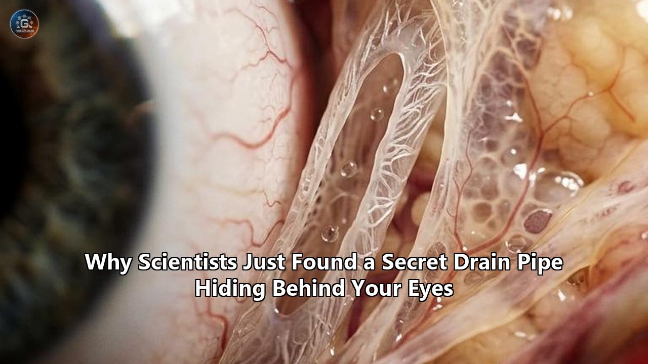
A team of researchers from the University of British Columbia and the University of Toronto published evidence in Translational Vision Science & Technology revealing an undocumented lymphatic clearance network operating at the posterior pole of the mammalian eye. Designated the Posterior Ocular Lymphatic Outflow (POLO) pathway, this vascular circuit directly links the suprachoroidal space beneath the retina to the deep cervical lymph nodes of the neck. For more than a century, classical anatomical doctrine maintained that the posterior interior of the eye lacked lymphatic drainage entirely, functioning as an immunologically segregated, self-contained sphere.
The experimental verification—led by Dr. Neeru Gupta, head of UBC’s Department of Ophthalmology and Visual Sciences, and Dr. Yeni H. Yücel, director of ophthalmic pathology at the University of Toronto and scientist at the Keenan Research Centre for Biomedical Science at St. Michael's Hospital—fundamentally overturns that assumption. By delivering albumin-conjugated imaging tracers into the suprachoroidal space of adult murine models, the investigators observed these tracers rapidly entering distinct, podoplanin- and VEGFR-3-positive lymphatic vessels within the choroid. Within 20 minutes, the tracer molecules exited the globe, navigated the nasal orbital soft tissue, and accumulated inside the ipsilateral cervical lymph nodes.
This discovery provides the missing physiological route for clearing fluid and metabolic waste from the retina, one of the most energetically demanding tissues in the human body. It also forces an immediate re-evaluation of the mechanisms driving the primary causes of irreversible blindness, including age-related macular degeneration and normal-tension glaucoma.
                 POSTERIOR OCULAR LYMPHATIC OUTFLOW (POLO)
                 =========================================

   +-------------------------------------------------------------+
   |                        RETINA                               |
   |   (High metabolic activity, shedding photoreceptor waste)   |
   +-------------------------------------------------------------+
                                  |
                                  v
   +-------------------------------------------------------------+
   |                     SUPRACHOROIDAL SPACE                    |
   |              (Interstitial fluid & waste pool)              |
   +-------------------------------------------------------------+
                                  |
                                  v
   +-------------------------------------------------------------+
   |                  CHOROIDAL LYMPHATICS                       |
   |      (Podoplanin+, VEGFR-3+, Prox1+ blind-ended channels)   |
   +-------------------------------------------------------------+
                                  |
                                  v
   +-------------------------------------------------------------+
   |                     NASAL ORBITAL TISSUE                    |
   |                (Retrobulbar extraocular exit)               |
   +-------------------------------------------------------------+
                                  |
                                  v
   +-------------------------------------------------------------+
   |                 DEEP CERVICAL LYMPH NODES                   |
   |    (Systemic waste clearance & neuro-immune processing)     |
   +-------------------------------------------------------------+
Viewed as a case study in modern biological discovery, the identification of the POLO pathway illustrates a broader pattern occurring across the neurosciences. For generations, anatomical science operated under rigid assumptions of physical compartmentalization, asserting that delicate central nervous tissues maintained their health through isolation rather than systemic integration. The identification of this hidden conduit demonstrates how stubborn dogmas form, why advanced multimodal imaging is required to dismantle them, and how recognizing unrecognized physiological drainage channels alters our approach to chronic neurodegenerative disease and targeted drug delivery.
Anatomy's Century-Old Blind Spot: The Dogma of the Closed Globe
To comprehend why the POLO pathway remained hidden until now, one must examine the entrenched theoretical architecture that governed ophthalmology throughout the twentieth century. Since the late nineteenth century, medical textbooks described the eye as a closed, rigid globe dependent almost exclusively on an anterior fluid loop.
According to this conventional framework, aqueous humor is continuously secreted by the ciliary body into the posterior chamber, flows forward through the pupil into the anterior chamber, and drains out through two specific pathways: the trabecular meshwork into Schlemm's canal (the conventional route) and the interstitial spaces of the ciliary muscle into the suprachoroidal space (the uveoscleral route). For decades, every clinical discussion regarding ocular pressure homeostasis, fluid regulation, and intraocular dynamics began and ended with this anterior cycle.
   CLASSICAL (ANTERIOR-ONLY) MODEL:
   Ciliary Body ---> Anterior Chamber ---> Trabecular Meshwork / Schlemm's Canal
                                       ---> Uveoscleral Outflow (Passive Stagnation)
   [Posterior Segment assumed completely devoid of active lymphatic clearance]

   REVISED (INTEGRATED) MODEL:
   Ciliary Body ---> Anterior Chamber ---> Trabecular Meshwork / Schlemm's Canal
                                       ---> Uveoscleral Outflow
                                                    |
   Suprachoroidal Space <---------------------------+
          |
          v
   POLO Pathway (Choroidal Lymphatics) ---> Nasal Orbit ---> Cervical Lymph Nodes
The posterior segment of the eye—housing the vitreous humor, the neural retina, the retinal pigment epithelium (RPE), and the highly vascular choroid—was treated as an anatomically distinct compartment devoid of lymphatic vessels. The absence of lymphatics was not viewed as an unresolved question; it was treated as an essential evolutionary design principle.
This view was deeply intertwined with the concept of ocular immune privilege, formalised by Sir Peter Medawar in the 1940s. Medawar observed that foreign tissue grafts placed into the anterior chamber of the eye survived far longer than those placed in other vascularized bodily sites. Researchers inferred that the interior of the eye maintained this privilege by severing connections with the peripheral immune system. The apparent lack of lymphatic drainage vessels was identified as the physical mechanism underpinning this isolation: without afferent lymphatic conduits, intraocular antigens could not readily reach regional lymph nodes to provoke an immune attack.
This conceptual model turned the absence of evidence into evidence of absence. When early twentieth-century anatomists conducted histopathological examinations of the choroid, they observed an intricate vascular meshwork composed of fenestrated choriocapillaris, larger feeding arteries, and collecting choroidal veins organized into Haller's and Sattler's layers. Because the choroid was already saturated with blood vessels, any clear, thin-walled, non-blood-bearing lumina were routinely dismissed as tissue processing artifacts, dilated capillary segments drained of erythrocytes during fixation, or empty interstitial spaces between collagen bundles.
Dogma functioned as an active perceptual filter. Generations of pathologists looked directly at the choroidal lymphatic vessels without recognizing what they were seeing, because prevailing physiological models declared that such vessels could not exist.
The first substantial crack in this paradigm appeared in 2009, when Dr. Gupta and Dr. Yücel challenged the consensus by identifying an anterior "uveolymphatic" pathway. Their research demonstrated that aqueous fluid draining through the uveoscleral route did not merely seep passively into scleral venous channels; it was actively collected by lymphatic vessels located in the ciliary body and directed toward the cervical lymph nodes.
Yet even after this discovery, the posterior pole of the eye remained walled off in the anatomical imagination. The retina and the posterior choroid, where severe neurodegenerative conditions such as age-related macular degeneration (AMD) originate, were still regarded as non-lymphatic territory. Scientists assumed that posterior metabolic byproducts were cleared entirely by transscleral diffusion or by crossing the blood-retinal barrier back into the bloodstream.
This assumption left a glaring physiological paradox unaddressed: how could a tissue with one of the highest metabolic demands in the entire body survive without an active, dedicated clearance system to sweep away macromolecular waste?
Mapping the Invisible: The Multimodal Breakthrough
The identification of the POLO pathway required dismantling this dogma not with theoretical debate, but with an exhaustive experimental protocol designed to eliminate every alternative explanation. The fundamental reason these channels evaded detection for decades is that lymphatic capillaries possess extremely thin endothelial walls devoid of pericytes and smooth muscle. Unlike blood vessels, which are conspicuously filled with red blood cells that naturally absorb and scatter light, lymphatic vessels are translucent, non-erythrocyte-bearing channels that readily collapse when tissue is harvested, fixed, and sectioned for classical light microscopy.
To overcome these physical limitations, the research team employed an integrated, multimodal imaging methodology combining live, non-invasive whole-animal imaging with molecular immunofluorescence.
               EXPERIMENTAL WORKFLOW: MULTIMODAL MAPPING
               =========================================

  [Stage 1: Micro-Volume Delivery]
  Suprachoroidal injection of Galbumin (MRI) & CF770-BSA (Near-Infrared Fluorescent)
                             |
                             v
  [Stage 2: Dynamic In Vivo Tracking]
  Time-resolved MRI & Confocal Scanning Laser Ophthalmoscopy (cSLO)
  -> Dynamic detection of preferential tracer outflow toward nasal orbit
                             |
                             v
  [Stage 3: Whole-Body Lymphatic Clearance]
  In situ Head/Neck Near-Infrared Fluorescence Imaging at 20 min post-injection
  -> Direct accumulation verified in ipsilateral cervical lymph nodes
                             |
                             v
  [Stage 4: Structural & Molecular Confirmation]
  High-resolution Hyperspectral & Confocal Laser Microscopy
  -> Immunostaining with Prox1-tdTomato, Podoplanin (PDPN), and VEGFR-3
  -> Injected tracer confirmed inside the lumen of verified choroidal lymphatics
The experiment began with the delivery of nanoliter-scale, albumin-based tracers into the suprachoroidal space (SCS) of anesthetized adult mice. The researchers selected two distinct tracer modalities to enable simultaneous cross-scale observation:
- Galbumin, an albumin-gadolinium complex optimized for magnetic resonance imaging (MRI).
- CF770-BSA, a bovine serum albumin conjugated to a high-quantum-yield near-infrared fluorophore.
Albumin was deliberately chosen because its molecular weight (~66 kDa) and physical dimensions approximate the macromolecular waste products and proteins that accumulate in the extracellular matrix of the aging retina. Small, low-molecular-weight molecules can passively diffuse across vascular walls into the bloodstream, but larger macromolecules rely specifically on lymphatic convection for tissue clearance.
Following the micro-injection, the team deployed high-resolution time-resolved MRI alongside confocal scanning laser ophthalmoscopy (cSLO) to record tracer movement across the live eye and orbital soft tissue in real time. Rather than diffusing symmetrically or stagnating locally, the tracer swept posteriorly and followed an organized directional trajectory, exiting the rear of the globe with a distinct preference for the nasal orbit.
Within 20 minutes of suprachoroidal administration, macroscopic near-infrared fluorescence imaging detected the tracer miles downstream from its ocular origin—specifically concentrated within the ipsilateral deep cervical lymph nodes. The tracer did not appear in the contralateral lymph nodes, proving that this was not a systemic artifact secondary to generalized blood absorption and recirculation. Instead, it moved along a direct, ipsilateral lymphatic highway draining the posterior orbit.
Functional drainage tracking alone was insufficient; the team also had to demonstrate that the vessels conducting this fluid met the strict molecular definitions of true lymphatic endothelium. To accomplish this, the investigators performed hyperspectral imaging and high-resolution confocal microscopy on fixed choroidal whole-mounts and orbital cross-sections using a panel of validated lymphatic endothelial markers:
- Podoplanin (PDPN): A transmembrane sialoglycoprotein expressed selectively on lymphatic endothelial cells, but entirely absent from blood vascular endothelium.
- Vascular Endothelial Growth Factor Receptor 3 (VEGFR-3): The primary tyrosine kinase receptor driving lymphangiogenesis and maintaining lymphatic identity.
- Prox1-tdTomato Transgenic Reporter Mice: A genetic animal model wherein cells expressing Prospero homeobox protein 1—the master transcription factor dictating lymphatic specification—fluoresce with high-intensity red emission.
The resulting confocal reconstructions provided clear anatomical proof: the fluorescent albumin tracer localized directly within the lumina of thin-walled, irregular vessels that co-expressed Podoplanin, VEGFR-3, and Prox1. These channels were situated in the outer choroidal stroma adjacent to the suprachoroidal space, distinct from the adjacent CD31-positive, erythrocyte-packed choriocapillaris.
By showing that these vessels were structurally, molecularly, and functionally distinct from blood vessels, the researchers proved that the back of the eye possesses a functional lymphatic outflow circuit. The missing drain pipe had been found.
The Metabolic Burden: Why the Retina Requires a Drain
To understand why this discovery alters our grasp of ocular biology, one must consider the extreme metabolic stress under which the neural retina operates daily. Far from an inert sheet of biological film, the retina is an active extension of the central nervous system with an energetic demand that, per gram of tissue, rivals or exceeds that of the cerebral cortex.
The primary engines of vision—the rod and cone photoreceptor cells—conduct continuous, high-volume phototransduction. This process demands immense adenosine triphosphate (ATP) turnover to run ion pumps, regenerate visual pigments, and synthesize new outer segment discs.
                       THE RETINAL WASTE CYCLE
                       =======================

          +------------------------------------------------+
          |      Photoreceptor Outer Segment Turnover      |
          |  (10% shed every 24 hours under photon stress) |
          +------------------------------------------------+
                                   |
                                   v
          +------------------------------------------------+
          |    Retinal Pigment Epithelium (RPE) Uptake     |
          |  (Phagocytosis, lipid processing, catabolism)  |
          +------------------------------------------------+
                                   |
                                   v
          +------------------------------------------------+
          |           Basolateral Waste Extrusion          |
          |   (Oxidized lipids, proteins, lipofuscin)      |
          +------------------------------------------------+
                                   |
                                   v
          +------------------------------------------------+
          |          Transit Across Bruch's Membrane       |
          |        (Permeability declines with age)        |
          +------------------------------------------------+
                                   |
                     +-------------+-------------+
                     |                           |
            [PATHOLOGICAL STASIS]        [PHYSIOLOGICAL POLO DRAIN]
                     |                           |
                     v                           v
             Drusen Formation            Choroidal Lymphatics
         Complement Activation                   |
          Choroidal Stagnation                   v
          Neovascular AMD / Dry AMD       Deep Cervical Lymph Nodes
This relentless activity comes at a high biochemical cost. To prevent catastrophic photo-oxidative damage caused by incoming light, photoreceptors undergo continuous rejuvenation: every 24 hours, the outer 10% of their outer segment tips are shed. The single layer of Retinal Pigment Epithelial (RPE) cells directly beneath them must engulf, digest, and process this immense volume of shed outer segment membranes. An individual RPE cell phagocytoses and degrades millions of lipid- and protein-rich discs over a human lifetime.
Inevitably, this high-throughput catabolism generates copious metabolic byproducts:
- Incompletely degraded autofluorescent lipid-protein aggregates (lipofuscin).
- Chemically oxidized polyunsaturated fatty acids.
- Extracellular fluid continually driven across the blood-retinal barrier by hydrostatic and oncotic pressure differentials.
- Inflammatory signaling factors, shed complement components, and damaged structural proteins.
The RPE continuously extrudes these waste products across its basolateral membrane into the underlying extracellular matrix, directly toward Bruch's membrane and the choroid. For over a century, biomedical science presumed that this massive waste stream was cleared almost entirely through passive, diffusion-driven transit into the blood of the choriocapillaris.
However, fluid dynamicists and biophysicists have long pointed out that passive diffusion across a capillary basement membrane is fundamentally inadequate for removing large, hydrophobic protein aggregates or maintaining steady, bulk fluid clearance. In every other metabolically hyperactive organ, passive capillary reabsorption handles small solutes, while the lymphatic system is responsible for removing larger proteins, bulk interstitial fluid, and cellular debris.
By demonstrating that the choroid contains functional lymphatic vessels leading directly to the cervical lymph system, the POLO pathway discovery resolves this long-standing mass-balance dilemma. The eye is not a static optical vesicle relying on slow diffusion. It relies on a high-throughput convective clearance system that gathers fluid and macromolecular byproducts from the suprachoroidal space, directs them into the nasal orbit, and deposits them into the body's general lymphatic circulation for systemic disposal.
Disease Re-Evaluations: Glaucoma and Macular Degeneration
The discovery of the POLO pathway provides a fresh perspective on the underlying causes of the two leading drivers of irreversible global blindness: glaucoma and age-related macular degeneration (AMD).
Glaucoma: Beyond Intraocular Pressure and the Anterior Eye
In clinical ophthalmology, glaucoma has traditionally been managed almost exclusively through an anterior-centric lens. The standard medical objective is straightforward: lower intraocular pressure (IOP) by targeting the anterior eye drainage system.
Clinicians administer pharmacological agents that either suppress aqueous humor production in the ciliary body (such as beta-blockers or carbonic anhydrase inhibitors) or accelerate fluid outflow through the anterior eye drainage system via the trabecular meshwork or uveoscleral route (such as prostaglandin analogs or rho-kinase inhibitors). When medical management fails, incisional surgeries—such as trabeculectomies, deep sclerectomies, or the insertion of minimally invasive glaucoma surgery (MIGS) devices—are performed to physically enhance outflow from the anterior chamber.
                      THE DUAL-DRAIN PARADOX IN GLAUCOMA
                      ==================================

  TRADITIONAL VIEW:
  Elevated Anterior Resistance ---> High IOP ---> Optic Nerve Axon Damage
  [Fixation: Lower anterior outflow resistance at trabecular meshwork]

  EXPANDED TRANSLAMINAR VIEW:
  Intraocular Pressure (IOP) <==================> Intracranial Pressure (ICP)
                                       |
                             LAMINA CRIBROSA
                                       |
    [Anterior Drainage]                |             [Posterior POLO Drainage]
    Trabecular Meshwork                |             Choroidal Lymphatics
    Uveoscleral Outflow                |             Optic Sheath Lymphatics
           |                           |                       |
           +---------------------------+-----------------------+
                                       |
                                       v
                    Dynamic Pressure Equilibrium & Waste Clearance
                                       |
                    [POLO Sclerosis / Lymphatic Stagnation]
                                       |
                                       v
                   Translaminar Gradient Collapse & Neurotoxin
                   Accumulation in Normal-Tension Glaucoma (NTG)
Yet clinicians have long grappled with a frustrating clinical paradox: a large percentage of individuals with normal-tension glaucoma (NTG) continue to suffer progressive retinal ganglion cell axon loss and visual field decay despite maintaining consistently normal or aggressively lowered intraocular pressures. Conversely, many ocular hypertensive patients with elevated IOP never develop optic nerve atrophy.
The existence of a posterior eye drainage system directly operating through choroidal lymphatics offers a compelling physiological explanation for this discrepancy. The damage observed in glaucoma occurs at the lamina cribrosa, the structural sieve where retinal ganglion cell axons exit the eye to form the optic nerve. The mechanical stress experienced by these axons is determined not by IOP in isolation, but by the translaminar pressure gradient—the difference between the intraocular pressure inside the globe and the retrobulbar cerebrospinal fluid (CSF) pressure inside the optic nerve sheath.
The POLO pathway runs through the suprachoroidal space adjacent to the optic nerve head, interfacing with the posterior fluid dynamics that govern this junction. If the posterior choroidal lymphatic network becomes sclerosed, inflamed, or obstructed, several pathological events unfold:
- Fluid accumulates in the posterior interstitial space, altering the local translaminar pressure dynamics independently of anterior chamber pressure.
- Neurotoxic metabolites—including beta-amyloid, glutamate, and damaged extracellular matrix proteins—accumulate around the unmyelinated optic nerve axons rather than being cleared into the cervical lymph nodes.
- Retinal ganglion cells suffocate in their own metabolic debris, driving progressive neurodegeneration even when an anterior eye drainage system is functioning properly.
Intriguingly, retrospective analyses by Dr. Gupta and Dr. Yücel previously demonstrated that latanoprost, a standard topical prostaglandin analog used worldwide to lower IOP, actively stimulates lymphatic vessel outflow. Conversely, certain beta-adrenergic receptor blockers were found to reduce lymphatic clearance.
These clinical observations were poorly understood when scientists believed the eye lacked posterior lymphatics. Viewed through the reality of the POLO pathway, it becomes clear that existing ophthalmic drugs may have been altering posterior lymphatic clearance for decades without our knowledge.
Age-Related Macular Degeneration: The Drusen Clog
The implications for age-related macular degeneration (AMD) are equally profound. Affecting hundreds of millions of elderly individuals worldwide, AMD begins as "dry" AMD, defined by the slow accumulation of yellowish, extracellular deposits termed drusen beneath the RPE and along Bruch's membrane. Over years, these focal deposits expand and fuse, triggering chronic local inflammation, oxidative damage, and eventual death of the overlying RPE and photoreceptors (geographic atrophy). In its "wet" (neovascular) form, hypoxic stress stimulates abnormal choroidal neovascularization, wherein fragile, immature blood vessels penetrate Bruch's membrane, leak fluid and blood into the subretinal space, and destroy central vision.
                  CHOROIDAL REMODELING IN RETINAL PATHOLOGY
                  =========================================

  HEALTHY STATE:
  [Neural Retina] -> [RPE Layer] -> [Bruch's Membrane] -> [Choroidal Matrix]
                                                                |
                                             +------------------+------------------+
                                             |                                     |
                                    (Choriocapillaris)                   (POLO Lymphatics)
                                     Gas/Nutrient Exchange                Macromolecular Drain
                                             |                                     |
                                             v                                     v
                                       Venous Return                         Cervical Nodes

  DISEASE STATE (AMD / CHRONIC LYMPHATIC FAILURE):
  [Neural Retina] -> [RPE Stress] -> [DRUSEN ACCUMULATION]
                                             |
                                  (Bruch's Calcification)
                                             |
                                    [Choroidal Matrix]
                                             |
                                    (Lymphatic Sclerosis) <--- [Blocked Drain]
                                             |
                                             v
                           Trapped Interstitial Proteins & Fluid
                                             |
                                             +---> Complement Activation & Influx
                                             +---> VEGF-A Overexpression
                                             +---> Neovascularization / Fibrosis
For decades, the formation of drusen has been framed as an intrinsic failure of RPE cellular metabolism. The working hypothesis suggested that aging RPE cells simply lose the enzymatic capacity to fully digest phagocytosed photoreceptor disc membranes, resulting in the basolateral exocytosis of metabolic sludge that passively precipitates against Bruch's membrane.
However, looking at this pathology from a lymphatic drainage perspective shifts the diagnostic framework:
- Drusen is composed of vitronectin, amyloid proteins, apolipoproteins, complement regulatory proteins, and lipid crystals. These are the classic constituents of stagnant lymphatic fluid observed in chronic peripheral lymphedema.
- Bruch's membrane is immediately adjacent to the newly identified choroidal lymphatic network.
- In other organs, when terminal lymphatic capillaries fail to clear the interstitial space, macromolecules precipitate within the extracellular matrix, triggering sterile, complement-mediated inflammation and tissue fibrosis.
The accumulation of drusen in AMD may not merely be a consequence of an overactive metabolic tap, but the direct result of a clogged drain. If aging, genetic susceptibility, or vascular risk factors cause progressive atrophy or sclerosis of the choroidal lymphatic vessels of the POLO pathway, the high macromolecular waste stream coming from the RPE can no longer be swept into the cervical lymph nodes.
Trapped in the interstitial space, these proteins oxidize, aggregate, and form the focal deposits recognized clinically as drusen. Over time, this stagnant pool of debris provokes chronic complement activation, recruits reactive macrophages, and triggers the inflammatory cascade that drives AMD progression.
The Broader Pattern: The Cranial Fluid Revolution
The discovery of the POLO pathway is not an isolated ophthalmic curiosity. Rather, it represents the latest milestone in an ongoing structural revolution that has spent the last fifteen years dismantling the nineteenth-century view of cranial fluid dynamics.
For more than a century, classical medical education maintained that the entire central nervous system—the brain, spinal cord, and sensory extensions like the eye—operated without lymphatic circulation. The brain and retina were taught as privileged sanctuaries shielded behind continuous, impermeable cellular seals: the blood-brain barrier and the blood-retinal barrier. Waste removal from the brain was believed to occur by slow diffusion into the cerebrospinal fluid, followed by passive drainage into the superior sagittal sinus through small dural projections known as arachnoid granulations.
        THE TIMELINE OF CRANIAL FLUID DOGMA COLLAPSE (2012-2026)
        ========================================================

  2012: The Glymphatic System Discovered
  Maiken Nedergaard and colleagues identify peri-arterial CSF bulk flow
  driven by astrocytic AQP4 water channels to clear brain amyloid-beta.
                            |
                            v
  2015: Meningeal Lymphatics Rediscovered
  Jonathan Kipnis and Kari Alitalo independently map functional,
  endothelial lymphatic vessels lining the dural sinuses of the cranium.
                            |
                            v
  2020: The Ocular Glymphatic Route Traced
  Lu Chen, Maya Koronyo-Hamaoui, and teams observe perivenous waste efflux
  from the retina traversing the glial lamina via AQP4-dependent transport.
                            |
                            v
  2024: Yale Eye-Brain Lymphatic Axis Mapped
  Eric Song and colleagues discover compartmentalized ocular-cervical
  lymphatics along the optic nerve sheath coordinating central nervous immunity.
                            |
                            v
  2026: The POLO Pathway Revealed (TVST Study)
  Neeru Gupta and Yeni Yücel demonstrate functional choroidal lymphatics
  draining the posterior eye to deep cervical lymph nodes within 20 minutes.
The systematic dismantling of this model began in 2012, when Dr. Maiken Nedergaard and her colleagues at the University of Rochester demonstrated the existence of the glymphatic system. Nedergaard's team proved that the brain relies on a convective, perivascular fluid transport network: arterial pulsations drive subarachnoid CSF into the brain along peri-arterial spaces, through the water channels of astrocytic end-feet (Aquaporin-4, or AQP4), across the interstitium, and out along perivenous pathways, washing away toxic proteins such as amyloid-beta and tau while we sleep.
Three years later, in 2015, the second pillar of cranial isolation collapsed. Independent research groups led by Dr. Jonathan Kipnis at the University of Virginia and Dr. Kari Alitalo at the University of Helsinki proved that the dura mater lining the cranium contains functional lymphatic vessels running parallel to the dural venous sinuses. These meningeal lymphatics absorb CSF and interstitial macromolecules from the brain, directing them straight into the deep cervical lymph nodes of the neck.
The eye, as an embryological outpouching of the diencephalon, followed the exact same historical trajectory:
- In 2020, researchers mapped an ocular glymphatic system in rodents, proving that vitreous fluid and amyloid-beta can be cleared across the lamina cribrosa into the perivenous spaces of the optic nerve through an AQP4-dependent mechanism.
- In 2024, a team led by Dr. Eric Song at Yale University published an investigation in Nature showing that the posterior compartment of the eye shares a direct lymphatic immune circuit with the brain. Immunizing mice via intravitreal injection induced protective immune responses against brain infections and intracranial tumors, driven by lymphatic channels running along the optic nerve sheath straight to the deep cervical nodes.
- Now, the UBC and University of Toronto discovery of the POLO pathway extends this cranial fluid network directly into the choroid itself.
                       THE CRANIAL DRAINAGE TRIAD
                       ==========================

                         [CENTRAL NERVOUS SYSTEM]
                                    |
            +-----------------------+-----------------------+
            |                                               |
            v                                               v
     [THE CRANIUM]                                    [THE GLOBE]
     - Glymphatic Perivascular Convection             - Ocular Glymphatic Efflux
     - Dural Meningeal Lymphatics                     - Uveolymphatic (Anterior)
     - Optic Nerve Sheath Conduits                    - POLO Pathway (Posterior)
            |                                               |
            +-----------------------+-----------------------+
                                    |
                                    v
                     [DEEP CERVICAL LYMPH NODES]
                                    |
                                    v
                   [SYSTEMIC VENOUS RECIRCULATION]
When evaluated alongside these earlier breakthroughs, a clear physiological principle emerges: the central nervous system and its sensory organs are not, and have never been, closed, self-contained systems.
The body does not preserve critical neural tissues through total physical isolation. Instead, it relies on continuous, high-volume fluid turnover. Whether dealing with the visual cortex, the olfactory bulb, the optic nerve, or the choroid beneath the retina, fluid and metabolic waste are systematically funneled into a single master drainage terminal: the deep cervical lymph nodes. The identification of the POLO pathway closes the anatomical loop, proving that the eye and the brain share a unified, continuous fluid and waste-management architecture.
Pharmacological Disruption: Rethinking Targeted Therapeutics
The discovery of the POLO pathway has immediate, disruptive consequences for pharmacology, drug formulation, and clinical ocular surgery. For decades, the greatest obstacle in medical ophthalmology has been the challenge of drug delivery: how to maintain therapeutic concentrations of a drug in the posterior segment of the eye without requiring frequent, risky intravitreal injections directly through the sclera into the vitreous body.
In recent years, the pharmaceutical industry embraced the suprachoroidal space (SCS) as a target for drug delivery. By using specialized microneedles—such as those developed for the commercial delivery of suprachoroidal triamcinolone acetonide to treat macular edema—clinicians inject pharmacological suspensions into the virtual space between the sclera and choroid. The long-standing theoretical justification for SCS delivery was that injected drugs would diffuse circumferentially around the posterior globe, bathing the choroid, RPE, and outer retina in high drug concentrations while minimizing systemic drug exposure.
Pharmacokinetic models assumed that clearance from this space was slow, driven primarily by passive transscleral seepage into periocular tissues.
                     SUPRACHOROIDAL PHARMACOKINETICS
                     ===============================

  ORIGINAL CLINICAL ASSUMPTION:
  SCS Micro-Injection ---> Local Circumferential Spread ---> Prolonged Retinal Depot
  [Presumption: Slow, passive clearance guarantees high local drug retention]

  REALITY POST-POLO DISCOVERY:
  SCS Micro-Injection ---> Rapid Lymphatic Uptake via Choroidal Vessels
                                     |
                                     v
                       POLO Drainage Outflow (20 minutes)
                                     |
                                     v
                       Ipsilateral Cervical Lymph Nodes
                                     |
                                     +---> Rapid local drug clearance (reduced duration)
                                     +---> Systemic off-target immune presentation
                                     +---> Potential neutralizing antibody production
The POLO pathway discovery complicates this pharmacokinetic model. The researchers injected their albumin-based tracers directly into the suprachoroidal space—the exact anatomical compartment utilized for SCS therapeutics. The tracers did not form a long-lasting, passive reservoir; instead, they were actively taken up by choroidal lymphatic capillaries and cleared to the neck's lymph nodes within 20 minutes.
This rapid clearance forces an immediate re-evaluation of ophthalmic therapeutics across several key domains:
1. Suprachoroidal Small Molecules and Biological Formulations
Clinicians have often noted that small-molecule suspensions delivered into the SCS clear much faster than initial in vitro diffusion models predicted. The POLO pathway identifies the active exit route responsible for this rapid loss.
To achieve sustained drug presence in the posterior segment, drug developers will need to engineer macromolecular carriers or formulations that resist rapid lymphatic uptake. Alternatively, formulations could be paired with localized, transient lymphatic inhibitors to deliberately delay clearance and extend the therapeutic window.
2. Retinal and Choroidal Gene Therapies
The ocular gene therapy field relies heavily on adeno-associated virus (AAV) vectors delivered to the retina to treat inherited retinal dystrophies, such as Leber congenital amaurosis and retinitis pigmentosa. When these vectors are injected into the eye, a significant percentage of patients develop intraocular inflammation or generate neutralizing antibodies against the viral capsid, which can blunt the treatment's efficacy or prevent successful re-administration.
The POLO pathway explains how these viral capsids exit the eye. Rather than remaining isolated behind the blood-retinal barrier, vectors delivered to the suprachoroidal space enter choroidal lymphatics and travel to regional lymph nodes, presenting capsids directly to dendritic cells and T-lymphocytes.
Conversely, researchers can leverage this mechanism to their advantage:
- For applications requiring therapeutic gene expression within the eye, clinicians could co-administer transient lymphatic-blocking agents (such as VEGFR-3 neutralizing antibodies) to trap the AAV vectors locally and suppress systemic anti-capsid immunity.
- For clinical conditions requiring rapid systemic immunization against central nervous system pathogens or neuro-oncological targets, the POLO pathway provides a direct, accessible route to generate robust protective immunity across the entire eye-brain axis.
3. Therapeutic Lymphangiogenesis
Perhaps the most intriguing therapeutic possibility involves targeted lymphangiogenesis. In conditions like dry AMD and chronic uveitis, where the primary disease driver is the buildup of fluid, proteins, and cellular debris, clinicians have lacked a mechanism to enhance clearing.
Now that researchers know the identity of these receptors—specifically VEGFR-3—it becomes possible to design targeted therapeutics, such as engineered VEGF-C variants, that selectively stimulate the growth, dilation, and pumping capacity of the POLO pathway vessels. Stimulating posterior ocular lymphatics could clear accumulated drusen, drain subretinal fluid, and relieve metabolic stress on the retina.
Lessons for Medical Science
The discovery of the POLO pathway serves as a case study in how biomedical science progresses, stalls, and ultimately corrects itself. Examining the decades-long failure to locate this drainage route yields foundational lessons that apply far beyond ophthalmology.
       EPISTEMOLOGICAL PRINCIPLES DERIVED FROM THE CASE STUDY
       ======================================================

  +-----------------------+--------------------------------------------------+
  | PRINCIPLE             | MANIFESTATION IN POLO DISCOVERY                  |
  +-----------------------+--------------------------------------------------+
  | 1. Negative Dogma     | Treating "absence of evidence" as functional     |
  |    as Cognitive Bias  | evolutionary proof of ocular isolation.          |
  +-----------------------+--------------------------------------------------+
  | 2. Methodological     | Overcoming histological collapse using live      |
  |    Blind Spots        | multi-wavelength tracers and molecular markers.  |
  +-----------------------+--------------------------------------------------+
  | 3. The Fallacy of     | Overcoming the artificial clinical divide        |
  |    Compartmental      | between the anterior segment and the posterior   |
  |    Specialization     | retina/choroid.                                  |
  +-----------------------+--------------------------------------------------+
  | 4. Evolutionary       | Biology rarely tolerates stagnant fluid; high-   |
  |    Mass-Balance       | throughput metabolic engines require active      |
  |    Imperatives        | bulk convection, not passive diffusion.          |
  +-----------------------+--------------------------------------------------+
The Inherent Vulnerability of Negative Facts
In medical science, few concepts are as resilient as a "negative fact"—the broad consensus that a biological structure or process does not exist. Once early twentieth-century anatomists looked for lymphatics in the choroid using rudimentary techniques and failed to find them, that absence was codified as a foundational biological rule. Subsequent generations of researchers were trained to view the eye as an organ completely devoid of posterior lymphatics.
This negative assumption created a severe confirmation bias: when investigators encountered unexplained fluid movements, unexpected tracer clearance, or atypical immune responses, they routinely invented complex, ad-hoc physiological explanations—such as speculative transscleral bulk flow or altered venous uptake—rather than questioning the foundational dogma itself.
The lesson is clear: in biological systems, stating that a structure or pathway does not exist is merely a claim about the limits of our current measurement tools, not an immutable law of nature.
Methodological Synergy as a Prerequisite for Discovery
The POLO pathway could not have been verified using a single experimental technique. Had the researchers relied solely on classical histology, the collapsed lymphatic capillaries would have remained indistinguishable from connective tissue. Had they used only MRI or near-infrared tracer tracking, critics would have argued that the fluid movement was a non-physiological artifact caused by the physical pressure of the injection needle.
The breakthrough required methodological synergy:
- Functional imaging (live MRI and cSLO) proved that macromolecular fluid moves rapidly toward the nasal orbit in living animals.
- Whole-body macroscopic imaging proved that this fluid reaches downstream lymphatic nodes.
- High-resolution molecular immunohistochemistry (co-localization of Podoplanin, VEGFR-3, and Prox1) proved that the conduit itself is lined with true lymphatic endothelium.
Real progress in structural biology rarely comes from incremental variations of a legacy technique; it emerges when distinct diagnostic technologies converge to examine a single question from multiple scales simultaneously.
The Danger of Compartmental Specialization
Modern medicine is divided into increasingly narrow clinical sub-specialties. In ophthalmology, this created a sharp divide between "anterior segment" specialists (who focused on the cornea, aqueous humor, cataracts, and glaucoma) and "posterior segment" specialists (who focused on the vitreous, retina, choroid, and macula).
This operational divide blinded both sides to unified physiological processes:
- Anterior segment surgeons focused entirely on trabecular and uveoscleral outflow, assuming fluid dynamics ended at the ciliary body.
- Posterior segment retinologists focused entirely on retinal photochemistry and the blood-retinal barrier, assuming posterior fluid was static.
This artificial separation delayed the recognition of a fluid clearance network that spans the entire globe. In living biology, there are no clean borders: an organ operates as an integrated whole, and fluid dynamics must always be evaluated across the complete anatomical system.
Research Horizons: Unresolved Questions and Human Translation
While the mapping of the POLO pathway provides a vital missing piece of ocular anatomy, it also opens a broad frontier of urgent scientific questions that require immediate investigation.
                       POLO PATHWAY: RESEARCH ROADMAP
                       ==============================

  [1. HUMAN VALIDATION & ANATOMICAL CORRELATION]
  - Map human donor globes across pediatric, adult, and geriatric cohorts.
  - Establish whether human choroidal lymphatics decline with advanced age.
                            |
                            v
  [2. DYNAMIC QUANTIFICATION & BIOPHYSICAL MODELING]
  - Measure volumetric flow rates, driving pressure heads, and pulsatile mechanics.
  - Determine whether ocular pulse or choroidal vascular expansion drives pumping.
                            |
                            v
  [3. PATHOPHYSIOLOGICAL MECHANISMS IN HUMAN DISEASE]
  - Quantify POLO sclerosis in donor eyes with geographic atrophy (dry AMD).
  - Assess lymphatic involvement in Normal-Tension Glaucoma (NTG).
  - Investigate Spaceflight-Associated Neuro-Ocular Syndrome (SANS) in microgravity.
                            |
                            v
  [4. THERAPEUTIC TARGETING & PHARMACOLOGICAL ENGINEERING]
  - Screen for localized lymphangiogenic drugs (selective VEGF-C/D agonists).
  - Design suprachoroidal formulations that avoid or exploit POLO clearance.
The immediate next priority is direct validation in human eyes. The foundational study conducted by Dr. Gupta, Dr. Yücel, and their collaborators was executed in murine models. While mouse and human eyes share fundamental structural blueprints, there are major differences in choroidal thickness, mechanical scleral rigidity, and the geometry of the lamina cribrosa.
Pathology teams must now replicate these findings in human donor globes across diverse age cohorts. Researchers need to determine whether human choroidal lymphatic density remains stable throughout life, or if it undergoes progressive, age-related degeneration that directly correlates with the clinical onset of macular degeneration or glaucoma.
Simultaneously, biomedical imaging engineers face the challenge of developing non-invasive imaging technologies capable of visualizing the POLO pathway in living human patients. Murine experiments rely on invasive micro-injections and ex vivo dissection, techniques that cannot be directly applied to human clinical diagnostics.
Translating these findings into the clinic will require adapting advanced human diagnostic tools:
- Ultra-high-field 7-Tesla clinical orbital MRI paired with human-grade lymphatic contrast agents.
- Optical coherence tomography angiography (OCT-A) calibrated to recognize non-erythrocyte-bearing, low-flow channels within the outer choroidal stroma.
- Adaptive optics scanning laser ophthalmoscopy (AOSLO) configured to track fluorescent lymphatic tracers in real time.
Beyond standard terrestrial medicine, the discovery of the POLO pathway provides a compelling new model for space medicine. During prolonged stays aboard the International Space Station, a significant percentage of astronauts develop Spaceflight-Associated Neuro-Ocular Syndrome (SANS), characterized by optic disc edema, globe flattening, choroidal folds, and hyperopic refractive shifts. The exact cause of SANS remains one of NASA's most persistent physiological mysteries.
In microgravity, the normal head-to-foot hydrostatic pressure gradient disappears, triggering a permanent cephalad fluid shift that elevates venous and lymphatic pressures in the head and neck. If the cervical lymph nodes experience elevated back-pressure, fluid clearance through the POLO pathway and retrobulbar optic sheath lymphatics will stagnate. This would cause fluid retention in the choroid, leading to tissue expansion, choroidal folding, and mechanical deformation of the globe. The Canadian Space Agency was notably among the direct financial supporters of the UBC and University of Toronto study, underscoring the deep relevance of this research to astronaut health.
Finally, the molecular pharmacology of the POLO pathway must be mapped in detail. What physiological signals trigger the opening and closing of these choroidal lymphatic channels? Do they respond to circadian rhythms, dilating during sleep in tandem with the brain's glymphatic system? Can their baseline flow be modulated through topical eye drops, or do they require targeted suprachoroidal infusions?
Addressing these questions will occupy teams of vision scientists, pharmacologists, and neurobiologists for decades to come. By identifying this missing drainage network behind the retina, the team has fundamentally altered our map of the eye, offering researchers a fresh opportunity to understand, protect, and restore human sight.
Reference:
- https://scitechdaily.com/the-eye-has-a-hidden-cleanup-system-and-scientists-just-discovered-it/
- https://www.thenews.com.pk/latest/1419507-scientists-discover-hidden-drainage-route-at-the-back-of-the-eye
- https://ophthalmology.utoronto.ca/news/researchers-discover-eyes-hidden-cleanup-system
- https://tvst.arvojournals.org/article.aspx?articleid=2818359
- https://www.papalocal.com/news/6022/20261011135102-cf6d
- https://www.news-medical.net/news/20260717/Study-identifies-new-waste-clearance-system-in-the-eye.aspx
- https://pmc.ncbi.nlm.nih.gov/articles/PMC13390728/
- https://pubmed.ncbi.nlm.nih.gov/42466938/?utm_source=Firefox&utm_medium=rss&utm_campaign=pubmed-2&utm_content=1D73V0aa8O89K7mDwzzJaHnQMp1tPV7x0odKw_sYtGgLq1Nkw0&fc=20240423213649&ff=20260718045428&v=2.20.0
- https://theophthalmologist.com/issues/2026/articles/september/the-posterior-outflow-puzzle/
- https://www.flobstar.com/article/scientists-discover-hidden-eye-drainage-system-linked-to-blindness
- https://www.sciencedaily.com/releases/2026/10/261008005254.htm
- https://www.sciencedaily.com/releases/2026/10/261008005254.htm
- https://optometry.berkeley.edu/an-eye-opening-look-at-the-glymphatic-system/
- https://pmc.ncbi.nlm.nih.gov/articles/PMC13473625/
- https://nzoptics.co.nz/live-articles/researchers-discover-retinas-hidden-cleanup-system/
- https://pmc.ncbi.nlm.nih.gov/articles/PMC11172116/
- https://www.facebook.com/medicalxpress/posts/a-previously-unknown-drainage-pathway-at-the-back-of-the-eye-appears-to-route-fl/1518509413641446/
- https://phoenixmicron.com/newly-discovered-eye-brain-immune-connection/
- https://research2reality.com/health-medicine/eye-drainage-system-lymphatic-pathway/
- https://www.nacionale.com/en/articles/scientists-discover-hidden-cleansing-system-in-eye