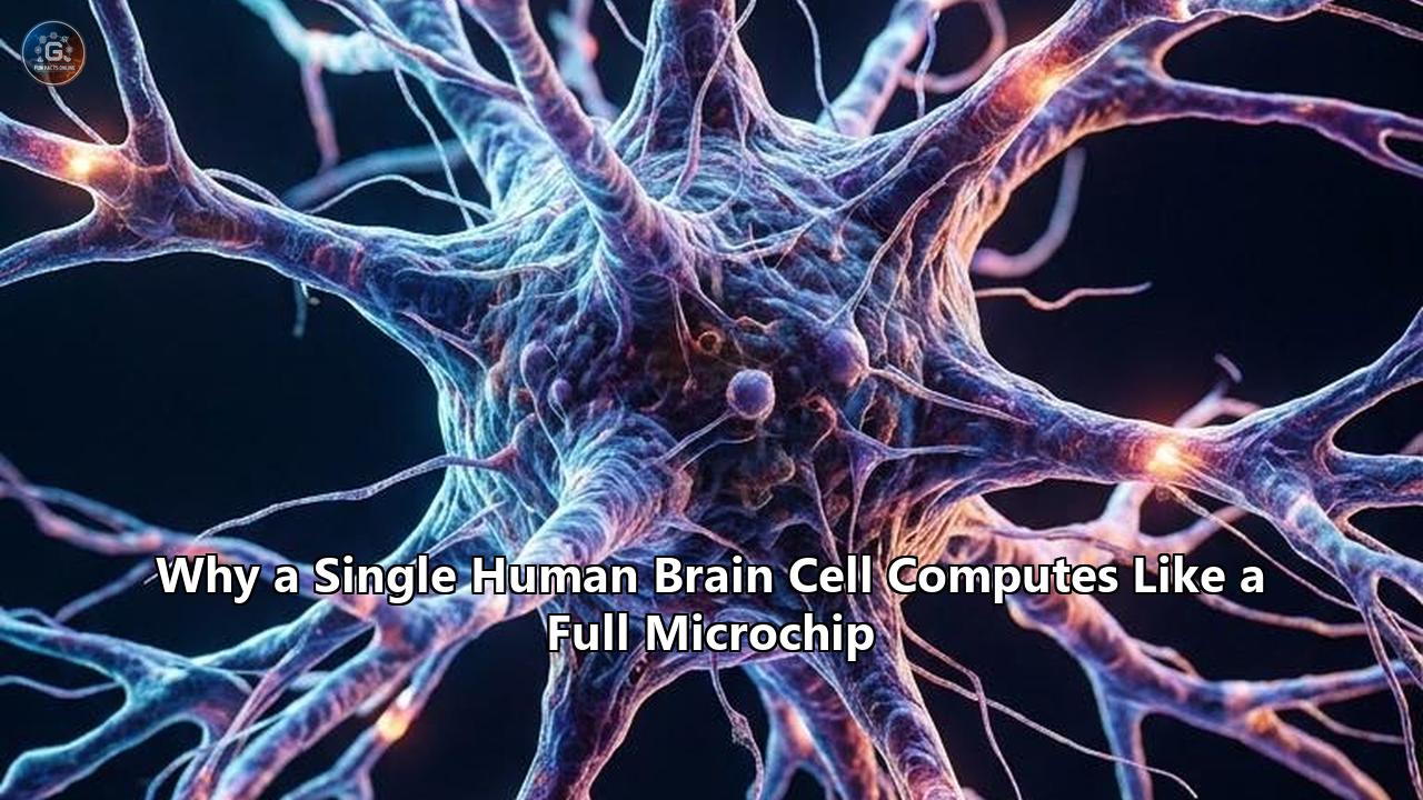
A peer-reviewed investigation published in the Proceedings of the National Academy of Sciences (PNAS) by researchers at the Hebrew University of Jerusalem and Vrije Universiteit Amsterdam demonstrates that a single human cortical neuron possesses the computational capacity of an entire multi-layered artificial deep neural network. By applying machine-learning algorithms to high-resolution biophysical models of human brain tissue, the research team—led by neurobiologists Idan Segev and Mickey London alongside doctoral researchers Ido Aizenbud and Daniela Yoeli—proved that replicating the input-output transformation of an individual human pyramidal cell requires a temporally convolutional deep neural network running five to eight layers deep.
This quantitative finding displaces the century-old assumption that the neuron functions merely as a binary point-processor—a simple threshold gate that sums inputs and fires an all-or-none spike. Instead, an individual human neocortical cell operates as a hierarchical, multi-stage computing engine containing hundreds of independent, non-linear computational subunits.
The empirical benchmarks for human brain cell computing fundamentally reset how neuroscientists calculate the processing bandwidth of the human central nervous system. Where previous mathematical models estimated that the 86 billion neurons in the human brain could generate roughly $10^{15}$ operations per second (1 petaflop) based on point-neuron assumptions, accounting for the internal depth of human dendritic trees elevates the theoretical throughput of the organ to between $10^{18}$ and $10^{21}$ operations per second (1 to 1,000 exaflops). The entire biological system achieves this scale while drawing an invariant power budget of just 20 Watts.
+---------------------------------------------------------------------------------------------------+
|               SINGLE HUMAN NEURON COMPUTATIONAL EQUIVALENCE SUMMARY                               |
+------------------------------------+----------------------------------+---------------------------+
| Metric                             | Classical Point-Neuron Model     | Biophysical Human Model   |
+------------------------------------+----------------------------------+---------------------------+
| Equivalent ANN Architecture        | 1 Linear Node (Perceptron)       | 5–8 Layer Deep CNN        |
| Equivalent Artificial Neurons      | 1 Node                           | ~1,000 Nodes              |
| Independent Processing Subunits    | 1 (Soma)                         | 300–600 (Dendritic Spines)|
| Total Synaptic Inputs per Cell     | 1,000–5,000                      | 30,000–40,000             |
| Boolean Logic Capability           | Linearly Separable (AND/OR)      | Linearly Non-Separable    |
|                                    |                                  | (XOR/XNOR)                |
| Power Consumption per Cell         | N/A                              | ~0.23 Nanowatts (2.3e-10 W)|
| Energy per Synaptic Operation      | N/A                              | 1–10 Femtojoules (1e-15 J)|
+------------------------------------+----------------------------------+---------------------------+
The 1,000-to-1 Equivalence: Deconstructing the PNAS Complexity Benchmark
To evaluate the mathematical complexity of living neurons, the Hebrew University team engineered an algorithmic testing framework called the Functional Complexity Index (FCI). The researchers harvested viable human cortical tissue resected during neurosurgical procedures in Amsterdam, performed whole-cell patch-clamp electrophysiology to record real-time voltage trajectories across human layer 2/3 (L2/3) and layer 5 (L5) pyramidal cells, and created 3D anatomical reconstructions containing exact compartmental morphologies.
                                 [ SYNAPTIC INPUTS ]
                       30,000+ distinct spatiotemporal signals
                                          │
                                          ▼
   +-----------------------------------------------------------------------------+
   | LAYER 1 (Distal Tuft): Local NMDA/dCaAP Spikes [Sub-millisecond Non-linear]  |
   +-----------------------------------------------------------------------------+
                                          │
                                          ▼
   +-----------------------------------------------------------------------------+
   | LAYER 2 (Apical Trunk): Impedance Mismatches & Active Attenuation           |
   +-----------------------------------------------------------------------------+
                                          │
                                          ▼
   +-----------------------------------------------------------------------------+
   | LAYER 3 (Oblique Dendrites): Independent Branch Arithmetic (AND, OR, XOR)   |
   +-----------------------------------------------------------------------------+
                                          │
                                          ▼
   +-----------------------------------------------------------------------------+
   | LAYER 4 (Basal Subtree): Spatial Pattern Recognition & Temporal Filtering   |
   +-----------------------------------------------------------------------------+
                                          │
                                          ▼
   +-----------------------------------------------------------------------------+
   | LAYER 5 (Soma & Axon Initial Segment): Global Thresholding & Spike Output    |
   +-----------------------------------------------------------------------------+
                                          │
                                          ▼
                                   [ AXONAL SPIKE ]
                             Millisecond temporal precision
The team trained feed-forward and temporally convolutional deep artificial neural networks (TCNs) to replicate the somatic voltage outputs of these biological models when stimulated by dynamic, Poisson-distributed barrages of synaptic inputs.
- Model Fidelity Metrics: A simple 1-layer artificial neural network (the standard algorithmic unit used in large language models and modern deep learning) failed entirely to predict human neuron spiking, scoring a variance explained ($R^2$) below 0.20.
- Network Depth Requirements: Achieving an $R^2 > 0.95$ prediction accuracy required a deep network architecture ranging from 5 to 8 convolutional layers, incorporating tens of thousands of artificial weights per single biological cell.
- Cross-Species Divergence: Applying the same quantitative benchmark to rodent pyramidal neurons revealed that rat cortical neurons required significantly shallower networks (typically 3 to 4 layers) to emulate their behavior.
"People often think of a neuron as a simple switch that either turns on or off," said lead investigator Idan Segev of the Edmond and Lily Safra Center for Brain Sciences. "What we show is that a single human neuron is itself an extraordinarily sophisticated computing device. The new deep learning network that we propose is built from artificial neurons whereby each of them is already five to eight layers deep."
The primary driver of this dimensional difference is the physical geometry and channel density of the human dendritic tree. Human layer 2/3 pyramidal neurons feature total dendritic arbor lengths exceeding 12,000 to 15,000 micrometers ($\mu$m), compared to just 3,000 to 5,000 $\mu$m in rodents. Furthermore, human dendrites receive upwards of 30,000 to 40,000 excitatory and inhibitory synaptic connections per cell, whereas rodent counterparts rarely exceed 8,000 to 10,000 inputs.
Anatomy as Architecture: Dendritic Cable Physics and Conductance Equations
The operational complexity of human neurons is governed by biophysical cable properties that prevent signals from linearly pooling together. Under classic 1D linear cable theory developed by Wilfrid Rall, the decay of membrane voltage ($V$) along a passive cylindrical dendritic branch of radius $a$, intracellular axial resistivity $r_i$, and membrane resistance $r_m$ is defined by:
$$\lambda = \sqrt{\frac{a \cdot r_m}{2 \cdot r_i}}$$
$$\tau_m \frac{\partial V}{\partial t} = \lambda^2 \frac{\partial^2 V}{\partial x^2} - V$$
Where:
- $\lambda$ represents the electrotonic length constant (typically 200 to 800 $\mu$m in biological dendrites).
- $\tau_m = r_m \cdot c_m$ represents the passive membrane time constant (ranging from 10 to 30 milliseconds).
In human pyramidal cells, the physical length of distal dendritic branches significantly exceeds $\lambda$. As an electrical signal travels from a distal spine toward the cell body (soma), it experiences massive electrotonic attenuation, dropping in amplitude by up to 80% to 95%. Consequently, distal synapses cannot influence somatic action potential generation through linear summation.
       DISTAL DENDRITIC COMPARTMENTS               PROXIMAL / AXONAL COMPARTMENT
   [Local Spine 1] ──┐
                     ├──> [Local NMDA Spike] ──┐
   [Local Spine 2] ──┘   (Non-linear boost)    │
                                               │
   [Local Spine 3] ──┐                         ├──> Electrotonic ──> [Axon Initial Segment]
                     ├──> [Local dCaAP Spike]  │    Attenuation       (Threshold: -50 mV)
   [Local Spine 4] ──┘    (Graded calcium)     │      Filter                   │
                                               │                               ▼
   [Local Spine 5] ──┐                         │                       Somatic Action
                     ├──> [Local Sub-linear] ──┘                          Potential
   [Local Spine 6] ──┘    (Current shunting)
To bridge this physical separation, human dendrites rely on active, voltage-gated ion conductances embedded directly within the dendritic membrane. When multiple excitatory synapses within a single branch cluster (within 10 to 30 $\mu$m of each other) fire within a narrow temporal window ($\Delta t < 5\text{ ms}$), the local membrane potential depolarizes beyond the threshold required to unblock voltage-sensitive magnesium ($\text{Mg}^{2+}$) ions from $N$-methyl-$D$-aspartate (NMDA) receptor channels:
$$I_{\text{NMDA}}(V, t) = g_{\text{max}} \cdot s(t) \cdot \frac{V_m(t) - E_{\text{rev}}}{1 + \frac{[\text{Mg}^{2+}]_o}{3.57} \cdot \exp(-0.062 \cdot V_m(t))}$$
This creates a regenerative dendritic NMDA spike—a sustained 20 to 50 millivolt (mV) local plateau potential lasting between 50 and 200 milliseconds.
Because this regenerative depolarization remains localized to that specific dendritic subunit, each individual branch acts as an independent analog-to-digital processing threshold. An apical dendritic tree with 40 distinct collateral branches functions as a 40-node hidden computational layer before its integrated outputs converge at the main bifurcation point.
Non-Linear Dendritic Logic: How Single Cells Solve the XOR Paradox
For more than half a century, machine learning architecture was defined by Marvin Minsky and Seymour Papert's 1969 proof that a single artificial perceptron cannot solve linearly non-separable problems—most notably the Exclusive-OR (XOR) boolean logic gate. Computing an XOR function historically required at least two network layers with multiple interconnected nodes:
                XOR LOGIC GATE TRUTH TABLE
       +---------+---------+--------------------+
       | Input A | Input B | Output (A XOR B)   |
       +---------+---------+--------------------+
       |    0    |    0    |         0          |
       |    1    |    0    |         1          |
       |    0    |    1    |         1          |
       |    1    |    1    |         0          |
       +---------+---------+--------------------+
A research initiative led by Albert Gidon and Matthew Larkum at Humboldt University of Berlin, published in Science, demonstrated that individual human layer 2/3 neocortical dendrites natively execute XOR computations without requiring multi-cellular networks.
The mechanism behind this operational capability is a biological class of electrical signal: the dendritic Calcium-mediated Action Potential (dCaAPs).
Membrane Potential (mV)
  ^
  |          dCaAP Amplitude Envelope
30|                 /-----\
  |                /       \
10|               /         \
  |   Threshold  /           \        Suppression / Dampening
 0|   Stimulus  /             \       Under Over-Activation
  |  (Input A) /               \      (Input A + Input B)
-20|           /                 \           /-------\
  |          /                   \         /         \
-50|  -------/                     \-------/           \-------
  |__________________________________________________________> Time (ms)
      Stimulus Intensity: Medium           Stimulus Intensity: High
      Logical Output: 1                    Logical Output: 0
Unlike classical somatic action potentials—which adhere strictly to an all-or-none binary firing regime governed by voltage-gated sodium ($Na_v$) channels—human dCaAPs are non-linear, graded responses driven by L-type calcium channels ($Ca_v$) modulated by potassium conductances ($g_K, g_{BKca}$):
- Subthreshold Stimulation: When zero or weak inputs arrive (Inputs: 0,0), local depolarization is insufficient, and no dendritic event occurs (Output: 0).
- Optimal Threshold Stimulation: When Input Path A or Input Path B delivers approximately 20 to 25 synchronous synaptic events, the local dendritic membrane depolarizes past threshold, triggering a maximum-amplitude dCaAP of +30 to +40 mV, which propagates to the soma to fire an axonal spike (Output: 1).
- Suprathreshold Co-Activation: When Input Path A and Input Path B fire simultaneously (50 combined synaptic inputs), the overwhelming local charge accumulation rapidly recruits high-conductance, calcium-activated potassium channels ($I_{K,\text{Ca}}$). The resulting massive potassium efflux rapidly repolarizes the membrane, paradoxically quenching the dCaAP amplitude. The damped signal fails to reach the somatic threshold, and the neuron remains silent (Output: 0).
A singular segment of human dendritic membrane less than 200 $\mu$m long resolves the XOR calculation natively through localized channel kinetics. Across an entire human cortical neuron harboring dozens of active apical branches, hundreds of linearly non-separable calculations occur in parallel across the analog dendritic tree prior to somatic spike emission.
Thermodynamic Audit: 20 Watts vs. 30 Megawatts
The energetic efficiency of biological information processing becomes starkly apparent when calculating the thermodynamics of human brain cell computing against modern silicon-based microchips.
+---------------------------------------------------------------------------------------------------+
|               THERMODYNAMIC AND SCALING BENCHMARKS                                                |
+------------------------------------+----------------------------------+---------------------------+
| Parameter                          | Human Central Nervous System     | Frontier Supercomputer    |
+------------------------------------+----------------------------------+---------------------------+
| Total Operating Power              | 20 Watts                         | 22.7 Megawatts (2.27e7 W) |
| Computational Substrate            | Carbon / Aqueous Electrolyte     | Silicon / Metal CMOS      |
| Equivalent Throughput              | 1 to 1,000 ExaFLOPS equivalent   | 1.206 ExaFLOPS (FP64)     |
| Energy per Elementary Operation    | 1–10 Femtojoules ($10^{-15}$ J)  | 1–5 Picojoules ($10^{-12}$ J)|
| Active Processing Elements         | 86 Billion Neurons               | 37,888 AMD MI250X GPUs    |
| Interconnect Count                 | ~100–150 Trillion Synapses       | Slingshot-11 Network Fabric|
| Volumetric Density                 | $1.3 \times 10^3\text{ cm}^3$    | 680 $m^3$ (74 Racks)      |
| Architecture Type                  | Colocated Memory & Processing    | Von Neumann Architecture  |
+------------------------------------+----------------------------------+---------------------------+
The human brain runs on approximately 20 Watts of continuous metabolic power, derived from the hydrolysis of roughly $5.7 \times 10^{21}$ molecules of adenosine triphosphate (ATP) per second. Distributed across approximately 86 billion neurons, each individual cell operates on an average baseline power allocation of:
$$P_{\text{neuron}} = \frac{20\text{ W}}{8.6 \times 10^{10}} \approx 2.33 \times 10^{-10}\text{ W} = 0.233\text{ Nanowatts}$$
A biological synapse consumes between 1 and 10 femtojoules ($10^{-15}\text{ Joules}$) per neurotransmitter release event. This metabolic cost accounts for:
- Presynaptic vesicular exocytosis and glutamate recycling ($1.6 \times 10^5\text{ ATP}$ per vesicle).
- Reversing postsynaptic ion influx ($Na^+, Ca^{2+}$) via $Na^+/K^+$-ATPase pumps ($1.2 \times 10^7\text{ ATP}$ per action potential).
- Local dendritic protein synthesis and spine structural maintenance.
In contrast, cutting-edge semiconductor nodes (e.g., 3-nanometer and 4-nanometer FinFET/GAA architectures) executing floating-point arithmetic (FP8 or FP16 matrix multiplication) consume approximately 1 to 5 picojoules ($10^{-12}\text{ Joules}$) per mathematical operation at the silicon chip level—three orders of magnitude higher than a biological synapse.
When factoring in data movement across high-bandwidth memory (HBM3e) and inter-rack communication across copper and optical interconnects, the operational energy surges to 20 to 50 picojoules per memory access due to the classic von Neumann memory wall.
To match the exaflop processing milestone achieved by Oak Ridge National Laboratory's Frontier supercomputer (1.206 ExaFLOPS at 22.7 Megawatts), silicon hardware requires more than one million times more energy than the biological brain. If every human neuron operates with the algorithmic richness of an 8-layer deep neural network, the aggregate biological throughput reaches the zettaflop scale ($10^{21}\text{ operations per second}$) while maintaining its fractional 20-Watt footprint.
Silicon vs. Wetware: Comprehensive Component-by-Component Benchmark
The mechanical disparity between silicon microprocessors and biological pyramidal neurons stems from structural differences in physical layout, carrier dynamics, and compute-memory colocalization.
+---------------------------------------------------------------------------------------------------+
|               COMPONENT-BY-COMPONENT ARCHITECTURAL AUDIT                                          |
+----------------------+------------------------------------+---------------------------------------+
| Feature              | Advanced Silicon Microprocessor    | Human Cortical Pyramidal Neuron       |
+----------------------+------------------------------------+---------------------------------------+
| Fundamental Unit     | Field-Effect Transistor (MOSFET)   | Voltage-Gated Ion Channel Cluster     |
| Unit Feature Size    | 2 nm to 5 nm (Gate pitch ~45 nm)   | 0.5 nm (Ion pore selectivity filter)  |
| Operating Carrier    | Electrons / Holes in conduction band| Hydrated Ions ($Na^+, K^+, Ca^{2+}, Cl^-$)|
| Signal Transport     | Drift / Diffusion in solid silicon | Electro-diffusion via Nernst-Planck   |
| Primary Clock Speed  | 2.0 GHz to 5.5 GHz ($10^{-9}\text{ s}$)| 1 Hz to 200 Hz ($10^{-2}\text{ s}$)   |
| Dynamic Range        | Discrete Binary (0.0 V vs 0.85 V)  | Hybrid Analog-Digital (-75 mV to +40 mV)|
| Noise Tolerance      | Deterministic / Noise Margin Guarded| Stochastic / Stochastic Resonance Utilized|
| Memory Integration   | Separated Register/SRAM/DRAM Caches| Colocated via Synaptic Spine Plasticity|
| Wiring Dimension     | Quasi-2D (12–17 Metal Layers)      | True 3D Fractal Dendritic Arborization|
| Computational Style  | High-Frequency Serial / SIMD       | Massively Parallel Asynchronous Event |
+----------------------+------------------------------------+---------------------------------------+
Signal Propagation and Carrier Transport Mechanics
A silicon microchip relies on the rapid transit of electrons through solid crystalline silicon channels modulated by electrostatic gates. Electron drift velocity in silicon saturates at roughly $10^7\text{ cm/s}$, allowing sub-nanosecond switching times:
$$\tau_{\text{gate}} \approx \frac{L_{\text{channel}}}{v_{\text{sat}}} \approx \frac{10\text{ nm}}{10^7\text{ cm/s}} = 10^{-13}\text{ seconds}$$
Conversely, an individual neuron relies on the physical transit of hydrated ions ($Na^+, K^+, Ca^{2+}, Cl^-$) across lipid bilayers dissolved in aqueous salt solutions. Ion flux through an individual selective channel pore ($10^7\text{ ions/sec}$) is dictated by the Nernst-Planck electro-diffusion equation:
$$J_i = -D_i \left( \frac{\partial C_i}{\partial x} + \frac{z_i F}{R T} C_i \frac{\partial \phi}{\partial x} \right)$$
Where:
- $J_i$ is ion flux density.
- $D_i$ is the ionic diffusion coefficient in water ($\sim 10^{-5}\text{ cm}^2/\text{s}$).
- $C_i$ is concentration, $z_i$ is ion valence, $F$ is Faraday's constant, and $\phi$ is electrical potential.
Because biological transport relies on thermal diffusion and electrostatic drift in fluid, the temporal velocity of electrical signals along unmyelinated dendritic cables ranges between 0.1 and 2.0 meters per second, compared to $2 \times 10^8\text{ meters per second}$ (two-thirds the speed of light) for electromagnetic signals traveling along copper traces on printed circuit boards.
Despite clock frequencies that are 10,000,000 times slower than microchips, the biological cell offsets its temporal latency through spatial multiplexing. While an advanced GPU routes data through an execution pipeline bound by a rigid digital bus width (e.g., 4096-bit memory interfaces), a single human neuron continuously processes 30,000 parallel analog inputs across hundreds of dendritic domains simultaneously.
                     SILICON COMPUTATION (VON NEUMANN)
     ┌────────────────┐         Memory Bus          ┌────────────────┐
     │ Central Memory │ <=========================> │ Execution Core │
     │ (DRAM / HBM)   │   High Latency / High Energy│ (ALU / Vector) │
     └────────────────┘                             └────────────────┘

              BIOLOGICAL COMPUTATION (CO-LOCATED COMPUTE-MEMORY)
     ┌───────────────────────────────────────────────────────────────┐
     │              Continuous Dendritic Cable Substrate             │
     │  [Synaptic Memory] ───> [Dendritic Non-Linear Logic] ────────┤
     │  (AMPA/NMDA Weight)       (dCaAP / Branch Computation)        │
     └───────────────────────────────────────────────────────────────┘
Compute-in-Memory vs. Von Neumann Bottlenecks
In classical silicon architecture, processing and storage are physically partitioned. Executing a mathematical operation requires shuttling floating-point weights from DRAM across physical wires into on-die register files, running arithmetic logic units (ALUs), and writing back the result. Data movement accounts for up to 80% to 90% of total processor thermal dissipation.
In contrast, human brain cell computing integrates processing directly within the storage element. Synaptic weight memory is stored chemically via the physical volume of the dendritic spine head ($0.01\text{ to }0.8\,\mu\text{m}^3$) and the absolute count of postsynaptic $\alpha$-amino-3-hydroxy-5-methyl-4-isoxazolepropionic acid (AMPA) receptor ion channels (varying from 10 to 200 channels per synapse):
$$w_i \propto N_{\text{AMPA}} \cdot \gamma_{\text{single-channel}}$$
When a presynaptic action potential triggers glutamate release, the current entering the spine instantly executes an analog vector-matrix multiplication directly inside the local branch volume:
$$I_{\text{local}}(t) = \sum_{j=1}^{K} w_j \cdot x_j(t)$$
Because storage and arithmetic are physically unified, the neuron eliminates data transfer overhead, processing complex real-world information with minimal thermodynamic dissipation.
Recalculating Cognitive Bandwidth: From Petaflops to Zettaflops
Quantifying the aggregate information processing capacity of the human brain has historically relied on the simplified assumption that an individual neuron operates as a single floating-point accumulator.
                  THE EVOLUTION OF BRAIN COMPUTE ESTIMATES
Throughput
  (OPS)
    ^
10^21|                                                     [BIOPHYSICAL MODEL]
    |                                                     Dendritic Deep-Tree
10^18|                                  [EXAFLOP SCALE]   1 to 1,000 ExaFLOPS
    |                                   Frontier Super-   (10^18 - 10^21 OPS)
10^15|          [POINT-NEURON MODEL]    computer
    |          Single Perceptron Units (1.2 x 10^18 FLOPS)
10^12|          0.1 to 1 PetaFLOP
    |          (10^14 - 10^15 OPS)
    +------------------------------------------------------------------------> Timeline / Model Complexity
Under this classical point-neuron model:
- Neuron Count: $8.6 \times 10^{10}$ cells
- Average Firing Rate: 1 to 5 Hz (action potentials per second)
- Average Synaptic Fan-In: $10^3$ to $10^4$ connections per cell
- Calculation: $(8.6 \times 10^{10}\text{ neurons}) \times (10^3\text{ connections}) \times (5\text{ operations/sec}) \approx 4.3 \times 10^{14}\text{ OPS}$ (~0.43 PetaFLOPS).
Integrating the findings of the Aizenbud et al. and Gidon et al. studies requires recalculating the baseline computational scale of the central nervous system:
- Subunit Multiplexing Factor ($M_s$): Each human pyramidal neuron contains between 300 and 600 semi-independent dendritic processing compartments capable of generating autonomous local non-linearities (NMDA spikes, dCaAPs, and branch shunts).
- Deep Neural Network Equivalent Factor ($D_L$): Simulating the time-dependent continuous input-output mapping of an individual human cell requires a 7-layer convolutional deep neural network containing approximately $10^3$ artificial processing units with continuous dynamic states.
- Temporal Integration Window ($T_{\text{res}}$): Dendritic spike integration operates at a sub-millisecond temporal resolution ($dt \approx 0.1\text{ to }1.0\text{ ms}$), enabling continuous spatiotemporal cross-correlation of incoming spike sequences.
Applying these biophysical variables:
$$\text{Total Human Cortical Bandwidth} = N_{\text{neurons}} \times M_s \times D_L \times F_{\text{bandwidth}}$$
$$\text{Capacity} = (8.6 \times 10^{10}) \times (500\text{ subunits}) \times (1,000\text{ nodes}) \times (100\text{ effective evaluations/sec})$$
$$\text{Capacity} \approx 4.3 \times 10^{18}\text{ to }10^{21}\text{ Effective Biological Operations per Second (1 to 1,000 ExaFLOPS)}$$
Synaptic Channel Capacity and Information Theory
From a Shannon information-theoretic perspective, the information transmission capacity ($C$) of an individual biological synapse is not constrained to a single binary bit (0 or 1).
Due to stochastic neurotransmitter vesicle release, temporal facilitation, short-term depression, and post-tetanic potentiation, a single synapse transmits information dynamically:
$$C = W \log_2 \left( 1 + \frac{S}{N} \right)$$
Electrophysiological reconstructions from the Salk Institute and the University of Texas at Austin show that dendritic spines dynamically modulate surface area across roughly 26 distinct discrete size states. This gives individual synapses a dynamic storage capacity between 4.1 and 4.7 bits of usable memory per connection.
Multiplying across the human brain's 150 trillion synapses yields a total structural memory capacity exceeding 0.6 Petabytes of non-volatile, self-addressable storage distributed directly within the computational fabric itself.
Neuromorphic Engineering: Translating Dendritic Complexity into Silicon
The realization that single biological neurons compute like microchips is forcing a structural reassessment in the semiconductor industry. For the past four decades, neuromorphic computing architectures—including Intel's Loihi, IBM's TrueNorth, and SpiNNaker—relied heavily on the leaky integrate-and-fire (LIF) point-neuron model:
$$\tau_m \frac{dv(t)}{dt} = -(v(t) - v_{\text{rest}}) + R_m \cdot I(t)$$
While LIF neuromorphic chips drastically reduced power compared to general-purpose GPUs, they lacked the computational richness of biological neural circuits, requiring vast numbers of physical silicon cores to perform complex temporal pattern recognition.
  TRADITIONAL NEUROMORPHIC CORE (LIF)            NEXT-GEN DENDRITIC NEUROMORPHIC CORE
         Point-Neuron Architecture                      Multi-Stage Tree Architecture
           ┌─────────────────┐                       ┌──────────────┐ ┌──────────────┐
  Inputs ─>│ Leaky Integrate │─> Spike Output        │Dendritic Ckt1│ │Dendritic Ckt2│
           │    and Fire     │                       └──────┬───────┘ └──────┬───────┘
           └─────────────────┘                              │                │
                                                            ▼                ▼
                                                     ┌───────────────────────────────┐
                                                     │Non-Linear Multi-Stage Combiner│
                                                     └──────────────┬────────────────┘
                                                                    │
                                                                    ▼
                                                            [Spike Output]
To bridge this operational gap, chip architects are developing neuromorphic hardware incorporating multi-compartment dendritic circuits:
- Intel Loihi 2: Features a fully programmable microcode pipeline allowing engineers to define custom multi-compartment dendritic topologies, supporting non-linear dendritic voltage integration and resonant resonant-and-fire equations.
- BrainScaleS-2 (Heidelberg University): Employs mixed-signal analog CMOS circuitry that runs $1,000\times$ to $10,000\times$ faster than biological real time. It implements analog dendritic compartments capable of emulating local NMDA-like plateau potentials and active dendritic shunting directly in continuous silicon circuits.
- Memristive Crossbar Arrays: Researchers are pairing non-volatile Phase Change Memory (PCM) and Oxide-based Resistive RAM ($HfO_x$ ReRAM) to mimic dendritic branch non-linearities. By engineering devices that exhibit negative differential resistance (NDR), engineers reproduce the calcium-mediated XOR functionality of human dendrites on a single solid-state electronic component.
+---------------------------------------------------------------------------------------------------+
|               NEUROMORPHIC HARDWARE GENERATIONAL EVOLUTION                                        |
+----------------------+--------------------+-----------------------+-------------------------------+
| System               | Generation / Year  | Neuron Model Implemented| Dendritic Architecture      |
+----------------------+--------------------+-----------------------+-------------------------------+
| IBM TrueNorth        | 1st Gen (2014)     | Digital Point LIF     | Flat (No Dendritic Processing)|
| Intel Loihi 1        | 1st Gen (2018)     | Digital Point LIF     | Flat (1 Compartment)          |
| SpiNNaker-1          | 1st Gen (2013)     | ARM Software LIF      | Flat (Point Neuron)           |
| Intel Loihi 2        | 2nd Gen (2021)     | Programmable Multi-Cpt| Multi-Compartment Tree Graphs |
| BrainScaleS-2        | 2nd Gen (2022)     | Analog Hodgkin-Huxley | Active Non-linear Dendrites   |
| 3D Dendro-Memristor  | Emerging (2025/26) | Analog dCaAP / XOR    | 3D Nanowire Dendritic Arbors  |
+----------------------+--------------------+-----------------------+-------------------------------+
Benchmarks demonstrate that replacing flat point-neuron algorithms with multi-compartment dendritic architectures allows spiking neural networks to solve complex temporal classification tasks—such as radar gesture recognition, biosignal arrhythmia detection, and audio acoustic processing—with 60% to 85% fewer physical neurons and up to a $100\times$ reduction in operating latency.
Methodological Milestones and the Next Quantitative Frontiers
The discovery that human cortical neurons operate as deep computational systems opens significant analytical challenges across computational neuroscience, bio-inspired computing, and brain mapping.
+---------------------------------------------------------------------------------------------------+
|               EXPERIMENTAL COMPLEXITY & DATA SCALING ROADMAP                                      |
+------------------------------------+----------------------------------+---------------------------+
| Research Frontier                  | Baseline Metric (Current State)  | Projected Next Milestone  |
+------------------------------------+----------------------------------+---------------------------+
| Multi-Site Dendritic Patch Clamping| 2–3 Simultaneous Pipettes/Neuron | 5+ Multi-Site Robotic Patches|
| Connectomic Reconstruction Volume  | 1 $mm^3$ (1.4 Petabytes Data)    | Full Human Cortical Column|
| Biophysical Model State Variables  | 20 Conductance Channels ($g_i$)  | 50+ Epigenetic/Phosphorylated|
| Spatial Voltage Imaging Resolution | 5 ms frame rate (Widefield)      | Sub-millisecond Holographic|
| Neuromorphic Density               | $10^6$ Neurons per die           | $10^9$ Dendritic Subunits/die|
+------------------------------------+----------------------------------+---------------------------+
Three major experimental frontiers define the path forward:
1. High-Density Multi-Site Electrophysiology in Living Human Tissue
Electrophysiological analysis of human dendrites remains constrained by physical accessibility. Obtaining viable, healthy human cortical tissue relies on surgical resections from epilepsy or glioma operations.
Simultaneously patching the minute terminal branches of human layer 2/3 apical tufts ($0.2\text{ to }0.5\,\mu\text{m}$ in diameter) requires dual or triple whole-cell pipette micromanipulation under high-magnification infrared differential interference contrast (IR-DIC) microscopy.
Automating this procedure via robotic multi-patch clamp systems and high-speed two-photon voltage-sensitive dye (VSD) imaging with sub-millisecond temporal resolution remains critical to mapping the remaining active conductances across human cortical layers.
2. Microscale Connectomics and Petabyte-Scale Datasets
Validating how these individual biological microchips interconnect into cortical microcircuits requires ultra-deep nanoscale electron microscopy.
The connectomic dataset reconstructed by Harvard University and Google Research—mapping just 1 cubic millimeter of human temporal cortex—generated 1.4 petabytes of imaging data, identifying roughly 57,000 cells, 150 million synapses, and unprecedented axonal whorls. Expanding this structural analysis to map entire functional cortical columns will require exabyte-scale computational pipelines and automated machine-learning segmentation tools.
               CROSS-SECTION: 1 MM³ HUMAN CORTEX DATA PIPELINE
    [Surgical Resection] ──> [Serial Sectioning: 30 nm Slices] ──> [Multi-Beam EM Imaging]
                                                                           │
                                                                           ▼
    [1.4 Petabytes Raw Data] <── [Automated AI Segmentation] <── [Alignment & Stitching]
              │
              ▼
    [Connectomic Circuit Graph]
     • 57,000 Cells
     • 150,000,000 Synapses
     • Multi-stage Dendritic Arbors
3. Molecular and Epigenetic Sub-State Computation
The mechanical models established by Segev, London, and Larkum focus primarily on electrophysiological membrane dynamics governed by Hodgkin-Huxley channel mathematics.
However, an entirely separate tier of intracellular information processing resides beneath the electrical layer. Intracellular signaling cascades—such as calcium/calmodulin-dependent protein kinase II (CaMKII) activation, cyclic adenosine monophosphate (cAMP) messaging pathways, and local dendritic messenger RNA (mRNA) translation—operate as a biochemical computational network.
Integrating these enzymatic and molecular state variables into existing electrical models will expand the Functional Complexity Index of human brain cells even further.
       CELLULAR COMPUTATION IS A MULTI-TIERED PROCESSING HIERARCHY
   =========================================================================
   TIER 1: CHEMICAL / MOLECULAR (Seconds to Hours)
   • Intracellular signaling cascades (cAMP, CaMKII, MAPK)
   • Local mRNA translation at dendritic spine bases
   • Epigenetic chromatin remodeling and state storage
   -------------------------------------------------------------------------
   TIER 2: ANALOG DENDRITIC PROCESSING (Milliseconds to Seconds)
   • Local NMDA plateau potentials & dCaAP graded spikes
   • Electrotonic cable filtering and branch shunting
   • XOR / XNOR non-linear arithmetic across branch nodes
   -------------------------------------------------------------------------
   TIER 3: DIGITAL AXONAL OUTPUT (Sub-millisecond)
   • All-or-none somatic action potential firing
   • Rapid propagation along myelinated axonal pipelines
   =========================================================================
As neuromorphic chip architects systematically work to translate these multi-tier dendritic principles into solid-state silicon, the boundary between biological neural computation and microelectronics continues to narrow.
The empirical proof that a single human brain cell operates as a deeply layered computing engine confirms that human intellectual capacity is not simply an emergent feature of a large network, but a fundamental capability embedded within the biophysical structure of each individual cell.
Reference:
- https://www.thebrighterside.news/post/human-brain-cells-may-be-far-more-powerful-computers-than-scientists-thought/
- https://scitechdaily.com/a-single-human-neuron-is-far-more-powerful-than-scientists-thought/
- https://pubmed.ncbi.nlm.nih.gov/34380016/
- https://neurosciencenews.com/branching-dendrite-human-cognition-31015/
- https://aiimpacts.org/brain-performance-in-flops/
- https://www.reddit.com/r/MachineLearning/comments/191ol1n/d_human_brain_flops_estimate_is_it_lower_than_we/
- https://www.nist.gov/blogs/taking-measure/brain-inspired-computing-can-help-us-create-faster-more-energy-efficient
- https://www.facebook.com/EuropeanFriendsHUJ/posts/-what-makes-the-human-brain-uniquea-new-study-from-researchers-at-the-hebrew-uni/1499063015595679/
- https://pubmed.ncbi.nlm.nih.gov/31896716/
- https://www.eurekalert.org/news-releases/927574
- https://www.sciencedaily.com/releases/2026/08/260812015216.htm
- https://dendrites.gr/?p=113
- https://pmc.ncbi.nlm.nih.gov/articles/PMC11655932/
- https://www.reddit.com/r/MachineLearning/comments/1bvsb9o/d_how_many_compute_flops_is_one_brain/
- https://www.frontiersin.org/news/2018/10/02/cellular-neuroscience-brain-neurons-memory