On June 10, 2026, a study published in the journal Nature fundamentally rewrote the opening chapters of complex life on Earth. Led by Dr. Toni Gabaldón, an ICREA researcher at the Institute for Research in Biomedicine (IRB) Barcelona and the Barcelona Supercomputing Center (BSC-CNS), a team of computational biologists utilized the massive processing power of the MareNostrum supercomputer to perform what can only be described as molecular archaeology. By analyzing public genomic data spanning the entirety of terrestrial biodiversity, the researchers traced the genetic footprints of the First Eukaryotic Common Ancestor (FECA)—the ancient, single-celled precursor that lived approximately two billion years ago and eventually gave rise to all animals, plants, fungi, and humans.
For decades, the standard textbook explanation for the origin of complex cells rested on a simple, two-player endosymbiotic theory: an ancient archaeon swallowed a bacterium, which gradually evolved into the mitochondrion, giving the cell the energy required to develop complex internal structures.
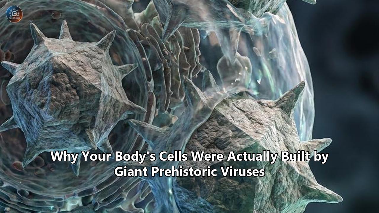
The June 2026 Nature paper revealed that this narrative is dramatically incomplete. Instead of a neat, two-domain fusion, the birth of the complex cell was a highly collaborative, multi-million-year epic featuring an unexpected third protagonist: giant viruses. The study demonstrated that key genomic innovations of early eukaryotes—long believed to be of bacterial or archaeal origin—were actually introduced via massive lateral gene transfers mediated by ancient giant viruses belonging to the phylum Nucleocytoviricota. These prehistoric giants acted as genetic shuttle buses, transferring massive chunks of DNA between coexisting microorganisms and directly shaping the ancestral eukaryotic genome.
This computational breakthrough coincided with a physical discovery made just months prior in early 2026. A research team led by Professor Masaharu Takemura at the Tokyo University of Science isolated a massive, previously unknown giant virus from Lake Ushiku in Ibaraki Prefecture, Japan. Named ushikuvirus, this giant DNA virus infects single-celled amoebae (Vermamoeba vermiformis) and displays a replication cycle that directly alters and disrupts the host cell's nuclear membrane. By demonstrating a physical and evolutionary bridge between giant viral replication compartments and the eukaryotic nucleus, ushikuvirus has provided physical corroboration for "viral eukaryogenesis"—the controversial theory that the very nucleus of your cells was originally a domesticated prehistoric virus.
Together, these twin discoveries of mid-2026 provide unparalleled evidence for the viral origin of human cells. Far from being mere pathogens or genetic hitchhikers, giant prehistoric viruses were the primary structural engineers, genetic architects, and evolutionary catalysts that built the complex cellular machinery making up the human body.
The Core Discoveries: From Supercomputers to Japanese Lakes
To comprehend the full scope of this paradigm shift, it is necessary to examine the two scientific pillars that have anchored this discovery: the computational reconstruction of the eukaryotic ancestor and the physical isolation of ushikuvirus.
The MareNostrum Analysis: Genomic Archaeology of the First Complex Cells
The study led by Dr. Toni Gabaldón at IRB Barcelona and the Barcelona Supercomputing Center tackled a problem that has plagued evolutionary biologists for a century. Because the transition from simple prokaryotic life (bacteria and archaea) to complex eukaryotic life occurred roughly two billion years ago, there are no physical fossils of the intermediate stages. The only remaining fossils are "written in genes"—scattered across the genomes of modern organisms.
[Prehistoric Microbial Ecosystem]
   ├── Archaea (Host Cytoplasm/Membrane)
   ├── Bacteria (Mitochondrial Ancestor)
   └── Nucleocytoviricota (Giant Viruses) ──► [Lateral Gene Transfer] ──► [First Eukaryotic Common Ancestor (FECA)]
Using the MareNostrum supercomputer, Gabaldón’s team built complex evolutionary trees for thousands of gene families. They analyzed how these genes traveled, mutated, and integrated across different domains of life. The supercomputer’s massive computational capacity allowed the team to look past the "noise" of horizontal gene transfers that occurred recently and isolate the earliest genetic integrations.
The results were stark:
- Multiple Bacterial Contributors: The ancestor of the mitochondrion (an alpha-proteobacterium) was not the only bacterial group to leave a mark. Multiple other bacterial lineages contributed genes involved in metabolism, membrane synthesis, and cellular structure prior to the endosymbiotic event.
- Giant Viruses as Genetic Vehicles: A substantial portion of the genes essential for eukaryotic cell division, DNA replication, and structural compartmentalization traced their ancestry directly back to ancient Nucleocytoviricota (giant viruses).
- The Vector Hypothesis: The researchers proposed that giant viruses, which possess genomes larger than many bacteria, acted as the primary vector for genetic exchange in prehistoric marine ecosystems. They infected various microorganisms, picked up host genes, shuffled them, and integrated them into the ancestral eukaryotic genome.
By mapping these ancient integration events, this supercomputing analysis provides the strongest genetic footprint yet for a viral origin of human cells, or at least their most defining components.
Ushikuvirus: The Living Missing Link of Lake Ushiku
While the Nature study provided the statistical and genomic framework, the physical mechanics of this ancient partnership were illuminated by the discovery of ushikuvirus in Japan.
For over two decades, Professor Masaharu Takemura of the Tokyo University of Science and Dr. Philip Bell of Macquarie University have championed the "viral eukaryogenesis" hypothesis. This theory suggests that the cell nucleus—the membrane-bound command center of eukaryotic life—evolved from an ancient, large DNA virus that established a permanent, symbiotic residency inside a prokaryotic host.
The discovery of ushikuvirus, published in the Journal of Virology, has supercharged this theory. This giant virus possesses several highly unusual characteristics:
- Host Enlargement: While most giant viruses eventually cause their host cells to burst and shrink, ushikuvirus causes its host amoeba, Vermamoeba vermiformis, to swell dramatically. Infected cells double in size on average, with some individual cells growing up to seven times their original volume.
- Prolonged Replication Cycle: The infection and replication cycle of ushikuvirus lasts an extraordinarily long 60 hours or more. During this time, the virus coexists with the host cell in a state that mimics a highly coordinated cellular process rather than a rapid, destructive infection.
- Nuclear Disruption and Compartmentalization: Unlike related giant viruses that either replicate entirely in the cytoplasm or quietly exploit the host's nucleus, ushikuvirus actively invades, disrupts, and restructures the host cell's nuclear membrane. It constructs a massive, membrane-bound "virus factory" that physically behaves like a synthetic nucleus, sequestering viral DNA replication away from the rest of the cell.
These features suggest that ushikuvirus represents a modern analogue of the ancient viral lineages that first taught prokaryotic hosts how to build large, membrane-enclosed genetic chambers.
Who Is Affected? A Paradigm Shift Across Disciplines
The revelation that prehistoric giant viruses laid the foundations of eukaryotic biology has triggered a cascading series of impacts across multiple scientific, medical, and technological domains.
┌────────────────────────────────────────────────────────────────────────┐
│                        FIELDS DEEPLY AFFECTED                          │
├───────────────────────────┬───────────────────────────┬────────────────┤
│   Evolutionary Biology    │    Biomedicine & Med      │   Biotech &    │
│      & Phylogenetics      │       Therapeutics        │ Synthetic Bio  │
├───────────────────────────┼───────────────────────────┼────────────────┤
│ • Abandoning 3-domain tree│ • Target HERV-W in MS     │ • Giant viral  │
│ • Networks of life models │ • "Viral mimicry" cancers │   vector systems│
│ • Viruses as "alive"      │ • Autoimmune etiology     │ • Organelle    │
│                           │                           │   engineering  │
└───────────────────────────┴───────────────────────────┴────────────────┘
1. Evolutionary Biologists and Taxonomists
For generations, biological classification has relied on the Three-Domain System proposed by Carl Woese in 1977, which divides all life into Bacteria, Archaea, and Eukarya. Under this system, viruses were excluded entirely, classified as non-living, parasitic genetic anomalies that existed outside the tree of life.
The 2026 discoveries have demolished this paradigm. Evolutionary biologists are now forced to abandon the concept of a clean, bifurcating tree of life in favor of a deeply reticulated, network-based model. In this new taxonomy:
- Viruses are recognized as primary genetic reservoirs and active participants in the evolutionary lineage of complex organisms.
- The boundary between "living" cellular organisms and "non-living" viral particles has blurred, as giant viruses contain genes for protein synthesis, energy metabolism, and cellular structural maintenance that were once thought to be exclusive to cells.
- The origin of complex life is no longer viewed as a rare, accidental endosymbiotic mutation, but as an inevitable consequence of viral-mediated genetic shuffling across diverse microbial communities.
2. Immunologists and Pathologists
Medical researchers studying human health and disease are experiencing a profound shift in how they view both human physiology and the nature of pathogens.
For decades, the immune system was understood as a defense network designed to eliminate foreign viral invaders. Now, immunologists must reckon with the fact that the human body's most basic immune signaling pathways are themselves derived from ancient viral machinery.
Furthermore, because our genomes are packed with thousands of endogenous viral genes, many chronic, unexplained inflammatory diseases—such as Multiple Sclerosis, Amyotrophic Lateral Sclerosis (ALS), and various cancers—are being re-evaluated not as attacks from external pathogens, but as the accidental "awakening" of the prehistoric viral ghosts residing inside our own DNA.
3. Neurologists and Cognitive Scientists
The brain has long been viewed as a highly specialized, human-specific organ governed by complex synaptic networks. However, the discovery that our capacity for memory, learning, and higher cognitive function relies on a domesticated retroviral gene called Arc has forced cognitive scientists to reframe the physical basis of human consciousness.
Neurologists are now studying how the brain uses virus-like capsids to send genetic messages between neurons. This changes our fundamental understanding of synaptic transmission and opens up entirely new avenues of research into neurodegenerative and neurodevelopmental disorders, which may be caused by the failure of these internal viral communication pathways.
What Changes? Rewriting the Blueprints of the Human Body
The realization that complex life is fundamentally viral in origin changes how we understand the basic anatomy and physiology of human cells. When we explore the viral origin of human cells, we must look directly at the defining architectural feature of eukaryote life: the nucleus.
1. The Architectural Template: Is the Cell Nucleus a Domesticated Virus?
The primary difference between a simple bacterium and a human cell is the presence of a nucleus: a double-membrane-bound vault that protects our DNA, regulates gene expression, and separates the process of transcription (reading DNA into RNA) from translation (building proteins from RNA).
For years, the origin of this complex chamber was a complete mystery. However, the viral eukaryogenesis hypothesis, supported by the discovery of giant viruses like Mimivirus, Medusavirus, and ushikuvirus, suggests that the nucleus did not evolve from within the cell; it was imported.
           [ARCHAEAL HOST CELL]
 ┌────────────────────────────────────────┐
 │   Cytoplasm                            │
 │                                        │
 │     [GIANT PREHISTORIC VIRUS]          │
 │     ┌────────────────────────┐         │
 │     │  Viral Factory         │         │
 │     │  • Double membrane     │         │
 │     │  • Compartmentalized   │ ────────┼─► Evolves into the
 │     │    DNA replication     │         │   Eukaryotic Cell Nucleus
 │     │  • Lacks ribosomes     │         │
 │     └────────────────────────┘         │
 │                                        │
 └────────────────────────────────────────┘
Consider the striking similarities between the eukaryotic cell nucleus and the "virus factories" constructed by giant Nucleocytoplasmic Large DNA Viruses (NCLDVs) inside host cells:
| Feature | Eukaryotic Cell Nucleus | Giant Virus Factory (e.g., Mimivirus, Ushikuvirus) |
|---|---|---|
| Membrane Structure | Double-membrane bound with nuclear pores. | Double-membrane bound, often constructed from host ER/nuclear membranes, possessing pore-like structures. |
| Ribosome Exclusion | Ribosomes are excluded from the interior; transcription is strictly separated from translation. | Ribosomes are excluded; DNA replication and transcription occur inside, while translation occurs in the surrounding cytoplasm. |
| DNA Topology | Linear chromosomes with telomeres (specialized end caps). | Linear double-stranded DNA genomes, often with terminal inverted repeats resembling telomeres. |
| Genetic Processing | Utilizes 5' mRNA capping, polyadenylation, and sophisticated RNA polymerase systems. | Encodes its own mRNA capping enzymes, polyA polymerases, and independent RNA polymerase machinery. |
According to the model proposed by Takemura and Bell, an ancient, complex DNA virus (akin to a prehistoric relative of poxviruses or giant mimiviruses) infected a single-celled archaeal host. Instead of destroying the host, the virus established a persistent, non-lethal infection, constructing a stable, membrane-enclosed "factory" to protect its own genome. Over evolutionary timescales, this viral factory absorbed the host's primary metabolic genes, gradually usurped control of the host cell, and transitioned into what we now recognize as the cell nucleus.
2. The Genetic Legacy: Endogenous Viruses in the Human Genome
If you look at the raw sequence of human DNA, the traditional view that we are purely "human" disappears.
Only about 1.5% to 2% of the human genome consists of traditional, protein-coding genes that define our physical traits (such as eye color, muscle structure, or digestive enzymes). In contrast, at least 8% to 10% of our genome is composed of complete or fragmented sequences of Human Endogenous Retroviruses (HERVs). If we include other retrovirus-like elements, such as retrotransposons (LINEs and SINEs), nearly half of the human genome is made up of genetic elements of viral origin.
These endogenous retroviruses represent the remnants of ancient, historic infections. Millions of years ago, exogenous retroviruses (similar to modern HIV) infected the germline cells (sperm or egg) of our prehistoric mammalian ancestors. When those infected cells successfully fertilized and produced offspring, the viral DNA was integrated permanently into the host genome, becoming a heritable trait passed down through Mendelian genetics.
Over evolutionary time, most of these viral sequences accumulated mutations that disabled their ability to produce infectious, cell-bursting viruses. However, instead of being discarded as useless "junk DNA," many of these viral genes were co-opted—or exapted—by our ancestors to perform biological functions that are now indispensable to human survival.
3. Physiological Innovations: Three Viral Systems That Define Humanity
Without three specific instances of prehistoric viral domestication, the human race as we know it could not exist.
A. The Placenta and Live Birth: Syncytin-1 and -2
One of the most profound evolutionary leaps in animal history was the transition from laying eggs to bearing live young. This transition required the development of the placenta, a highly complex organ that allows a pregnant mother to supply nutrients and oxygen to her developing fetus while simultaneously carrying away metabolic waste.
However, the placenta presents a massive biological paradox: because the fetus carries genetic material from both the mother and the father, it is technically an immunological "foreign object". Under normal circumstances, the mother's immune system should recognize the fetal tissues as an invader and launch a deadly autoimmune attack.
The structural barrier that prevents this attack is the syncytiotrophoblast—a seamless, multi-nucleated single layer of fused cells that forms the outermost barrier of the placenta, separating maternal blood from fetal tissues. This layer acts as a physical filter that allows nutrients through but prevents maternal immune cells from crossing over and destroying the fetus.
[Maternal Blood Stream]
       │
 ──────┼─────────────────────────────────────────────
       │ ◄─── Syncytiotrophoblast (Fused Cell Barrier)
       │      • Created by SYNCYTIN-1 & SYNCYTIN-2 (Retroviral Env)
       │      • Prevents maternal immune cell crossing
 ──────┼─────────────────────────────────────────────
       │
[Fetal Blood Stream / Placenta]
This cell fusion is facilitated entirely by two proteins of retroviral origin: Syncytin-1 (encoded by the ERVWE1 gene) and Syncytin-2 (encoded by the ERVFRDE1 gene).
These syncytin genes are derived from the env (envelope) genes of ancient endogenous retroviruses that infected our primate ancestors roughly 25 to 40 million years ago. In an active retrovirus, the envelope glycoprotein's sole job is to bind to a host cell receptor and force the viral envelope to fuse with the host cell's membrane, allowing the virus to enter.
Mammals co-opted this exact membrane-fusing mechanism. Instead of using the env protein to fuse a virus to a cell, placental mammals express the protein on the surface of placental cells (cytotrophoblasts), forcing them to fuse with one another to create the seamless, protective syncytial barrier.
This viral hijacking was so successful that it happened multiple times independently across mammalian history. Rodents captured their own unique set of retroviral envelope genes (syncytin-A and syncytin-B), carnivores captured syncytin-Car1, and ruminants captured syncytin-Rum1. Without prehistoric viral infections, placental mammals—including humans—could not give birth.
To prevent this fusogenic process from running wild and destroying the structural integrity of the placenta, human cells also co-opted another retroviral gene from the HERV-H family called Suppressyn (SUPYN). Suppressyn binds to the exact same cell-surface receptor as Syncytin-1 (the neutral amino acid transporter ASCT2). By occupying this receptor, Suppressyn acts as an evolutionary check-and-balance, blocking excess cell fusion and protecting the cell from being invaded by external, modern retroviruses that attempt to exploit the same pathway.
B. Memory, Cognition, and Thought: The Arc Gene
Our cells are not just physically constructed by prehistoric viral processes; our thoughts, memories, and behaviors are governed by them.
For decades, neuroscientists knew that a gene called Arc (Activity-regulated cytoskeleton-associated protein) was essential for long-term memory consolidation, learning, and synaptic plasticity. Mice born without the Arc gene are unable to form lasting memories; they can learn a task in the short term, but by the next day, the memory is entirely gone.
In 2018, studies published in the journal Cell revealed the startling truth: Arc is actually an evolutionary domesticated retrotransposon Gag protein.
           [NEURON A]
  ┌───────────────────────────┐
  │  Arc gene transcribed     │
  │  translates Arc protein   │
  │                           │
  │    [Arc Capsid Assembly]  │
  │    ┌─────────────────┐    │
  │    │  Arc Protein    │    │
  │    │   (capsid)      │    │
  │    │   • Encapsulates │    │
  │    │     Arc mRNA    │    │
  │    └────────┬────────┘    │
  └─────────────┼─────────────┘
                │  (Released in Extracellular Vesicle)
                ▼
           [NEURON B]
  ┌─────────────┼─────────────┐
  │             ▼             │
  │    [Arc Capsid Entry]     │
  │    • Endocytosed          │
  │    • Translates mRNA locally
  │    • Drives Synaptic      │
  │      Plasticity / Memory  │
  └───────────────────────────┘
In an active retrovirus, the Gag gene encodes the physical capsid—the protein shell that wraps around the viral genetic material, protects it, and transports it between host cells.
The Arc gene operates exactly like a retrovirus:
- When a neuron is stimulated by an experience or learning event, it transcribes the Arc gene.
- The resulting Arc proteins self-assemble into hollow, icosahedral virus-like capsids.
- These capsids selectively encapsulate Arc mRNA inside themselves.
- The neuron then packages these Arc capsids into extracellular vesicles and releases them into the synaptic cleft.
- Neighboring recipient neurons absorb these capsids via endocytosis, open them up, and translate the enclosed Arc mRNA into functional proteins locally at the synapse.
This is not a traditional cellular communication pathway; it is an active, ongoing viral infection occurring inside your brain every time you form a memory. Eukaryotes repurposed an ancient viral delivery system to allow neurons to perform high-speed, localized genetic transfers across synapses, enabling the complex synaptic scaling and remodeling that underpins human intelligence.
C. The Host Defense: Innate Immunity and the Interferon Pathway
Our primary weapon against modern viral infections is our innate immune system, specifically the type I and type III interferon responses. When a human cell detects a viral invasion (such as the presence of foreign double-stranded RNA in the cytoplasm), it releases signaling proteins called interferons, which trigger neighboring cells to shut down protein translation and activate hundreds of interferon-stimulated genes (ISGs) to halt the spread of the virus.
This entire survival mechanism is built from domesticated viral components. Many of our primary intracellular viral sensors, as well as the enhancers and promoters that regulate the transcription of interferon-stimulated genes, are derived from ancient long terminal repeats (LTRs) left behind by ancient endogenous retroviruses. Over millions of years of co-evolution, our cells hijacked the promoters of prehistoric viruses—which were designed to rapidly turn on viral replication in response to cellular stress—and rewired them to turn on our host defense genes instead.
We use the genetic switches of dead prehistoric viruses to fight off the living viruses of the modern world.
Short-Term Consequences: Redefining Pathology and Therapeutics
The validation of the viral origin of human cells is not just an academic milestone; it has immediate, real-world consequences for how we diagnose, treat, and understand human disease.
1. Autoimmune Diseases: Waking the Sleeping Giants
For decades, autoimmune diseases like Multiple Sclerosis (MS) have baffled medical science. In MS, the body's own immune system launches a devastating attack on the central nervous system, specifically targeting and stripping away the protective myelin sheath that wraps around nerve fibers. The root cause of this autoimmune trigger has long remained elusive.
The confirmation of the viral origin of human cells has directed therapeutic attention toward the Human Endogenous Retrovirus (HERV) families.
                [ENVIRONMENTAL TRIGGER]
              (e.g., Epstein-Barr Virus)
                          │
                          ▼
             [Epigenetic Repression Fails]
                          │
                          ▼
           [HERV-W Envelope Protein Reactivated]
                          │
                          ▼
             [Toll-Like Receptor 4 (TLR4)]
                    (on Microglia)
                          │
                          ▼
            [Oligodendrocyte Precursor Cells]
                  (OPC Differentiation Blocks)
                          │
                          ▼
                [Demyelination of Neurons]
                  (Multiple Sclerosis)
We now know that patients with Multiple Sclerosis exhibit abnormal activation of a dormant retrovirus called HERV-W (specifically the Multiple-sclerosis-associated retrovirus, or MSRV) and HERV-H within their central nervous system. Under normal healthy conditions, the human body uses epigenetic silencing—such as DNA methylation and histone deacetylation—to keep these ancient viral sequences permanently locked down.
However, when a patient experiences severe cellular stress, oxidative damage, or infection with certain common modern viruses (such as the Epstein-Barr Virus), this epigenetic containment can fail. When the HERV-W promoter is reactivated, the cell begins producing the ancient viral envelope protein (pHERV-W env).
This reactivated viral protein has devastating consequences:
- TLR4 Activation: The pHERV-W envelope protein is highly immunogenic and binds directly to Toll-Like Receptor 4 (TLR4) expressed on the surface of microglia and macrophages.
- Pro-inflammatory Cascade: This binding triggers a massive, chronic inflammatory response, releasing neurotoxic cytokines like TNF-alpha, IL-1-beta, and IL-6.
- Blockade of Remyelination: Most importantly, the reactivated viral protein blocks the differentiation of Oligodendrocyte Precursor Cells (OPCs) into mature myelin-producing oligodendrocytes.
With the repair cells frozen in an immature state, the ongoing inflammation strips the myelin away from the axons, leading to the progressive physical and cognitive decline characteristic of MS.
This biochemical realization unveils a practical medical dimension to what once seemed a purely academic debate over the viral origin of human cells. It has led to clinical trials of monoclonal antibodies specifically designed to bind to and neutralize the pathogenic HERV-W envelope protein in MS patients, offering a targeted treatment that leaves the patient’s normal immune system intact.
2. Oncology and the "Viral Mimicry" Revolution
In cancer biology, the viral origin of our cells is being exploited to create a therapeutic technique known as viral mimicry.
Cancer cells are notoriously adept at hiding from the human immune system. They do this by actively suppressing their own antigen presentation machinery and turning off genes that would normally alert the immune system to their presence, creating "cold" tumors that immunotherapies cannot find.
To bypass this defense, researchers are using epigenetic drugs—specifically DNA methyltransferase inhibitors (DNMTis) like decitabine or 5-aza-2-deoxycytidine—to selectively demethylate the cancer cell's DNA.
                     [CANCER CELL]
                ┌─────────────────────┐
                │   Silenced HERVs    │
                └──────────┬──────────┘
                           │ (Treat with Epigenetic Drug: DNMTis)
                           ▼
                ┌─────────────────────┐
                │   Reactivated ERVs   │
                │ • Transcription of  │
                │   ancient dsRNA     │
                └──────────┬──────────┘
                           │
                           ▼
                ┌─────────────────────┐
                │ Intracellular Sensors│
                │  • MDA5 & PKR       │
                └──────────┬──────────┘
                           │
                           ▼
                ┌─────────────────────┐
                │ Interferon Response │
                │  • Type I & III IFNs │
                │  • Cell stops trans-│
                │    lation           │
                └──────────┬──────────┘
                           │ (Tumor turns "HOT")
                           ▼
             [Immune System Destroys Tumor]
When a cancer cell is treated with these inhibitors, the epigenetic locks holding our ancient endogenous retroviruses silent are broken. The cancer cell begins to express thousands of ancient viral genes that have been dormant for millions of years.
Because these reactivated viral sequences often produce double-stranded RNA (dsRNA), the cell's internal viral defense sensors—such as MDA5 and PKR—are triggered. The cancer cell is tricked into thinking it has just been hit by a massive, lethal viral infection.
This triggered state of "viral mimicry" changes the tumor microenvironment:
- Interferon Release: The cancer cell is forced to secrete massive amounts of type I and type III interferons.
- Immune Recruitment: These interferon signals alert the surrounding immune system, recruiting natural killer (NK) cells and cytotoxic CD8+ T-cells directly to the tumor.
- Antigen Upregulation: The tumor cells express high levels of viral antigens on their surface, making them visible targets.
By turning the tumor's own viral ancestry against itself, this technique transforms "cold" tumors into highly immunogenic "hot" targets, allowing standard immune checkpoint inhibitors to find and destroy cancer cells that were previously invisible.
Long-Term Consequences: Synthetic Biology and the Future of Life
Looking beyond immediate medical applications, the conceptual and physical validation of our viral origins will fundamentally alter the future of human biotechnology, genetic engineering, and synthetic biology over the next century.
1. The Era of Giant Viral Vector Engineering
Modern gene therapies rely heavily on viral vectors, such as Adeno-Associated Viruses (AAVs) or lentiviruses, to deliver corrective genes into human cells. However, these modern vectors possess a massive physical limitation: carrying capacity. An AAV has a packaging limit of approximately 4.7 kilobases of DNA, which makes it impossible to deliver large genes, complex regulatory regions, or multi-gene synthetic biological circuits in a single treatment.
The discovery and characterization of giant prehistoric viruses (such as ushikuvirus, Mimivirus, and Pandoravirus) provide a blueprint for a new class of synthetic biology delivery systems.
┌────────────────────────────────────────────────────────────────────────┐
│                        VECTOR SYSTEM COMPARISON                        │
├───────────────────────────────────┬────────────────────────────────────┤
│       Standard AAV Vector         │        Giant Viral Vector          │
│         (Modern Biotech)          │         (Future Biotech)           │
├───────────────────────────────────┼────────────────────────────────────┤
│ • Payload capacity: ~4.7 kb       │ • Payload capacity: ~1,500 kb      │
│ • Single gene insertion only      │ • Entire metabolic pathways        │
│ • No complex regulatory circuits  │ • Complete gene editing circuits   │
│ • Simple capsid structure         │ • Synthetic organelle scaffolds    │
└───────────────────────────────────┴────────────────────────────────────┘
Giant viruses possess double-stranded DNA genomes that can exceed 1.5 million base pairs—larger than the genomes of many free-living bacteria. By engineering non-pathogenic, synthetic variants of these giant viral architectures, biotechnologists can create delivery systems with vast payload capacities.
In the future, instead of inserting a single corrective gene into a patient, a single giant viral vector could deliver:
- Complete Metabolic Pathways: Inserting entire synthetic metabolic circuits to correct complex, multi-gene genetic disorders.
- Multiplexed Gene Editing Machineries: Delivering a complete suite of CRISPR-Cas systems, guide RNAs, and template DNAs required to edit hundreds of loci simultaneously.
- Synthetic Organelle Blueprints: Introducing engineered cellular compartments, modeled after giant virus factories, to insulate toxic metabolic reactions or construct entirely new synthetic functions within human cells.
2. Organelle Engineering and Synthetic Eukaryogenesis
In synthetic biology, a primary goal is the creation of artificial cells designed to produce biofuels, synthesize complex pharmaceutical compounds, or perform environmental cleanup. Until now, synthetic biologists have been largely restricted to engineering prokaryotes like E. coli because building complex, compartmentalized eukaryotic cells from scratch was too difficult.
Understanding the viral origins of the eukaryotic cell nucleus changes this paradigm. Instead of trying to build a cell nucleus through complex membrane-folding pathways, synthetic biologists can utilize the natural machinery of giant viruses.
By introducing synthetic giant viral genomes into simple prokaryotes, researchers can force the host cells to construct their own highly stable, double-membrane-bound compartments that mimic a cell nucleus. These synthetic "virus factories" can be engineered to isolate specific genetic processes away from the host cell's cytoplasm, protecting synthetic genomes from host degradation enzymes and creating a highly controlled, compartmentalized platform for advanced synthetic biology.
3. A Philosophical Shift in the Tree of Life
Ultimately, the deepest impact of this discovery is philosophical. For centuries, humanity has viewed itself as a distinct, sovereign organism at the pinnacle of biological complexity. We have viewed viruses as external, parasitic enemies—malicious anomalies that exist merely to exploit and destroy living things.
The validation of our viral origins dismantles this anthropocentric view. It reveals that we are not separate from the viral world; we are a continuation of it.
Our cells are built from viral components. The vault that protects our thoughts is a domesticated virus factory. Our ability to remember, to learn, and to love is governed by retroviral capsids running back and forth across our synapses. Our very entry into this world is mediated by viral fusion proteins in the womb.
                         [HUMAN COMPLEXITY]
                                 │
     ┌───────────────────────────┼───────────────────────────┐
     ▼                           ▼                           ▼
[The Vault]                [The Thoughts]              [The Genesis]
Cell Nucleus                  Arc Capsids                Syncytins
  Derived from               Transmitting messages       Fusing cells
  Prehistoric Giant           to form long-term          to build the
  Viral Factories             memories                   mammalian placenta
This realization forces us to abandon our defensive, pathogen-centric view of virology. Viruses are no longer seen merely as sources of death and disease, but as the primary genetic engineers of terrestrial complexity—the planetary circulation system that moves genetic information across species barriers, driving the evolution of all complex life. We are not just hosts to ancient viral hitchhikers; we are the physical embodiment of their long-term survival strategy.
Technical Appendix: Evolutionary Timeline of Viral Integrations
To map how the human cell was constructed, we can trace a chronological timeline of key viral events that shaped our evolutionary lineage over two billion years:
[Timeline of Key Evolutionary Viral Integrations]

2 Billion Years Ago ──► Giant DNA Viruses (Nucleocytoviricota) infect ancestral host
                         └─► Establishes the Cell Nucleus template (Viral Eukaryogenesis)

1.5 Billion Years Ago ─► Lateral Gene Transfer from Giant Viruses introduces
                         └─► Genes for DNA replication & division machinery

450 Million Years Ago ─► Ancestral Ty3/Gypsy retrotransposon integrates into early vertebrates
                         └─► Evolves into the Arc Gene, enabling synaptic plasticity

40-80 Million Years Ago ─► Endogenous Retroviruses infect primitive mammals
                         └─► Co-opts Syncytin genes (e.g., Syncytin-Car1) for placental birth

25-40 Million Years Ago ─► Primates infected with HERV-W and HERV-FRD
└─► Co-opts Syncytin-1 & -2, refining human gestation- ~2 Billion Years Ago: The Nucleus Acquisition (Viral Eukaryogenesis)
Event: A complex, double-stranded DNA virus belonging to the ancestral Nucleocytoviricota establishes a persistent infection inside an archaeal cell.
Outcome: The viral factory, which excludes ribosomes and replicates DNA within a double-membrane boundary, stabilizes and eventually becomes the cell nucleus.
- ~1.5 Billion Years Ago: Supercomputing-Verified Lateral Gene Transfers
Event: Continued infections by giant viruses across diverse microbial communities facilitate the transfer of genes essential for early eukaryotic structure, cell division, and chromosome maintenance.
Outcome: These viral genes are permanently integrated, establishing the common genetic framework of the First Eukaryotic Common Ancestor (FECA).
- ~450 Million Years Ago: The Genesis of Cognitive Memory
Event: An ancient Ty3/gypsy retrotransposon integrates into the genome of early ancestral vertebrates.
Outcome: The transposon's Gag gene is co-opted, losing its parasitic transposition capability and evolving into the Arc gene. This enables high-speed, localized, intercellular RNA transport between neurons, laying the physical foundation for complex vertebrate learning and memory consolidation.
- ~40 to 80 Million Years Ago: The First Mammalian Placenta
Event: Early primitive mammals are infected by ancestral retroviruses containing membrane-fusing env genes.
Outcome: Distinct lineages independently domesticate these viral envelope proteins, creating syncytin genes. This allows the development of a fused multi-nucleated cellular barrier (syncytiotrophoblast), initiating the transition from egg-laying monotremes to placental mammals.
- ~25 to 40 Million Years Ago: Primate-Specific Gestation Refinement
Event: Ancestral primates are infected by two distinct retroviruses, integrating the HERV-W and HERV-FRD families into their germline.
Outcome: The co-option of Syncytin-1 and Syncytin-2 replaces or refines older mammalian syncytin systems. This creates the highly specialized, long-duration placental barrier that characterizes primate and eventually human pregnancy.
Looking Forward: Unresolved Questions in Viral Eukaryogenesis
As research continues in the wake of the 2026 breakthroughs, several major questions remain at the frontier of biology:
- How many other cellular organelles have viral roots? While the nucleus has the strongest case for viral origin, some researchers suggest that other membrane-bound eukaryotic structures, such as the endoplasmic reticulum or peroxisomes, may also have evolved from ancient viral compartments or interactions.
- What is the full catalog of co-opted viral genes? With the computational frameworks established by the MareNostrum supercomputer, researchers are systematically scanning the human genome to identify other uncharacterized viral elements that may control undiscovered physiological pathways.
- Can we permanently silence pathogenic HERVs? While co-opted viral genes like Syncytin are essential, the pathological reactivation of HERV-W in MS and other autoimmune conditions remains a major health crisis. Scientists are currently developing CRISPR-based epigenetic editing tools to permanently lock down these viral elements without disrupting the beneficial, domesticated ones.
The answers to these questions will not only deepen our understanding of our prebiotic past but will continue to redefine the boundaries of human medicine, biotechnology, and our collective place in the living world.
Reference:
- https://www.eurekalert.org/news-releases/1131233
- https://www.sciencedaily.com/releases/2026/02/260219040814.htm
- https://www.express.co.uk/news/science/2154720/incredible-giant-virus-linked-life-japan-ushikuvirus
- https://scientificinquirer.com/2026/01/09/new-giant-virus-discovery-in-japan-adds-clues-to-the-origin-of-complex-life/
- https://thedebrief.org/this-giant-virus-previously-unknown-to-science-may-help-unlock-the-mystery-of-lifes-origins/
- https://en.wikipedia.org/wiki/Viral_eukaryogenesis
- https://www.quantamagazine.org/did-viruses-create-the-nucleus-the-answer-may-be-near-20201125/
- https://www.epiphanyasd.com/2019/09/treatable-human-endogenous-retroviruses.html
- https://pmc.ncbi.nlm.nih.gov/articles/PMC5884693/
- https://pubmed.ncbi.nlm.nih.gov/33013805/
- https://pmc.ncbi.nlm.nih.gov/articles/PMC7494782/
- https://www.researchgate.net/publication/11822844_Viral_Eukaryogenesis_Was_the_Ancestor_of_the_Nucleus_a_Complex_DNA_Virus
- https://www.semanticscholar.org/paper/Evidence-supporting-a-viral-origin-of-the-nucleus.-Bell/0a3d6196bde4552aea991598739bf6ff61aaaf49
- https://www.researchgate.net/figure/The-molecular-mimicry-hypothesis-in-MS-and-the-expression-of-pathogenic-HERV-W-env_fig3_393262934
- https://pmc.ncbi.nlm.nih.gov/articles/PMC10697888/
- https://www.mdpi.com/2079-7737/10/3/238
- https://www.pnas.org/doi/10.1073/pnas.0406509102
- https://pmc.ncbi.nlm.nih.gov/articles/PMC9367772/
- https://www.nanion.de/news/how-ancient-retroviruses-shaped-our-placenta-and-may-help-fight-cancer/
- https://static1.squarespace.com/static/619e578d05554c46e9a4382c/t/67c74fad6ff5526397d6bdaa/1741115311448/Arc+Review-main.pdf
- http://blog.jordan.matelsky.com/365papers/172/
- https://pdfs.semanticscholar.org/6b90/f1ff98083d7efaeb546953dd512dc68279b4.pdf
- https://pmc.ncbi.nlm.nih.gov/articles/PMC6459794/
- https://www.intechopen.com/chapters/49403
- https://www.researchgate.net/publication/322412523_The_Neuronal_Gene_Arc_Encodes_a_Repurposed_Retrotransposon_Gag_Protein_that_Mediates_Intercellular_RNA_Transfer