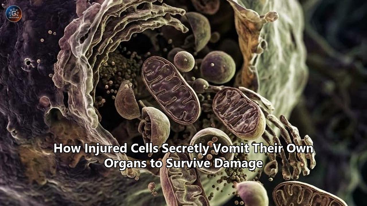
A team of gastrointestinal pathologists and cell biologists at Washington University School of Medicine in St. Louis and Baylor College of Medicine has documented a biological escape hatch used by severely injured cells: when pushed to the brink of death, mature cells can violently purge their internal organelles into the surrounding tissue space to downsize, survive, and rapidly rebuild damaged organs.
The discovery, published in Cell Reports by lead author Dr. Jeffrey W. Brown and senior author Dr. Jason C. Mills, establishes a previously unrecognized cellular purging mechanism termed cathartocytosis—derived from the Greek root for cleansing or violent evacuation.
┌──────────────────────────────────────────────────────────────────────────┐
│                     THE CATHARTOCYTOSIS ESCAPE HATCH                     │
├──────────────────────────────────────────────────────────────────────────┤
│                                                                          │
│   [ Mature, Differentiated Cell ]                                        │
│   • Packed with heavy secretory machinery (ER, mitochondria, granules)   │
│   • Optimized for specialized metabolic work, not rapid division         │
│                                │                                         │
│                      ACUTE TISSUE DAMAGE                                 │
│                                ▼                                         │
│   [ The Emergency Crossroads ]                                           │
│   ├── Slow Autophagy (Lysosomal bottleneck; high ATP demand; 36-72 hrs)  │
│   ├── Programmed Suicide (Apoptosis; permanent loss of tissue mass)      │
│   └── CATHARTOCYTOSIS (Bulk organelle extrusion; 8-24 hrs)               │
│                                │                                         │
│                                ▼                                         │
│   [ Rapid Structural Downsizing ]                                        │
│   • Jettisons ~66% of cytoplasmic volume into luminal/extracellular space│
│   • Bypasses lysosomal enzymatic breakdown limits                        │
│                                │                                         │
│                                ▼                                         │
│   [ Paligenic Reprogramming ] ──────────► [ Proliferative Repair State ] │
│                                           • Divides to replace dead cells│
│                                           • Tradeoff: DAMP/Immune hazard │
└──────────────────────────────────────────────────────────────────────────┘
Using high-resolution electron microscopy, live-cell kinetic tracking, and chemical injury models in mice, the researchers observed differentiated gastric cells dumping up to two-thirds of their physical mass—including endoplasmic reticulum networks, zymogen secretory granules, and mitochondria—directly into the gastric lumen within 8 to 24 hours of injury. By executing this bulk internal ejection, the cells rapidly strip away their energy-intensive specialized machinery, reverting to primitive, stem-like progenitor cells capable of dividing and regenerating the epithelial barrier.
"After an injury, the cell's job is to repair that injury," said Dr. Brown, an assistant professor of medicine in the Division of Gastroenterology at WashU Medicine. "But the cell's mature cellular machinery for doing its normal job gets in the way. So, this cellular cleanse is a quick way of getting rid of that machinery so it can rapidly become a small, primitive cell capable of proliferating and repairing the injury."
The identification of cathartocytosis forces a fundamental re-evaluation of how mammalian tissues handle survival crises. For decades, cell biology taught that cells confront severe stress through a binary choice: quietly recycle their internal parts through lysosomal degradation (autophagy) or dismantle themselves entirely via programmed cell death (apoptosis).
Cathartocytosis introduces an aggressive third path—a high-speed, messy shortcut that prioritizes immediate survival and regeneration over internal metabolic cleanliness. However, this survival strategy carries dangerous biological tradeoffs. The uncontained release of intact organelles and intracellular debris exposes the extracellular environment to damage-associated molecular patterns (DAMPs) and reactive enzymes, fueling chronic inflammation, tissue metaplasia, and potentially driving the early stages of gastric and pancreatic cancers.
The Biomechanical Anatomy of Cellular Purging
To understand how cathartocytosis functions, researchers tracked gastric chief cells—the massive, highly differentiated factories of the stomach lining responsible for synthesizing and secreting digestive enzymes like pepsinogen. In their baseline state, chief cells are biological heavyweights: dense with rough endoplasmic reticulum (ER), packed with large secretory vesicles, and supported by extensive mitochondrial networks designed to sustain continuous protein synthesis.
When the stomach lining experiences acute chemical or toxic trauma—conditions designed to mirror infection by Helicobacter pylori or acute ulceration—these mature cells face an urgent logistical barrier. They cannot divide while weighed down by their complex secretory apparatus, yet waiting for the internal lysosomal system to enzymatically dismantle every individual lipid, protein, and membrane structure takes days.
STAGES OF CATHARTOCYTIC ORGANELLE EJECTION
──────────────────────────────────────────────────────────────────────────
Stage 1 (0 to 4 Hours)   : Cytoskeletal Realignment & Apical Polarization
                            Microtubule and actin networks dissociate from
                            basolateral domains, shifting secretory granules
                            and damaged organelles toward the apical membrane.

Stage 2 (4 to 8 Hours)   : Plasma Membrane Invagination & Pore Dilation
                            Deep invaginations form at the luminal surface;
                            specialized extrusion cavities open directly to
                            the extracellular space.

Stage 3 (8 to 24 Hours)  : Bulk Mass Ejection (Cathartocytosis)
                            Rough ER, mitochondria, and enzyme granules are
                            forcibly jettisoned; cell sheds ~66% of its volume.

Stage 4 (24 to 48 Hours) : Progenitor Transition & Proliferative Expansion
                            The shrunken cell activates embryonic transcriptional
                            programs (Sox9, CD44) to initiate mitosis.
──────────────────────────────────────────────────────────────────────────
Electron microscopy revealed that within hours of injury, chief cells undergo rapid cytoskeletal remodeling. Rather than forming double-membrane autophagosomes to envelope damaged components for internal destruction, the cells drive large swaths of cytoplasm toward the apical plasma membrane facing the stomach cavity. Specialized membrane invaginations open wide pores, allowing the cell to literally vomit its internal components into the luminal void.
By the 24-hour mark, the cells appear visibly altered. Their volume shrinks dramatically, their specialized architecture vanishes, and their cytoplasmic interior is stripped down to bare essentials: a preserved nucleus, basic cytoskeletal scaffolding, and the minimum metabolic baseline needed to support cell division.
This violent evacuation represents the initial, physical phase of paligenosis, a multi-step regenerative program first identified in 2018 by Dr. Mills. In paligenosis, mature, differentiated cells systematically reverse their developmental specialization to act as emergency stem cells, stepping in to restore lost tissue architecture when resident stem cell pools are depleted or overwhelmed.
"Instead of doing this slow process where you have to break down every lipid, sugar, and protein—even organelles—they can just get rid of the stuff," Dr. Brown noted regarding the speed of the evacuation. "And in the stomach, it'll just get absorbed later on."
Comparing Competing Cellular Responses to Damage
The discovery of cathartocytosis highlights an intense evolutionary competition between different cellular survival strategies. When toxic, mechanical, or inflammatory damage strikes, cells deploy distinct molecular pathways to stabilize the tissue. Each strategy exhibits distinct tradeoffs in speed, energy consumption, immunological consequences, and regenerative potential.
┌────────────────────────────────────────────────────────────────────────────────────────────────────────────────────────┐
│                                COMPREHENSIVE CELLULAR CRISIS RESPONSE SPECTRUM                                         │
├───────────────────┬────────────────────────────┬─────────────────────────────┬───────────────────────────┬─────────────┤
│ Response Strategy │ Primary Molecular Drivers  │ Kinetic Speed               │ Energetic Cost            │ Fate        │
├───────────────────┼────────────────────────────┼─────────────────────────────┼───────────────────────────┼─────────────┤
│ Cathartocytosis   │ Cytoskeletal pores,        │ Fast (8–24 hours)           │ Low (Passive bulk         │ Survival &  │
│                   │ Paligenic induction        │                             │ clearance)                │ Division    │
├───────────────────┼────────────────────────────┼─────────────────────────────┼───────────────────────────┼─────────────┤
│ Canonical         │ ATG5, ATG12, LC3-II,       │ Slow (36–72+ hours)         │ High (Enzymatic synthesis │ Survival    │
│ Autophagy         │ Lysosomal hydrolases       │                             │ & acidification)          │ (Stasis)    │
├───────────────────┼────────────────────────────┼─────────────────────────────┼───────────────────────────┼─────────────┤
│ Exophergenesis    │ Tubulin-dependent buds,    │ Intermediate (12–36 hours)  │ Moderate (Targeted        │ Survival    │
│                   │ Phagocytic receptors       │                             │ membrane packaging)       │ (Conserved) │
├───────────────────┼────────────────────────────┼─────────────────────────────┼───────────────────────────┼─────────────┤
│ Apoptosis         │ Caspase-3/9, Cytochrome c, │ Rapid (2–6 hours)           │ Moderate (ATP-dependent   │ Programmed  │
│                   │ BAX/BAK pore formation     │                             │ execution)                │ Death       │
├───────────────────┼────────────────────────────┼─────────────────────────────┼───────────────────────────┼─────────────┤
│ Cellular          │ p53/p21, p16INK4a,         │ Indefinite / Permanent      │ Chronic High              │ Permanent   │
│ Senescence        │ SASP secretory machinery   │                             │ (Secretory phenotype)     │ Arrest      │
└───────────────────┴────────────────────────────┴─────────────────────────────┴───────────────────────────┴─────────────┘
1. Cathartocytosis vs. Canonical Autophagy
For nearly three decades, macroautophagy (often referred to simply as autophagy) was considered the exclusive pathway by which cells dismantled their own organelles. Governed by conserved autophagy-related (ATG) protein complexes, canonical autophagy encloses cytoplasmic contents in a de novo double membrane (the autophagosome), which then traffics along microtubules to fuse with an acidic lysosome. Within the autolysosome, dozens of acid hydrolases break proteins, nucleic acids, and phospholipids down into basic monomers (amino acids, free fatty acids, nucleotides) for metabolic reuse.
CANONICAL AUTOPHAGY                          CATHARTOCYTOSIS
[ Damaged Organelles / Mass ]                [ Damaged Organelles / Mass ]
             │                                            │
   De Novo Membrane Isolation                   Direct Polarized Transport
   (Requires ATG5-ATG12-LC3)                    (Actin/Microtubule dependent)
             ▼                                            ▼
     [ Autophagosome ]                         Apical Membrane Cavitation
             │                                            │
   Lysosomal Fusion & Acidification                       ▼
   (ATP-dependent V-ATPase pumps)               [ BULK LUMINAL DISCHARGE ]
             ▼                                  (Unencapsulated/Pore Extrusion)
[ Slow Enzymatic Degradation ]                            │
• High energetic/enzymatic demand                         ▼
• 36-72 hours to clear cytoplasm                [ Immediate Size Reduction ]
• Zero extracellular DAMP leakage               • Rapid transition (8-24 hrs)
                                                • DAMP leakage / Immune trigger
While autophagy preserves nutrients and keeps damaged material quarantined, it exhibits severe operational bottlenecks during acute crisis:
- Enzymatic Capacity Limits: Lysosomal degradation is rate-limited by the abundance and turnover rate of specific enzymes (such as cathepsins and lipases). When a large cell must shed 60% to 70% of its volume, the lysosomal system becomes physically overwhelmed.
- Energy Consumption: Operating thousands of lysosomal proton pumps (V-ATPases) to maintain an internal pH between 4.5 and 5.0 consumes vast stores of cellular ATP—energy that a severely damaged cell cannot afford to waste.
- Temporal Lag: Enzymatic recycling of complex membrane systems like the rough ER requires several days. In an open wound or infected tissue, waiting 72 hours to begin tissue regeneration exposes the host to mucosal collapse and fatal sepsis.
Cathartocytosis bypasses this entire enzymatic assembly line. By treating unwanted cytoplasmic components as bulk refuse rather than precious nutrients, the cell exchanges biochemical efficiency for raw speed. In gastrointestinal tissues, this calculation is evolutionary optimized: the expelled proteins and lipids are not entirely lost to the organism; they are emptied into the gut lumen, where luminal digestive enzymes break them down, allowing downstream intestinal enterocytes to reabsorb them.
2. Cathartocytosis vs. Exophergenesis and Migrasomes
Cathartocytosis is not the only mechanism by which cells push internal components across the plasma membrane. Over the last ten years, researchers have identified several specialized vesicle-based extrusion pathways, most notably exophers and migrasomes.
                      ORGANELLE DISPOSAL PATHWAYS
                                   │
         ┌─────────────────────────┴─────────────────────────┐
         ▼                                                   ▼
[ Vesicle-Enclosed Extrusion ]                     [ Bulk Naked Evacuation ]
         │                                                   │
  ┌──────┴───────────────────┐                               ▼
  ▼                          ▼                        CATHARTOCYTOSIS
EXOPHERS                 MIGRASOMES                   • Large membrane cavities
• 1–50 µm diameter       • Tetraspanin-4 enriched     • Unencapsulated organelle
• Membrane-bound         • Migrating cells              purging
• Neurons & heart cells  • Mitochondria disposal      • Rapid cellular downsizing
• Targeted to phagocytes • Retracting fibers • Epithelial reprogramming- Exophers: First discovered in Caenorhabditis elegans neurons by Dr. Monica Driscoll and later documented in mammalian cardiomyocytes, exophers are massive, membrane-bound extracellular vesicles measuring between 1 and 50 micrometers in diameter. Stressed neurons and heart cells load dysfunctional mitochondria, neurotoxic protein aggregates (such as huntingtin or alpha-synuclein), and damaged lysosomes into these giant membrane bubbles, pinching them off intact into the extracellular space.
Unlike cathartocytosis, exophergenesis is a quarantined process. The expelled organelles remain enclosed inside a stable phospholipid bilayer, displaying specific "eat-me" signals (like phosphatidylserine) that recruit resident macrophages via receptors like MERTK. This prevents toxic mitochondrial contents from directly contacting the parenchymal microenvironment.
- Migrasomes and Mitocytosis: Discovered during cell migration studies, migrasomes are specialized vesicular structures that form at the tips of retracting cellular fibers as a cell moves. Through a process called mitocytosis, migrating cells package damaged, low-membrane-potential mitochondria into these tetraspanin-enriched structures, leaving them behind like a biological trail of breadcrumbs to prevent mitochondrial stress from halting cell motility.
Cathartocytosis differs fundamentally from both exophers and migrasomes in scale, geometry, and intent. It does not package discreet packets of waste into membrane-bound vesicles destined for macrophage phagocytosis, nor is it dependent on cell migration. It is a centralized, non-vesicular mass evacuation executed by stationary epithelial cells, utilizing dynamic apical membrane cavities to clear out the bulk of the cytoplasm in one sustained event.
The Molecular Circuitry of Paligenic Transformation
Cathartocytosis is not an isolated malfunction; it is the physical engine driving paligenosis. Research across multiple organ systems—including the stomach, pancreas, liver, and kidneys—demonstrates that paligenosis follows a tightly coordinated, three-stage molecular trajectory that allows fully differentiated, post-mitotic cells to re-enter the cell division cycle.
Understanding this pathway explains how cathartocytic purging serves as an indispensable prerequisite for effective cellular damage repair in tissues where resident stem cell populations have been wiped out by injury.
THE THREE-PHASE ARCHITECTURE OF PALIGENOSIS
══════════════════════════════════════════════════════════════════════════

PHASE 1: AUTOLYTIC & CATHARTOCYTIC CLEARANCE (0 TO 24 HOURS)
──────────────────────────────────────────────────────────────────────────
• Immediate shutoff of mTORC1 (Mechanistic Target of Rapamycin Complex 1)
• Upregulation of DDIT4 / REDD1 stress proteins
• Massive actin/tubulin reorganization driving apical cathartocytosis
• Selective preservation of nuclear envelope and minimal bioenergetic machinery

PHASE 2: STEMNESS & METABOLIC ACTIVATION (24 TO 48 HOURS)
──────────────────────────────────────────────────────────────────────────
• Re-activation of mTORC1 in an amino acid-independent manner
• Transcriptional upregulation of progenitor markers: CD44, Sox9, Mist1 suppression
• High expression of embryonic transcription factor networks
• Srebp1-driven lipid biogenesis to rebuild primitive membranes

PHASE 3: CELL CYCLE ENTRY & REGENERATIVE MITOSIS (48+ HOURS)
──────────────────────────────────────────────────────────────────────────
• Cyclin D1 / CDK4 activation driving transit across the G1/S restriction point
• DNA replication and high-frequency symmetric cell division
• Clonal expansion to resurface damaged tissue bed
• Redifferentiation into mature lineages once tissue integrity is restored
══════════════════════════════════════════════════════════════════════════
The initial trigger for paligenosis involves the rapid, profound suppression of mTORC1 (mechanistic target of rapamycin complex 1), the master nutrient sensor and metabolic engine of the cell. In healthy, fully differentiated cells, high mTORC1 activity drives continuous protein translation, structural maintenance, and specialized secretory output.
Upon acute damage, stress-inducible factors such as DDIT4 (DNA damage-inducible transcript 4) rapidly repress mTORC1. This sudden metabolic shutdown halts normal cellular translation and releases the brake on structural remodeling, clearing the path for the cytoskeletal contractions that execute cathartocytosis.
Once the bulk organelle payload has been expelled and the cell shrinks, Phase 2 begins. The downsized cell reactivates mTORC1 through an alternative, damage-responsive circuit. This second wave of mTORC1 activation does not restore differentiated protein synthesis; instead, it fuels the expression of stem-cell transcription factors, specifically Sox9 and CD44, while simultaneously suppressing lineage-defining transcription factors such as Mist1 (BHLHA15).
With its specialized machinery jettisoned and its genome reprogrammed, the cell enters Phase 3: active, symmetric cell division. The former secretory factory divides repeatedly to generate thousands of new cells, creating a cellular patch that covers the injured basement membrane. Once tissue architecture is stabilized and inflammatory cues subside, these newly generated cells receive spatial and biochemical signals that prompt them to redifferentiate into functional, mature tissue lineages.
The Danger Zone: DAMPs, Inflammation, and Metaplasia
While cathartocytosis offers an ultra-fast path to survival for an injured cell, its biological consequences for the surrounding tissue microenvironment are fraught with hazard. When an intact cell ejects un-degraded organelles, enzymes, and nucleic acids into the extracellular matrix or mucosal lumen, it unleashes a potent cocktail of proinflammatory molecules.
                     THE CATHARTOCYTIC TOXICITY CASCADE
                                      │
                         Cathartocytic Bulk Purge
                                      │
              ┌───────────────────────┼───────────────────────┐
              ▼                       ▼                       ▼
    [ Mitochondrial DNA ]     [ Lysosomal Enzymes ]   [ Zymogen Proteases ]
    (Unmethylated CpG motifs) (Cathepsins B, D, L)    (Pepsinogens, Hydrolases)
              │                       │                       │
              ▼                       ▼                       ▼
     cGAS-STING Activation     Extracellular Matrix     Direct Epithelial
     & TLR9 Recognition        Degradation & Necrosis   Barrier Erosion
              │                       │                       │
              └───────────────────────┼───────────────────────┘
                                      ▼
                        Severe Sterile Inflammation
                                      ▼
                     Chronic Macrophage/Neutrophil Influx
                                      │
                   ┌──────────────────┴──────────────────┐
                   ▼                                     ▼
      [ SPEM Metaplastic Shift ]              [ Oncogenic Transformation ]
      (Spasmolytic Polypeptide Metaplasia)    (Gastric/Pancreatic Adenocarcinoma)
The Immunological Shockwave
Mitochondria are evolutionary descendants of ancient alphaproteobacteria that entered eukaryotic cells via endosymbiosis billions of years ago. As a result, mitochondrial components look profoundly alien to the mammalian immune system:
- Mitochondrial DNA (mtDNA): Rich in unmethylated CpG dinucleotide motifs, extracellular mtDNA binds directly to Toll-Like Receptor 9 (TLR9) on resident macrophages and dendritic cells, triggering massive transcription of pro-inflammatory cytokines such as IL-1β, TNF-α, and IL-6. If mtDNA leaks into the cytosol of neighboring cells, it triggers the cGAS-STING pathway, driving a potent type I interferon response.
- Formyl Peptides: Mitochondrial proteins retain N-formylated methionine residues at their amino termini, which act as high-affinity chemoattractants for human neutrophils via formyl peptide receptors (FPR1).
- Cardiolipin: This unique phospholipid, normally sequestered exclusively within the inner mitochondrial membrane, triggers the NLRP3 inflammasome when exposed extracellularly.
In addition to mitochondrial DAMPs, cathartocytosis in gastrointestinal tissues ejects active or semi-active zymogens and acidic lysosomal proteases (such as Cathepsins B, D, and L). Outside the controlled environment of the intracellular vesicle, these enzymes degrade extracellular matrix proteins, cleave cell-adhesion molecules like E-cadherin on adjacent surviving cells, and cause localized tissue collateral damage.
The Link to Cancer and Metaplasia
If an injury is acute and isolated—such as a single toxic insult—the rapid regenerative benefits of cathartocytosis outweigh the temporary inflammatory flare. The extruded debris is washed downstream or cleared by infiltrating phagocytes, the surviving cells divide, and normal tissue architecture is restored.
However, when damage becomes chronic—such as lifelong Helicobacter pylori infection, chronic alcohol consumption, autoimmune gastritis, or chronic pancreatitis—cathartocytosis runs continuously.
"That expedience isn't free, though," noted senior author Dr. Jason C. Mills, professor of medicine and pathology at Baylor College of Medicine. "Releasing all that waste so quickly could cause new problems, like chronic inflammation and increased cancer risk."
Under sustained inflammatory barrage, cells undergoing repeated rounds of cathartocytosis and paligenosis become locked in their primitive, dedifferentiated state. In the stomach, this persistent regenerative phenotype is known clinically as Spasmolytic Polypeptide-Expressing Metaplasia (SPEM). SPEM cells divide continuously in an environment saturated with inflammatory reactive oxygen species (ROS) produced by recruited neutrophils and macrophages.
This chronic collision of high proliferative drive, damaged DNA checkpoints, and extracellular oxidative stress creates an ideal incubator for oncogenic mutations. Clinical pathologists have established that SPEM is the primary pre-neoplastic lesion that progresses toward intestinal-type gastric adenocarcinoma—the fourth leading cause of cancer death worldwide.
Cross-Tissue Manifestations: A Universal Biological Imperative?
Although the Washington University and Baylor study focused on gastric chief cells, comparative analysis suggests that organelle purging and paligenic dedifferentiation operate across a broad spectrum of human tissues. Whenever large, metabolically specialized cells face sudden physical or chemical trauma, the imperative to downsize overrides internal degradation pathways.
┌────────────────────────────────────────────────────────────────────────────────────────────────────────┐
│                          ORGAN-SPECIFIC CELLULAR PURGING & PLASTICITY                                  │
├──────────────────┬───────────────────────┬────────────────────────────────┬────────────────────────────┤
│ Organ System     │ Specialized Cell Type │ Primary Extrusion Mechanism    │ Pathological Risk          │
├──────────────────┼───────────────────────┼────────────────────────────────┼────────────────────────────┤
│ Stomach          │ Gastric Chief Cells   │ Luminal Cathartocytosis        │ Atrophic Gastritis, SPEM,  │
│                  │                       │ (Unencapsulated organelle bulk)│ Gastric Adenocarcinoma     │
├──────────────────┼───────────────────────┼────────────────────────────────┼────────────────────────────┤
│ Pancreas         │ Pancreatic Acinar     │ Paligenic Degranulation        │ Chronic Pancreatitis,      │
│                  │ Cells                 │ & Autophagic Extrusion         │ Pancreatic Ductal (PDAC)   │
├──────────────────┼───────────────────────┼────────────────────────────────┼────────────────────────────┤
│ Liver            │ Mature Hepatocytes    │ Mitolysosome Exocytosis /      │ Liver Cirrhosis,           │
│                  │                       │ Ectosome Secretion             │ Hepatocellular Carcinoma   │
├──────────────────┼───────────────────────┼────────────────────────────────┼────────────────────────────┤
│ Heart / Brain    │ Cardiomyocytes /      │ Exophergenesis                 │ Fibrosis, Toxic Aggregate  │
│                  │ Cortical Neurons      │ (Membrane-bound exophers)      │ Spreading (PD, AD)         │
├──────────────────┼───────────────────────┼────────────────────────────────┼────────────────────────────┤
│ Kidney           │ Proximal Tubular      │ Extracellular Vesicle Shedding │ Acute Kidney Injury (AKI)  │
│                  │ Epithelial Cells      │ & Apical Blebbing              │ to CKD Transition          │
└──────────────────┴───────────────────────┴────────────────────────────────┴────────────────────────────┘
The Pancreas: Acinar-to-Ductal Metaplasia (ADM)
Pancreatic acinar cells, which synthesize and secrete massive quantities of digestive enzymes (amylase, trypsinogen, lipase), share striking structural and functional similarities with gastric chief cells. When exposed to chemical injury, ductal obstruction, or mutant KRAS oncogene expression, acinar cells shed their zymogen granules and dissolve their extensive rough ER networks.
This process, termed Acinar-to-Ductal Metaplasia (ADM), mirrors the paligenic sequence: the mature acinar cells downsize, downregulate Mist1, upregulate Sox9 and KRT19, and transform into duct-like progenitor cells that proliferate to patch the damaged lobule. If this regenerative circuit remains activated under persistent inflammation, ADM acts as the direct precursor to Pancreatic Ductal Adenocarcinoma (PDAC).
The Liver: Hepatocyte Plasticity
Mature hepatocytes are dense with metabolic machinery: extensive smooth ER for drug detoxification, peroxisomes for fatty acid oxidation, and dense arrays of mitochondria. During acute toxic insults (such as acetaminophen poisoning or ischemia-reperfusion injury), damaged hepatocytes deploy a combination of secretory autophagy and mitolysosome exocytosis to rapidly evacuate dysfunctional mitochondrial networks. This rapid mass unloading allows surviving hepatocytes to re-enter the cell cycle, driving the liver's capacity for rapid tissue mass restoration.
The Heart and Central Nervous System: Exopher Containment
In tissues where cells cannot readily divide—such as adult cardiac myocytes and cortical neurons—cellular purging takes a more conservative, quarantined form. Because heart muscle cells and neurons cannot simply undergo paligenesis and proliferate to regenerate lost tissue, they do not execute cathartocytosis.
Instead, they rely on exophergenesis, packing damaged mitochondria and neurotoxic aggregate proteins into sealed, membrane-bound exophers that are systematically transferred to adjacent glia or cardiac-resident macrophages. This prevents catastrophic loss of baseline cell volume while still excising toxic components that would otherwise trigger apoptotic cell suicide.
Methodological Breakthroughs: How the Purge Was Captured
For years, cell biologists routinely observed fragments of organelles, membranous debris, and digestive enzymes floating outside damaged cells in tissue culture and animal injury models. However, these observations were almost universally dismissed as artifactual—interpreted as the messy leftovers of necrotic cell lysis or accidental sample tearing during tissue fixation and histological sectioning.
TRADITIONAL INTERPRETATION               CATHARTOCYTOSIS DISCOVERY
┌───────────────────────────────┐       ┌───────────────────────────────┐
│ Extracellular organelle mass  │       │ Synchronized in vivo damage   │
│ observed in tissue histology  │       │ tracking (0 to 48 hours)      │
└──────────────┬────────────────┘       └──────────────┬────────────────┘
               ▼                                       ▼
┌───────────────────────────────┐       ┌───────────────────────────────┐
│ Dismissed as artifact:        │       │ Confirmed as ordered biology: │
│ • Necrotic lysis debris       │       │ • Preserved viable nuclei     │
│ • Mechanical section tearing  │       │ • Structured membrane pores   │
│ • Sample preparation errors   │       │ • Dynamic paligenic survival  │
└───────────────────────────────┘       └───────────────────────────────┘
The breakthrough by the Washington University and Baylor team hinged on three key methodological innovations:
- Synchronized In Vivo Injury Models: Rather than studying heterogeneous cell cultures, the investigators utilized a specialized mouse model using high-dose tamoxifen administration. In the murine gastric mucosa, this treatment induces synchronized, simultaneous injury across 100% of gastric chief cells. By synchronizing the damage response, the researchers converted what looked like occasional background noise into an undeniable biological event: every single chief cell was expelling its interior simultaneously.
- Time-Resolved Kinetic Electron Microscopy: The team tracked cell morphology at tight intervals (2, 4, 8, 12, 24, and 48 hours post-injury). Transmission electron microscopy (TEM) and serial block-face scanning electron microscopy (SBF-SEM) demonstrated that the cells spitting out their organelles were not dead or dying. Their plasma membranes remained intact except at controlled apical pore sites; their nuclear membranes were undamaged; their chromatin remained unfragmented (ruling out apoptosis); and their metabolic activity persisted.
- Subcellular Fluorescent Fate Mapping: By engineering transgenic mice expressing fluorescent markers targeted to distinct organelle compartments (e.g., mCherry targeted to the rough ER, GFP targeted to mitochondria, and tagged zymogen markers), the researchers visually confirmed the directional translocation of these organelles. Over a 24-hour window, the fluorescent signals migrated from the basolateral compartment to the apical pole, accumulated in extrusion cavities, and were discarded into the stomach lumen—while the parent cells remained viable and took up EdU (a thymidine analog), confirming active DNA synthesis and entry into the cell cycle.
"It's really giving us a better understanding of how gastric chief cells can reprogram themselves so quickly, and it's through a process that we didn't know existed before," said Dr. Richard DiPaolo, an immunologist and expert in gastrointestinal pathology at Saint Louis University who was not involved in the study.
Therapeutic Implications: Exploiting and Taming the Purge
The realization that cells can jettison their internal machinery to survive opens entirely new strategies for therapeutic development across regenerative medicine, infectious disease, and oncology.
┌────────────────────────────────────────────────────────────────────────────────────────────────────────┐
│                                   THERAPEUTIC INTERVENTION STRATEGIES                                  │
├────────────────────────────────┬───────────────────────────────────┬───────────────────────────────────┤
│ Clinical Objective             │ Molecular Strategy                │ Target Indications                │
├────────────────────────────────┼───────────────────────────────────┼───────────────────────────────────┤
│ Accelerate Acute Tissue Repair │ • Paligenosis Agonists            │ • Severe Gastric Ulcers           │
│                                │ • DDIT4 / Srebp1 Inducers         │ • Acute Radiation Sickness        │
│                                │ • Controlled mTORC1 cycling       │ • Ischemic Bowel Injury           │
├────────────────────────────────┼───────────────────────────────────┼───────────────────────────────────┤
│ Neutralize Extracellular       │ • DAMP Scavengers                 │ • Sepsis & Systemic Toxicity      │
│ Inflammatory Fallout           │ • Topical DNase I (mtDNA targets) │ • Severe Pancreatitis             │
│                                │ • TLR9 / FPR1 Receptor Antagonists│ • Inflammatory Bowel Disease      │
├────────────────────────────────┼───────────────────────────────────┼───────────────────────────────────┤
│ Halt Metaplastic Cancer        │ • Paligenosis Checkpoint Blockers │ • Gastric Cancer Prevention       │
│ Progression                    │ • Mist1 Transcriptional Stabilizers│ • Pancreatic Ductal Cancer       │
│                                │ • Sox9 / CD44 Targeted Inhibitors │ • Chronic *H. pylori* Gastritis   │
└────────────────────────────────┴───────────────────────────────────┴───────────────────────────────────┘
1. Accelerating Tissue Recovery After Acute Trauma
In acute trauma settings—such as massive radiation exposure, severe caustic chemical ingestion, acute mucosal ulceration, or ischemic reperfusion injury—the medical priority is rapid resurfacing of the epithelial barrier to prevent bacterial translocation and fatal sepsis.
By developing small-molecule therapeutics that selectively activate the paligenic switch (such as transient mTORC1 inhibitors paired with DDIT4 activators), clinicians could pharmacologically trigger cathartocytosis across surviving cells at the wound margins. This would accelerate cellular damage repair, driving injured cells into a proliferative state within hours rather than waiting days for slow endogenous repair pathways to take effect.
2. DAMP Neutralization Therapies
While promoting cathartocytosis can save wounded tissues, clinicians must also manage the toxic extracellular fallout. In conditions like severe acute pancreatitis, where acinar cells purge massive quantities of active enzymes and mitochondrial DNA, systemic inflammation can rapidly trigger acute respiratory distress syndrome (ARDS) and multi-organ failure.
Co-administering topical or localized DAMP scavengers—such as:
- Recombinant DNase I to dismantle inflammatory extracellular mtDNA,
- High-affinity TLR9 and FPR1 antagonists to blind patrolling immune cells to extruded debris, and
- Synthetic broad-spectrum protease inhibitors to block leaked cathepsins and zymogens—
could allow patients to benefit from rapid cathartocytic tissue closure while extinguishing the dangerous inflammatory shockwave.
COMBINED REGENERATIVE / DAMP-NEUTRALIZATION REGIMEN
══════════════════════════════════════════════════════════════════════════
[ Acute Mucosal Trauma / Chemical Burn ]
                 │
                 ├───────────────────────────────┐
                 ▼                               ▼
      [ Pro-Paligenic Pulse ]         [ Anti-DAMP Buffer ]
      • Transient mTORC1 inhibition   • Recombinant DNase I infusion
      • Accelerates cathartocytosis   • TLR9/cGAS pathway antagonists
      • Drives instant downsizing     • Broad-spectrum cathepsin block
                 │                               │
                 ▼                               ▼
      [ Rapid Barrier Resurfacing ]   [ Zero Sterile Inflammation ]
                 │                               │
                 └───────────────┬───────────────┘
                                 ▼
         [ Optimal Tissue Healing Without Fibrosis or Cancer ]
══════════════════════════════════════════════════════════════════════════
3. Cancer Interception in Chronic Inflammatory Diseases
In patients suffering from chronic inflammatory conditions like longstanding Helicobacter pylori gastritis, Barrett's esophagus, or chronic ulcerative colitis, the therapeutic objective is precisely the opposite: shut down uncontained paligenesis and cathartocytic purging before it drives malignant transformation.
Researchers are now screening for small molecules that stabilize lineage-specific transcription factors like Mist1, preventing mature cells from loosening their differentiated identity. By blocking the cellular escape hatch in chronically irritated tissues, cells burdened with oncogenic DNA mutations would be forced out of the regenerative loop and directed into canonical apoptosis—eliminating pre-cancerous clones before they establish invasive metaplastic fields.
Comparative Tradeoff Matrix: Structural and Mechanistic Breakdown
To illustrate how cathartocytosis fits into the broader architecture of cellular survival systems, the following matrix compares the mechanistic parameters across all major survival, recycling, and clearance modes.
┌────────────────────────────────────────────────────────────────────────────────────────────────────────────────────────┐
│                               MECHANISTIC BREAKDOWN OF CELLULAR QUALITY CONTROL & SURVIVAL                             │
├───────────────────┬───────────────────┬───────────────────┬───────────────────┬───────────────────┬────────────────────┤
│ Strategy          │ Cargo Destination │ Packaging Style   │ Speed of Process  │ ATP Consumption   │ Local Immune Impact│
├───────────────────┼───────────────────┼───────────────────┼───────────────────┼───────────────────┼────────────────────┤
│ Cathartocytosis   │ Extracellular /   │ Naked /           │ 8 to 24 hours     │ Very Low          │ Severe Sterile     │
│                   │ Mucosal Lumen     │ Open Cavities     │                   │                   │ Inflammation       │
├───────────────────┼───────────────────┼───────────────────┼───────────────────┼───────────────────┼────────────────────┤
│ Macroautophagy    │ Intracellular     │ Double-Membrane   │ 36 to 72+ hours   │ High (V-ATPase &  │ Silent             │
│                   │ (Lysosomes)       │ (Autophagosome)   │                   │ Hydrolase demand) │ (Immunotolerant)   │
├───────────────────┼───────────────────┼───────────────────┼───────────────────┼───────────────────┼────────────────────┤
│ Exophergenesis    │ Extracellular     │ Single Bilayer    │ 12 to 36 hours    │ Moderate          │ Controlled         │
│                   │ (Macrophage Fate) │ Membrane (1-50 µm)│                   │ (Vesicle Budding) │ (MERTK Cleared)    │
├───────────────────┼───────────────────┼───────────────────┼───────────────────┼───────────────────┼────────────────────┤
│ Mitocytosis       │ Extracellular     │ Migrasome Pocket  │ Dynamic           │ Moderate          │ Minimal Localized  │
│                   │ (Migration Trail) │ (Tetraspanin-4+)  │ (Motility-linked) │ (Cytoskeletal)    │ Exposure           │
├───────────────────┼───────────────────┼───────────────────┼───────────────────┼───────────────────┼────────────────────┤
│ Apoptosis         │ Phagocytosed      │ Apoptotic Bodies  │ 2 to 6 hours      │ Moderate          │ Anti-inflammatory  │
│                   │ (Neighbor/Macro)  │ (Enclosed)        │                   │ (Caspase cascade) │ Signaling          │
├───────────────────┼───────────────────┼───────────────────┼───────────────────┼───────────────────┼────────────────────┤
│ Necroptosis /     │ Extracellular     │ Explosive Lysis   │ 1 to 3 hours      │ Very Low          │ Extreme Pro-       │
│ Pyroptosis        │ (Unregulated)     │ (Complete rupture)│                   │ (Rupture phase)   │ inflammatory DAMPs │
└───────────────────┴───────────────────┴───────────────────┴───────────────────┴───────────────────┴────────────────────┘
Future Frontiers in Cellular Waste Dynamics
The confirmation that injured cells can purge their internal organs to survive damage shifts fundamental assumptions across cell biology and medicine. It demonstrates that mature, differentiated cells possess remarkable behavioral plasticity, executing emergency measures that challenge textbook definitions of cellular compartmentalization.
┌──────────────────────────────────────────────────────────────────────────┐
│                 CRITICAL UNRESOLVED QUESTIONS IN THE FIELD               │
├──────────────────────────────────────────────────────────────────────────┤
│                                                                          │
│  1. PORE ARCHITECTURE                                                    │
│     What specific fusion proteins and SNARE complexes assemble the       │
│     apical extrusion cavities that permit organelle ejection without     │
│     triggering full plasma membrane rupture?                             │
│                                                                          │
│  2. NUCLEAR RETENTION DYNAMICS                                           │
│     How does the shrinking cell physically anchor and insulate its       │
│     nucleus to prevent genomic DNA from being swept into the bulk        │
│     cytoplasmic purge?                                                   │
│                                                                          │
│  3. METABOLIC RESET THRESHOLDS                                           │
│     What quantitative ratio of preserved-to-ejected mitochondria         │
│     dictates whether a cell successfully divides or falls into necrotic  │
│     metabolic failure?                                                   │
│                                                                          │
│  4. SOLID PARENCHYMA DYNAMICS                                            │
│     How do cells in enclosed, non-luminal organs (e.g., kidneys, liver, │
│     muscles) execute cathartocytosis without inducing catastrophic local │
│     matrix necrosis?                                                     │
│                                                                          │
└──────────────────────────────────────────────────────────────────────────┘
Several critical questions now lead the research agenda:
- The Molecular Gatekeepers of the Purge: Researchers are actively hunting for the specific SNARE complexes, membrane-tethering proteins, and actin-myosin motor assemblies that physically build the extrusion cavities. Identifying the unique structural proteins that form these temporary purge gates will allow the development of precise pharmacological tools to turn cathartocytosis on or off at will.
- Nuclear Protection Mechanisms: When two-thirds of a cell's volume is forcibly squeezed into the extracellular space, the mechanical forces exerted on the remaining structures are immense. How the cell selectively anchors, cushions, and protects its nuclear envelope, genomic DNA, and primary centrosomes from being caught in the outward flow remains an active area of investigation.
- The Minimal Viable Organelle Set: Understanding how a purging cell calculates its stopping point is critical. What molecular sensor tells the cell that it has discarded enough specialized machinery to re-enter the cell cycle, while ensuring it retains the bare minimum number of functional mitochondria needed to sustain baseline ATP production?
- Parenchymal Adaptation in Closed Tissues: In mucosal linings (such as the stomach and intestines), expelling cytoplasmic waste into an open lumen provides a relatively safe disposal route. Investigating how closed parenchymal tissues—such as the renal cortex, the liver parenchyma, or cardiac tissue—handle cathartocytic events without triggering catastrophic localized necrosis will be vital for translating these findings into broad clinical therapies.
As intravital two-photon microscopy and single-cell spatial multi-omics continue to mature, scientists are now positioned to watch this biological process unfold inside living mammalian tissues in real time. The discovery that injured cells vomit out their insides to survive damage provides far more than a vivid biological curiosity: it delivers a mechanistic blueprint of how life reclaims function from the brink of destruction, illuminating the delicate balance between rapid regeneration and malignant collapse.
Reference:
- https://medicine.washu.edu/news/cells-vomit-waste-to-promote-healing-mouse-study-reveals/
- https://www.the-scientist.com/stomach-cells-vomit-waste-not-digest-it-to-mend-injuries-73569
- https://biology.mit.edu/biologists-discover-a-trigger-for-cell-extrusion/
- https://www.fightaging.org/archives/2024/06/cells-can-eject-damaged-mitochondria/
- https://www.sciencealert.com/injured-cells-can-vomit-waste-to-boost-healing-study-finds
- https://portlandpress.com/biochemsoctrans/article/50/6/1773/232226/Mitolysosome-exocytosis-a-novel-mitochondrial
- https://pmc.ncbi.nlm.nih.gov/articles/PMC4590602/
- https://pmc.ncbi.nlm.nih.gov/articles/PMC8339668/
- https://en.wikipedia.org/wiki/Exopher
- https://pubmed.ncbi.nlm.nih.gov/33016946/
- https://pmc.ncbi.nlm.nih.gov/articles/PMC9705561/
- https://www.barthgrant.com/research/proteostasis-aging-neurons-and-giant-extracellular-vesicles-exophers/
- https://www.sciencedaily.com/releases/2025/08/250830001207.htm
- https://elifesciences.org/articles/96813