A convergence of analytical biochemistry, molecular paleobiology, and physical chemistry has dismantled one of science’s most stubborn geochemical dogmas: the absolute theoretical lifespan of biological proteins.
For three decades, conventional geochemical kinetics held that peptide bonds—the chemical links that hold proteins together—could not physically endure in fossilized bone beyond a few hundred thousand to, at absolute thermodynamic limits in deep permafrost, roughly one million years. Under ambient surface conditions and neutral pH, the uncatalyzed half-life of a peptide bond in water is roughly 400 to 500 years. Beyond that, spontaneous hydrolysis breaks the amino acid chain into disconnected fragments.
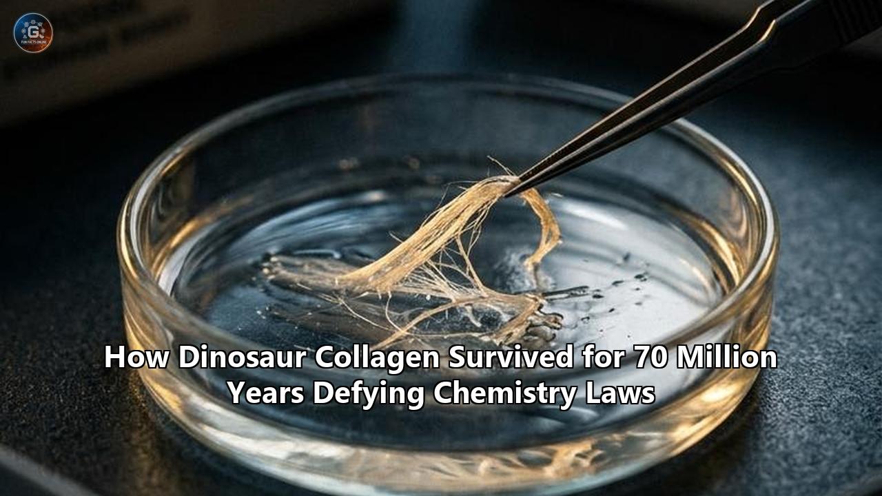
Yet research across multiple independent laboratories has proven that authentic vertebrate collagen can survive inside fossil bone from the Cretaceous and Jurassic periods, spanning 66 million to nearly 200 million years. The latest evidence, published in Analytical Chemistry by a consortium led by the University of Liverpool, confirmed the presence of endogenous collagen alpha-1 peptide fragments and quantified the diagnostic collagen amino acid hydroxyproline inside an exceptionally preserved 22-kilogram Edmontosaurus sacrum excavated from the Hell Creek Formation of South Dakota.
This empirical confirmation comes on the heels of a physical chemistry discovery published in ACS Central Science by researchers at the Massachusetts Institute of Technology. Led by Ronald Raines, the Firmenich Professor of Chemistry at MIT, the team identified a quantum-level stereoelectronic shield—an $n\to\pi^$ orbital interaction—that mechanically bars water molecules from attacking collagen’s peptide backbone.
                                COLLAGEN TRIPLE HELIX
                 ┌─────────────────────────────────────────────────┐
                 │  Gly - X - Y  ~  Gly - Pro - Hyp  ~  Gly - X - Y  │
                 └───────────────────────┬─────────────────────────┘
                                         │
        ┌────────────────────────────────┼────────────────────────────────┐
        │                                │                                │
        ▼                                ▼                                ▼
1. QUANTUM SHIELD               2. CHEMICAL TOASTING             3. MINERAL EMBEDDING
  • n → π* orbital overlap         • Maillard crosslinking          • Hydroxyapatite crystal
  • Pauli exclusion barrier        • AGE / ALE polymer crust          entrapment
  • Blocks water attack            • Hydrophobic shell              • Excludes liquid solvent
Combined with iron-mediated chemical fixation, oxidative diagenetic tanning, and mineral crystal entrapment, these findings explain the biophysical mechanisms that allow organic structures to survive deep geological time, rewriting textbooks on taphonomy, paleobiology, and protein degradation.
The Thermodynamic Wall: Why Chemistry Predicted Zero Preservation
To understand the magnitude of these findings, one must examine the kinetic calculations that paleontologists and geochemists have relied upon for half a century.
Proteins are polymers composed of L-amino acids linked sequentially by amide (peptide) bonds:
$$-\mathrm{C}(=\mathrm{O})-\mathrm{NH}-$$
In the presence of water, peptide bonds are thermodynamically unstable. The reaction:
$$\mathrm{Peptide} + \mathrm{H}_2\mathrm{O} \longrightarrow \mathrm{Amine} + \mathrm{Carboxylic\ Acid}$$
is exergonic, exhibiting a standard Gibbs free energy change ($\Delta G^\circ$) of approximately $-10\text{ to }-15\text{ kJ/mol}$ at physiological pH. Proteins do not fall apart instantaneously in living organisms only because the activation energy ($\Delta G^\ddagger$) for the uncatalyzed hydrolytic cleavage of an unactivated amide bond is extraordinarily high—approximately $85\text{ to }100\text{ kJ/mol}$.
Hydrolysis Reaction:

      O                            O
      ║                            ║
── R₁─C ── NH ─ R₂──  +  H₂O  ──► ── R₁─C ─ OH  +  H₂N ─ R₂──
   (Peptide Bond)                 (Free Carboxyl)  (Free Amine)
In 2000, paleochemist Matthew Collins and colleagues published landmark degradation models calculating that even in optimal, sterile, bone-buffered geochemical matrices at neutral temperatures (10°C to 15°C), the continuous cleavage of peptide bonds would reduce all sequenceable vertebrate proteins to single free amino acids within 1 million years. At higher ambient temperatures (20°C to 25°C), typical of the subtropical climates during the Mesozoic Era, that timeline drops below 100,000 years.
Beyond hydrolysis, proteins in post-mortem biological tissue face several destructive processes:
- Microbial and autolytic enzymatic degradation: Endogenous proteases and soil bacteria rapidly digest soft tissue within weeks to months of burial.
- Spontaneous deamidation: Asparagine and glutamine residues lose their amide groups, converting into aspartate and glutamate through a cyclic succinimide intermediate, destabilizing tertiary structures.
- Amino acid racemization: L-amino acids invert across their $\alpha$-carbon stereocenters to form D-enantiomers over tens of thousands of years, scrambling the structural templates necessary for protein maintenance.
- Geochemical leaching: Underground hydrologic flows continuously flush porous sedimentary rock, carrying away soluble organic fractions.
Because of these kinetic constraints, reports of surviving Mesozoic proteins were long dismissed by mainstream geochemistry as artifacts, fungal byproducts, modern laboratory contamination, or microbial biofilms.
The physical reality of dinosaur collagen survival across 70 million years required an uncharacterized chemical mechanism—a physical structural defense capable of defying standard Arrhenius decay rates.
The Quantum Shield: How $n\to\pi^$ Interactions Block Water Attack
The structural explanation for how collagen evades standard kinetic decay was resolved at the sub-atomic level by Ron Raines, Jinyi Yang, Volga Kojasoy, and Gerard Porter at MIT.
Collagen is the most abundant structural protein in animals, comprising approximately 30% of total vertebrate protein mass. It is built from repeating tripeptide sequences, classically denoted as:
$$\mathrm{Gly-X-Y}$$
where $\mathrm{X}$ is frequently proline and $\mathrm{Y}$ is frequently $(2S,4R)$-4-hydroxyproline (Hyp). Three parallel polypeptide strands, each wound into a left-handed polyproline II (PPII) helix, wrap around one another about a central common axis to form a right-handed triple-helical tropocollagen superhelix.
       Gly-X-Y REPEATING AMINO ACID POLYPEPTIDE CHAIN
 ──[ Gly ]──────[ Pro (X) ]──────[ Hyp (Y) ]──────[ Gly ]──
      │               │                │              │
      └── H-Bond ─────┴── n → π* ──────┴── H-Bond ────┘
The MIT group demonstrated that collagen’s hyper-stability is governed by a quantum mechanical orbital interaction known as an $n\to\pi^$ interaction. In an ordinary peptide backbone, the carbonyl oxygen atom ($\mathrm{C=O}$) possesses two nonbonding lone pairs of electrons ($n$). In the specific dihedral geometry imposed by collagen's tight triple-helix framework, one of these nonbonding electron pairs overlaps directly with the empty antibonding orbital ($\pi^$) of the carbonyl group of the adjacent peptide bond along the chain.
           PEPTIDE BACKBONE STEREOELECTRONIC SHIELDING

              O : (Nonbonding lone pair, n)
             //
          ── C
              \
               N ── C_alpha
              / \
             H   \       Empty Antibonding Orbital (π*)
                  \      ┌───────┐
                   ── C ═│= O    │
                         └───────┘
                             ▲
                             │ Nucleophilic Attack Blocked
                             │ by Pauli Exclusion
                           H₂O (Water Molecule)
This electron sharing creates an energetic barrier against chemical degradation:
- Orbital Delocalization: The overlap ($n\to\pi^$) delocalizes electron density from the carbonyl oxygen directly into the neighboring $\mathrm{C=O}$ carbon atom.
- Pauli Exclusion Principle in Action: Under quantum mechanics, two identical fermions cannot occupy the same quantum state simultaneously. The overlap fills the antibonding orbital region around the carbonyl carbon, occupying the precise spatial trajectory—known as the Bürgi-Dunitz angle ($\sim 107^\circ$)—that an attacking nucleophile must travel to hit the carbonyl carbon.
- Hydrolytic Impasse: When a water molecule ($\mathrm{H}_2\mathrm{O}$) approaches the peptide bond to initiate hydrolysis, its lone pairs are repelled by the electron density already filling that orbital space. Water cannot access the carbon atom to initiate the cleavage reaction.
To verify this physical barrier, the MIT researchers synthesized two structural mimics of collagen peptides: a trans conformer matching the natural triple helix and a rotated cis conformer where the peptide bond angle was forced out of alignment. Under identical experimental hydrolytic conditions, the trans conformer completely resisted water attack, while the cis conformer, lacking the $n\to\pi^$ overlap, was rapidly hydrolyzed.
Globular proteins such as hemoglobin, albumin, or enzymes feature localized alpha helices and beta sheets connected by flexible, unstructured loop regions. These loops lack continuous orbital overlap, exposing them to hydrolytic cleavage. Once a single loop in a globular protein is cut, the entire structure unravels.
Collagen is unique: it is composed entirely of continuous, unbroken triple helices from end to end. Because every single peptide bond in the sequence participates in this stereoelectronic electron-sharing network, the molecular rope features no unstructured weak links where hydrolysis can gain an initial foothold.
The Chemical Matrix: Iron-Mediated Crosslinking and Fenton Chemistry
While quantum-level orbital shielding explains why an isolated collagen triple helix resists water, it does not explain how biological tissue survives the destructive geochemical environment of an underground burial site. The broader puzzle of dinosaur collagen survival requires an understanding of early-stage chemical fixatives that stabilize tissue before mineralization occurs.
       FENTON-MEDIATED AUTO-FIXATION PROCESS

 Post-Mortem Cell Lysis ──► Hemoglobin Release
                                   │
                                   ▼
                             Fe²⁺ / Fe³⁺
                         (Free Iron Ions)
                                   │
                                   ▼  (Reaction with O₂ / H₂O₂)
                         FENTON REACTION
                                   │
                                   ▼
                         •OH (Hydroxyl Radicals)
                                   │
                                   ▼
                 Oxidative Protein Crosslinking
                                   │
                                   ▼
              INSOLUBLE, ENZYME-RESISTANT COLLAGEN MATRIX
Mary Schweitzer, a paleobiologist at North Carolina State University who pioneered the recovery of soft tissues from Mesozoic specimens, formulated the iron-mediated radical crosslinking hypothesis.
When a large animal dies, its vascular system breaks down, hemolyzing red blood cells and releasing hemoglobin into the surrounding interstitial tissue. Hemoglobin contains coordinated iron ($\mathrm{Fe}^{2+}$). In an oxygenated sediment environment, this free iron reacts with environmental oxygen and hydrogen peroxide via the Fenton reaction:
$$\mathrm{Fe}^{2+} + \mathrm{H}_2\mathrm{O}_2 \longrightarrow \mathrm{Fe}^{3+} + \cdot\mathrm{OH} + \mathrm{OH}^-$$
$$\mathrm{Fe}^{3+} + \mathrm{H}_2\mathrm{O}_2 \longrightarrow \mathrm{Fe}^{2+} + \cdot\mathrm{OOH} + \mathrm{H}^+$$
The resulting hydroxyl ($\cdot\mathrm{OH}$) and hydroperoxyl ($\cdot\mathrm{OOH}$) radicals are reactive oxidizing agents. Instead of destroying the structural matrix, they rapidly oxidize adjacent amino acid side-chains, forming covalent carbon-carbon and carbon-nitrogen crosslinks between adjacent collagen fibrils:
- Dityrosine crosslinks: Hydroxyl radicals abstract hydrogen from tyrosine residues, allowing the aromatic rings of neighboring chains to couple covalently into stable dityrosine bridges.
- Aldehyde formation: Lysine and hydroxylysine side-chains are oxidized into reactive aldehydes (allysine), which condense with nearby free amines to form irreversible crosslinks.
This iron-induced radical cascade works like tissue fixation with formaldehyde or glutaraldehyde. In experimental tests, Schweitzer's laboratory immersed modern ostrich blood vessels in concentrated hemoglobin solutions. While control vessels kept in water decayed into liquid sludge within three days, vessels treated with iron-rich hemoglobin remained structurally intact, flexible, and pliable at room temperature for more than two years.
Inside dinosaur cortical bones, micro-computed tomography and elemental mapping show high concentrations of iron nanoparticles (frequently in the form of goethite, $\alpha$-$\mathrm{FeO(OH)}$, or hematite, $\alpha$-$\mathrm{Fe}_2\mathrm{O}_3$) encrusting the vascular channels and osteocyte lacunae. This mineralized iron shell forms an antimicrobial and chemically inert barrier around soft tissue microstructures.
"Toasting" Deep Time: Advanced Glycation End-Products (AGEs)
A complementary chemical mechanism explaining non-mineralized tissue preservation was identified by Jasmina Wiemann (formerly at Yale University, now at Caltech) and Derek Briggs. Their work revealed that original Mesozoic soft tissues are preserved not as pristine, delicate peptides, but as chemically altered, hydrophobic polymers known as Advanced Glycation End-products (AGEs) and Advanced Lipoxidation End-products (ALEs).
DIAGENETIC "TOASTING" OF COLLAGEN FIBRILS

         Amine Groups (Lys/Arg)  +  Sugars/Lipid Aldehydes
                               │
                               ▼
                        Schiff Base
                               │
                               ▼
                       Amadori Product
                               │
                               ▼ (Oxidative Diagenesis)
                     AGE / ALE POLYMERS
            • N-, O-, and S-rich Heterocycles
            • Completely Insoluble in Water
            • Impervious to Microbial Hydrolases
            • Brownish, Chemically Inactive Crust
This transformation is driven by the Maillard reaction—the same fundamental chemical cascade that causes the browning of toasted bread or grilled meat.
During early diagenesis in oxidative sediment conditions, reactive carbonyl groups from decaying lipids and cellular carbohydrates condense with the unprotonated nucleophilic $\epsilon$-amino groups of basic amino acids, primarily lysine and arginine, within the collagen scaffold:
$$\mathrm{R-NH}_2 + \mathrm{R'-C(=O)H} \rightleftharpoons \mathrm{R-N=CH-R'} + \mathrm{H}_2\mathrm{O}\quad \text{(Schiff Base)}$$
The unstable Schiff base rearranges spontaneously into a more stable ketoamine known as an Amadori product. Over geological time, these Amadori intermediates undergo oxidative crosslinking, dehydration, and condensation reactions, forming complex, highly crosslinked $N$-, $O$-, and $S$-rich heterocyclic polymers.
Wiemann and her colleagues employed high-resolution in situ Raman microspectroscopy to analyze fossilized bones, blood vessels, and osteocytes across a wide range of fossil taxa. The spectra yielded distinct Raman signatures corresponding to:
- Cis- and trans-amides (retained from the original peptide architecture)
- Thioethers, ethers, and carbonyl bonds
- Alkyl-pyridine, pyrrole, and imidazole heterocyclic rings
These crosslinked AGE/ALE matrices are:
- Water-insoluble: They eliminate the solvent interactions required for hydrolysis to occur.
- Sterically unrecognizable to enzymes: Microbial collagenases and proteases cannot dock with the altered, crosslinked amino acid configurations.
- Chemically inert: The reactive terminal groups are locked into aromatic and heterocyclic rings, stabilizing the organic structure for hundreds of millions of years.
                     PRIMARY CHEMICAL PRESERVATION MECHANISMS
┌───────────────────────────┬──────────────────────────────────┬─────────────────────────────────────┐
│ Mechanism                 │ Principal Researchers / Group    │ Biophysical Function                │
├───────────────────────────┼──────────────────────────────────┼─────────────────────────────────────┤
│ n → π* Orbital Overlap    │ R. Raines, J. Yang (MIT)         │ Pauli exclusion prevents water from │
│                           │                                  │ attacking peptide carbonyls         │
│ Iron-Fenton Crosslinking  │ M. Schweitzer (NC State)         │ Radical-induced covalent bonding     │
│                           │                                  │ fixes vascular structures          │
│ AGE / ALE Polymerization  │ J. Wiemann, D. Briggs (Yale)     │ Maillard-type "toasting" creates     │
│                           │                                  │ an insoluble heterocyclic shield   │
│ Bioapatite Entrapment     │ J. Orgel (IIT), J. San Antonio   │ Nanoscale mineral packing limits    │
│                           │                                  │ molecular mobility and solvent access│
└───────────────────────────┴──────────────────────────────────┴─────────────────────────────────────┘
Nanoscale Armor: The Supramolecular Crystal Template
The survival of collagen cannot be separated from the nanoscale mineral scaffolding that encases it: bone bioapatite (carbonated calcium hydroxyapatite, $\mathrm{Ca}_{10}(\mathrm{PO}_4)_{6-x}(\mathrm{CO}_3)_x(\mathrm{OH})_{2}$).
Bone is an anisotropic biocomposite. Tropocollagen triple helices do not float freely; they are organized into higher-order cylindrical fibrils roughly 100 nm in diameter. Within each fibril, individual tropocollagen molecules are staggered laterally by a distance of approximately 67 nm (the $D$-periodic spacing), creating alternating "gap" and "overlap" zones along the longitudinal axis.
                  COLLAGEN FIBRIL D-PERIODIC PACKING (67 nm)
 ═════════════════════════════════════════════════════════════════════════
 ─── Tropocollagen 1 ───►          ─── Tropocollagen 2 ───►
 ═════════════════════════════════════════════════════════════════════════
            │  GAP ZONE  │ OVERLAP ZONE │
            │  [Mineral] │              │
            └────────────┴──────────────┘
            Bioapatite nanocrystals nucleate inside
            gap zones, locking the triple helices.
Joseph Orgel of the Illinois Institute of Technology and James San Antonio mapped surviving Mesozoic peptide sequences directly onto high-resolution 3D models of mammalian and avian collagen fibrils. Their structural mapping revealed two architectural principles:
          CROSS-SECTION OF A PACKED COLLAGEN MICROFIBRIL
                     ┌───────────────────┐
                     │   OUTER LAYER     │
                     │  (Degraded by     │
                     │   Environment)    │
                     │   ┌───────────┐   │
                     │   │   CORE    │   │
                     │   │ PROTECTED │   │
                     │   │ PEPTIDES  │   │
                     │   │ SURVIVED  │   │
                     │   └───────────┘   │
                     │  Acidic Residues  │
└───────────────────┘- Preferential Survival in Protected Cores: The eleven specific peptide sequences recovered from Tyrannosaurus rex and Brachylophosaurus canadensis fossils mapped exclusively to the interior core of the microfibril. Exposed surface residues on the exterior of the fibril were degraded, while the sterically compressed inner bundles remained protected.
- Exclusion of Acidic Residues: The surviving peptide regions contained fewer acidic amino acids (aspartate and glutamate) than the overall average for collagen. Acidic residues carry negative charges that attract polar water molecules and catalyze self-hydrolysis; their absence makes the interior core hydrophobic, repelling water and preserving the peptide bonds.
- Epitaxial Mineral Entombment: During bone formation, bioapatite crystal platelets nucleate specifically within the gap zones between tropocollagen ends. As bone mineralizes, these flat, nanocrystalline apatite sheets wrap around the collagen strands, forming a tight mineral envelope. This epitaxial encapsulation limits the conformational mobility of the protein backbone—preventing it from uncoiling into the extended, flexible states required for hydrolytic cleavage.
Under dry diagenetic conditions, this mineral-protein interface functions as a dehydration vault, locking water out of the central organic fibril core for tens of millions of years.
Thirty Years of Controversy: From Scepticism to High-Resolution Tandem Mass Spectrometry
The realization that Mesozoic proteins can endure into deep time is the culmination of three decades of debate in paleontology.
                           PALEOPROTEOMICS TIMELINE

 1990s                      2005-2007                   2018-2024                 2025-2026
 ┌─────────────────────────┐ ┌─────────────────────────┐ ┌───────────────────────┐ ┌────────────────────────┐
 │ First Soft Tissue Claims│ │ Schweitzer Identifies   │ │ Quantum & AGE Chemical│ │ Definitive Multi-Lab   │
 │ • Initial reports of    │ │ T. rex Soft Tissue      │ │ Models Confirmed      │ │ Consensus Established  │
 │   dinosaur bone organics│ │ • Demineralized vessels │ │ • MIT discovers n→π*  │ │ • Edmontosaurus sacrum │
 │   dismissed as modern   │ │   and osteocytes        │ │   orbital shield      │ │   hydroxyproline &     │
 │   contamination.        │ │ • T. rex collagen alpha │ │ • Raman AGE/ALE tests │ │   alpha-1 sequences    │
 │                         │ │   sequenced by MS.      │ │   clarify diagenesis. │ │   verified by MS.      │
 └─────────────────────────┘ └─────────────────────────┘ └───────────────────────┘ └────────────────────────┘
The dispute began in earnest in 2005 when Mary Schweitzer’s team published findings in Science detailing the extraction of flexible, transparent blood vessels, intact osteocytes with radiating filopodia, and pliable fibrous matrix from the cortical interior of a 68-million-year-old Tyrannosaurus rex femur (specimen MOR 1125) excavated from the Hell Creek Formation of Montana. In 2007, John Asara of Harvard Medical School used liquid chromatography-tandem mass spectrometry (LC-MS/MS) to sequence seven peptide fragments from the MOR 1125 extract, matching them to collagen alpha-1 (I) sequences of modern birds.
The discoveries faced intense pushback. Critics argued that:
- The pliable microstructures were recent bacterial biofilms (endolithic "slime") that had colonized the porous cavities of the fossil.
- The mass spectrometry signals were trace contaminants from modern laboratory consumables, bovine serum albumin, or latex gloves.
- Sequence similarities with birds were an artifact of software algorithms matching noisy spectra to common chicken sequences (Gallus gallus).
The paleoproteomics community responded by designing rigorous, multi-method extraction and testing protocols:
- In Situ Immunofluorescence and Antibody Binding: Antibodies raised specifically against modern avian collagen type I bind directly to the internal microstructures of demineralized dinosaur bone slices, displaying clear localization along vascular channels and osteocyte walls, while failing to react with surrounding sedimentary rock.
- Synchrotron X-ray and ATR-FTIR Microspectroscopy: Attenuated Total Reflectance Fourier-Transform Infrared spectroscopy detects the distinct vibrational bands of organic Amide I ($\sim 1650\text{ cm}^{-1}$, $\mathrm{C=O}$ stretch), Amide II ($\sim 1550\text{ cm}^{-1}$, $\mathrm{N-H}$ bend and $\mathrm{C-N}$ stretch), and Amide III ($\sim 1240\text{ cm}^{-1}$) inside untouched fossil cross-sections without prior chemical extraction.
- Detection of Hydroxyproline via Tandem Mass Spectrometry: Hydroxyproline (Hyp) is an amino acid synthesized almost exclusively as a post-translational modification in animal collagen; it is essentially absent from bacterial biofilms, soil fungi, and common laboratory contaminants. High-resolution triple-quadrupole mass spectrometry confirmed the stoichiometric presence of hydroxyproline inside Mesozoic fossil bone digests.
                CONFIRMATORY ANALYTICAL TOOLKIT
┌────────────────────────────────────────┬───────────────────────────────────────────┐
│ Analytical Technique                   │ Diagnostic Signature Verified             │
├────────────────────────────────────────┼───────────────────────────────────────────┤
│ LC-MS/MS & Tandem Mass Spectrometry    │ Exact amino acid sequences, diagnostic    │
│                                        │ hydroxyproline quantification            │
│ In Situ Raman Microspectroscopy        │ Heterocyclic AGE/ALE fingerprints,        │
│                                        │ cis/trans-amide vibrational bands        │
│ Cross-Polarized Light Microscopy (XPol)│ Birefringence of intact, aligned          │
│                                        │ collagen fibril arrays                    │
│ ATR-FTIR Spectroscopy                  │ Amide I, Amide II, and Amide III          │
│                                        │ infrared absorption peaks                 │
│ Transmission Electron Microscopy (TEM) │ 67-nanometer D-periodic structural        │
│                                        │ banding of collagen fibrils               │
└────────────────────────────────────────┴───────────────────────────────────────────┘
The Liverpool study on the Edmontosaurus sacrum verified dinosaur collagen survival by applying this multi-tiered analytical battery across independent laboratories.
Steve Taylor, chair of the Mass Spectrometry Research Group at the University of Liverpool's Department of Electrical Engineering & Electronics, underscored the finality of the data:
"This research shows beyond doubt that organic biomolecules, such as proteins like collagen, appear to be present in some fossils. Our results have far-reaching implications. Firstly, it refutes the hypothesis that any organics found in fossils must result from contamination. Secondly, it suggests that cross-polarized light microscopy images of fossil bones, collected for a century, should be revisited. These images may reveal intact patches of bone collagen, potentially offering a ready-made trove of fossil candidates for further protein analysis."
Mapping the Dinosaur Tree of Life: Molecular Phylogenetics
The survival of sequenceable collagen fragments opens a new window into evolutionary biology. While intact DNA remains constrained by its phosphodiester backbone—which hydrolyzes far faster than peptide bonds, setting a theoretical survival ceiling of roughly 100,000 to 1.5 million years in deep permafrost—collagen can preserve primary amino acid sequence data across tens of millions of years.
                ARCHOSAUR PHYLOGENETIC RECONSTRUCTION
                 (Derived from Collagen α-Chain Sequences)

                      ┌── Modern Birds (e.g., Gallus gallus)
             ┌────────┤
             │        └── Tyrannosaurus rex / Hadrosaurs
      ───────┤
             │
             └── Crocodilians (e.g., Alligator mississippiensis)
In animals, collagen alpha-chain sequences diverge at a predictable baseline rate over evolutionary time, serving as an effective molecular clock. By aligning recovered dinosaur peptides against proteomic libraries of living organisms, researchers can test phylogenetic hypotheses using molecular data rather than relying solely on comparative osteological morphology:
- Avian Sister-Group Confirmation: The initial peptide fragments recovered from Tyrannosaurus rex (including sequences like GVQGPPGPQGPR and GAPGPQGPGGAPGPK) matched modern galliforms (chickens) and ratites (ostriches) more closely than any extant reptilian clade, such as squamates or chelonians. This provided molecular confirmation of the theropod ancestry of birds.
- Hadrosaur Proteomics: Subsequent sequencing of collagen extracts from the hadrosaur Brachylophosaurus canadensis yielded peptides showing clear archosaurian affinities, clustering precisely at the base of the bird-crocodilian split.
- Resolving Disputed Morphological Phylogenies: For taxa where skeletonization is incomplete, highly crushed, or distorted by extreme taphonomic pressures, collagen barcoding via MALDI-TOF (ZooMS—Zooarchaeology by Mass Spectrometry) allows researchers to classify unidentifiable bone fragments, map taxonomic distributions, and trace anatomical transitions across the Mesozoic-Cenozoic boundary.
The Four-Tier Preservation Model
The survival of Mesozoic soft tissue is not the result of a single anomaly; it is the consequence of a four-tier chemical, structural, and mineralogical defense system. When these four barriers operate in concert, organic peptide bonds can remain intact for hundreds of millions of years:
                      THE FOUR-TIER PRESERVATION MODEL

  ┌────────────────────────────────────────────────────────────────────────┐
  │ TIER 1: QUANTUM-LEVEL STEREODEFENSE                                    │
  │ • n → π* orbital delocalization shields every peptide bond             │
  │ • Pauli exclusion blocks nucleophilic attack trajectories of H₂O       │
  └───────────────────────────────────┬────────────────────────────────────┘
                                      │
                                      ▼
  ┌────────────────────────────────────────────────────────────────────────┐
  │ TIER 2: MOLECULAR PACKING & HYDROPHOBICITY                             │
  │ • Dense triple-helix bundles hide nonpolar cores                       │
  │ • Acidic residues are excluded from interior fibril regions            │
  └───────────────────────────────────┬────────────────────────────────────┘
                                      │
                                      ▼
  ┌────────────────────────────────────────────────────────────────────────┐
  │ TIER 3: OXIDATIVE FIXATION & TOASTING                                  │
  │ • Hemoglobin-derived iron radicals catalyze covalent crosslinks        │
  │ • Maillard reactions transform external residues into AGE/ALE crusts    │
  └───────────────────────────────────┬────────────────────────────────────┘
                                      │
                                      ▼
  ┌────────────────────────────────────────────────────────────────────────┐
  │ TIER 4: BIOAPATITE EPITAXIAL ENCAPSULATION                             │
  │ • Nanocrystalline calcium phosphate encloses gap zones                 │
  │ • Rigid mineral housing prevents water ingress and uncoiling           │
└────────────────────────────────────────────────────────────────────────┘- Tier 1 (Sub-Atomic Level): The $n\to\pi^$ stereoelectronic orbital overlap between neighboring carbonyl groups creates an electron-density barrier that prevents incoming water molecules from initiating nucleophilic hydrolysis.
- Tier 2 (Supramolecular Level): The tight, helical packing of tropocollagen bundles sequesters non-polar, hydrophobic peptide sequences inside protected cores while burying self-catalyzing acidic residues.
- Tier 3 (Geochemical/Diagenetic Level): Iron-mediated Fenton reactions and Maillard glycoxidation reactions crosslink the outer surface of the collagen bundle into an insoluble, rot-resistant AGE/ALE polymer crust that microbial enzymes cannot digest.
- Tier 4 (Macro-Mineral Level): Bioapatite nanocrystals precipitate within the fibril gap zones, creating a rigid mineral envelope that eliminates conformational mobility and blocks solvent transport.
Applied Horizons: From Ancient Fossils to Next-Generation Materials
The implications of the chemistry underlying dinosaur collagen survival reach far beyond paleobiology. Deciphering how nature preserves delicate peptide chains against environmental decay is driving new research across multiple scientific disciplines:
1. Ultra-Resistant Synthetic Biomaterials
Current synthetic collagen scaffolds used in human medicine—such as artificial skin grafts, tendon repair matrices, and reconstructive surgery implants—suffer from rapid enzymatic degradation and hydrolytic breakdown inside the body. By leveraging the MIT findings on $n\to\pi^$ stereoelectronic shielding, chemical biologists are designing non-natural collagen mimetics with customized stereocenters (such as fluorinated prolines or synthetic acyl modifications). These engineered polypeptides maintain maximum orbital overlap, resulting in medical implants that resist host collagenase degradation and last years longer in vivo without provoking inflammatory immune responses.
                           APPLIED BIOMATERIAL DESIGN
 Natural Collagen Mimetic                        Engineered Mesozoic-Inspired Mimetic
 ────────────────────────                        ────────────────────────────────────
 • Vulnerable to thermal uncoiling               • Fluorinated Proline stereocenters
 • Susceptible to enzymatic cleavage             • Maximized n → π* orbital overlap
 • Half-life in vivo: weeks/months               • Impervious to hydrolytic breakdown
                                                 • Half-life in vivo: multi-year durability
2. High-Durability Industrial Polymers
Industrial plastics and green biopolymers are prone to hydrolytic breakdown and photo-oxidation. Material scientists are synthesizing peptide-based polymers that mimic the alternating crosslink architecture of Advanced Glycation End-products (AGEs). These bio-derived plastics offer high tensile strength, water-insolubility, and natural flame resistance without relying on petroleum feedstocks or toxic chemical stabilizers.
3. Deep-Time Astrobiological Biosignatures
As interplanetary rovers and sample-return missions analyze rocks on Mars and the icy moons of the outer Solar System, the discovery of peptide survival across deep geological time provides an essential baseline for detecting extraterrestrial life. Rather than searching exclusively for pristine, unstable biomolecules or fragile DNA sequences, instruments such as Raman spectrometers, laser desorption mass spectrometers, and sub-surface organic analyzers are calibrated to detect the durable, heterocyclic, oxidized crosslinked signatures (AGEs/ALEs) produced when complex protein networks fossilize in mineral matrices.
Upcoming Milestones and Unresolved Questions
With the fundamental chemistry of deep-time protein survival established, molecular paleobiology is moving to answer new empirical questions. The immediate research horizon is defined by several milestones:
- Expanding the Mesozoic Sample Array: Using cross-polarized light microscopy (XPol) and non-destructive Raman screening to evaluate thousands of fossil bones stored in museum archives worldwide, identifying candidate fossils with intact collagen birefringence for proteomic sequencing.
- Reconstructing the Jurassic and Triassic Proteome: Applying Orbitrap mass spectrometers to push deep-time protein sequencing past the 66-million-year Cretaceous boundary, targeting Jurassic sauropodomorphs and Triassic archosaurs to map the early evolutionary diversification of dinosaurs.
- Mapping the Limits of Non-Collagenous Proteins: Investigating whether other fibrous and structural proteins—such as beta-keratin in dinosaur feathers, elastin in fossilized vascular walls, or osteocalcin in bone matrix—possess similar stereoelectronic and oxidative mechanisms that protect them from total degradation.
- Standardizing Decontamination Protocols: Establishing universally adopted, multi-blinded analytical standards for ancient mass spectrometry to eliminate remaining skepticism regarding contamination and ensure reproducible peptide recovery across laboratories globally.
The survival of collagen across 70 million years is no longer a chemical impossibility. It is the predictable outcome of physical organic chemistry, structural biology, and geochemistry working in tandem. By uncovering the sub-atomic electron shields, iron-radical fixatives, and mineral envelopes that protected Mesozoic proteins, science has solved a long-standing paleontological mystery—revealing that the molecular building blocks of life are far more durable than previously imagined.
Reference:
- https://www.acs.org/pressroom/presspacs/2024/september/why-dinosaur-collagen-might-have-staying-power.html
- https://news.mit.edu/2024/mit-chemists-explain-why-dinosaur-collagen-survived-millions-years-0904
- https://www.discovermagazine.com/collagen-mysteriously-persists-in-dinosaur-bones-for-tens-of-millions-of-46579
- https://news.liverpool.ac.uk/2025/01/31/discovery-of-collagen-in-fossil-bone-could-unlock-new-insights-into-dinosaurs/
- https://blog.everythingdinosaur.com/blog/_archives/2025/02/04
- https://www.sci.news/paleontology/original-collagen-edmontosaurus-fossil-bone-13631.html
- https://www.earth.com/science/how-dinosaur-collagen-survived-the-test-of-time/
- https://connectsci.au/news/news-parent/1738/How-collagen-survives-for-millions-of-years
- https://www.researchgate.net/publication/368967876_A_chemical_framework_for_the_preservation_of_fossil_vertebrate_cells_and_soft_tissues
- https://dokumen.pub/remnants-of-ancient-life-the-new-science-of-old-fossils-9780691221151.html
- https://www.researchgate.net/publication/328841671_Fossilization_transforms_vertebrate_hard_tissue_proteins_into_N-heterocyclic_polymers
- https://pmc.ncbi.nlm.nih.gov/articles/PMC7439315/
- https://www.iit.edu/news/dinosaur-peptides-suggest-mechanisms-protein-survival
- https://pubmed.ncbi.nlm.nih.gov/21687667/
- https://www.sciencedaily.com/releases/2026/08/260811053042.htm
- https://www.facebook.com/groups/10150112797390640/posts/10168185054645640/