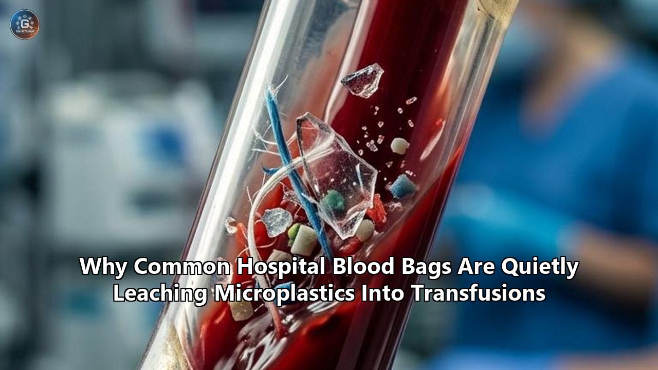
For decades, blood transfusions have stood as one of modern medicine’s most dependable lifelines, saving millions of lives each year during major surgeries, trauma interventions, and cancer therapies. Yet landmark laboratory analyses published in ACS Environment & Health and the British Journal of Haematology have revealed an unsettling hidden dynamic in transfusion medicine: standard medical-grade plastic storage bags actively shed microplastics into donated blood products prior to infusion.
The studies demonstrate that red blood cells and preservative solutions stored in standard polyvinyl chloride (PVC) bags absorb thousands of microscopic polymer fragments over time. During a standard 30-day cold storage cycle, microplastic concentrations inside tested blood containers increase seven- to 20-fold. Advanced Raman spectroscopy and scanning electron microscopy have confirmed that these microplastics do not simply float freely in the preservation fluid; they physically attach to red blood cell membranes, altering cell structure and accelerating red blood cell breakdown before the blood ever reaches a patient.
This physical shedding compounds a long-standing chemical issue in transfusion medicine: the leaching of softeners like di(2-ethylhexyl) phthalate (DEHP). While environmental microplastics are typically ingested or inhaled and partially filtered by the gastrointestinal system or lungs, microplastics from transfusions enter human circulation directly through intravenous lines. This direct entry bypasses the body's protective epithelial barriers, delivering plastic particles directly to vital organs and the vascular system.
Understanding why this occurs requires examining the chemistry of blood preservation, the physics of polymer degradation, and a complex medical trade-off that has challenged healthcare regulators for decades.
                  THE BLOOD BAG TRANSFUSION TIMELINE

  Day 0                 Day 10–20               Day 30–42
  [Donation] ---------> [Storage] ------------> [Infusion]
  • Donor blood         • Plasticizer (DEHP)    • Microplastics surge 7x–20x
    collected into        leaches into cell     • PVC microparticles bind to
    flexible PVC          membranes.              red blood cell walls.
    storage bag.        • Interior bag lining   • Intravenous delivery
                          begins shedding         bypasses gut and lung
                          microplastics (1-100μm).  protective barriers.
The 70-Year Paradox: Why Hospitals Rely on PVC
To understand how microplastics end up in preserved blood, one must look back to the mid-20th century. Prior to the 1950s, blood was collected, stored, and transfused using heavy glass bottles. While chemically inert, glass was brittle, difficult to transport, required complex venting during infusion, and carried a high risk of air embolism and microbial contamination.
The introduction of flexible plastic container systems in the late 1950s transformed transfusion medicine. Polyvinyl chloride emerged as the ideal base polymer because it was inexpensive, durable, transparent, and capable of withstanding both steam sterilization and deep freezing. However, pure PVC is naturally rigid and brittle, akin to a household drainage pipe. To make it soft, flexible, and squeeze-compatible for blood processing and centrifugation, manufacturers must blend raw PVC resin with liquid plasticizers, which make up 30% to 40% of the finished bag's total weight.
For nearly seven decades, the dominant plasticizer of choice has been di(2-ethylhexyl) phthalate. This choice created an unexpected biological compromise.
Phthalate molecules are not chemically bound (covalently bonded) to the PVC polymer backbone; instead, they sit loosely within the matrix of plastic polymer chains like oil trapped inside a sponge. Because DEHP is lipophilic (fat-soluble), it slowly diffuses out of the plastic and dissolves into the lipid-rich membranes of red blood cells.
Intriguingly, hematologists in the 1970s discovered that this chemical leaching served a crucial protective purpose: leached DEHP integrates into red blood cell membranes, increasing their flexibility and preventing them from rupturing during cold storage. Without DEHP leaching, stored red blood cells suffer high rates of hemolysis (cell destruction), releasing free hemoglobin that can damage patient kidneys upon transfusion.
This creates a central paradox in modern blood storage: the very mechanism that keeps stored red blood cells intact relies on a chemical softener leaching out of a degrading plastic container.
                 CHEMICAL VS. PHYSICAL LEACHING IN PVC BAGS

  +-----------------------------------------------------------------------+
  |  Polyvinyl Chloride (PVC) Wall Matrix                                 |
  |                                                                       |
  |   [DEHP Phthalate Molecules] -------> Leaches Chemically into Lipid   |
  |   (Loose chemical softeners)          Membranes of Red Blood Cells    |
  |                                       (Protects cell flexibility)     |
  |                                                                       |
  |   [PVC Polymer Microparticles] ----> Sheds Physically into Fluid      |
  |   (Fragmented wall particles)         Attaches to Red Blood Cells     |
  |                                       (Triggers inflammation & damage)|
  +-----------------------------------------------------------------------+
How Plastic Fragments Shed into Stored Blood
While the chemical leaching of DEHP has been studied for decades, recent research highlights a distinct physical process: the shedding of solid plastic fragments, ranging from nanometers to micrometers in size, directly into stored blood components.
Research tracking storage conditions reveals that microparticle formation occurs through three primary mechanisms:
1. Chemical Surface Degradation
Blood preservation media—such as Citrate-Phosphate-Dextrose-Adenine (CPDA-1) or Mannitol-Adenine-Phosphate (MAP)—are designed to keep red cells metabolically active during cold storage. Over weeks of contact, the chemical interaction between these nutrient solutions, blood lipids, and the inner wall of the PVC container slowly degrades the smooth lining of the plastic. Atomic force microscopy (AFM) of blood bag interiors shows progressive surface roughening over a standard 30- to 42-day shelf life, creating fragile micro-cracks along the container lining.
2. Physical Friction and Mechanical Stress
Blood products do not remain motionless throughout their lifecycle. After donation, blood bags are centrifuged at high speeds to separate whole blood into packed red cells, plasma, and platelets. The bags are subsequently handled, squeezed, stacked, and transported across regional cold-chain distribution networks. Scanning electron microscopy demonstrates that physical agitation dislodges weakened microscopic ridges along the interior plastic surface, shedding PVC particles into the liquid.
3. Temperature Cycles and Polymer Embrittlement
Blood products are held at strict refrigeration temperatures between 1°C and 6°C. Cold temperatures alter polymer flexibility, making microscopic surface features brittle. When bags undergo minor temperature variations during handling, the localized mechanical stress causes microscopic surface shedding.
                FACTORS ACCELERATING MICROPLASTIC SHEDDING

   +-----------------------+     +-----------------------+     +-----------------------+
   |  Chemical Erosion     |     |  Mechanical Stress    |     |  Temperature Shifts   |
   | Preservative solutions| +-> | High-speed centrifuge | +-> | Refrigeration cycle   |
   | (CPDA-1/MAP) degrade  |     | & transport shaking   |     | causes localized      |
   | interior polymer wall |     | dislodge fragments    |     | surface brittleness   |
   +-----------------------+     +-----------------------+     +-----------------------+
                                             |
                                             v
                               +---------------------------+
                               | Up to 20-Fold Increase in |
                               |  Microplastic Count Over  |
                               |      30 Days Storage      |
                               +---------------------------+
Laboratory measurements using Raman micro-spectroscopy reveal that these shed microplastics primarily consist of irregular PVC fragments measuring between 1 and 62 micrometers in diameter. High-resolution imaging captured these PVC microparticles physically adhering to the surface membranes of stored red blood cells.
This binding disrupts normal cell function. As microplastics attach to cell walls, red blood cells experience accelerated loss of adenosine triphosphate (ATP)—the essential energy molecule required to maintain cellular integrity and cell shape. This energy depletion hastens what hematologists refer to as a "storage lesion," causing red blood cells to age prematurely and lose deformability before they are transfused.
The Route of Exposure: Why Intravenous Injection Matters
The medical community evaluates environmental microplastic exposure across three main pathways: ingestion through food and water, inhalation through air, and dermal contact. In the gastrointestinal tract, the gut epithelium acts as a primary physical filter; the majority of ingested plastic particles larger than a few micrometers are excluded from systemic circulation and excreted. Particles that do penetrate the intestinal lining pass through the portal vein into the liver, where hepatic macrophages work to neutralize foreign debris.
Intravenous exposure through blood transfusions bypasses these natural defense systems entirely.
             EXPOSURE ROUTE COMPARISON: GASTROINTESTINAL VS. INTRAVENOUS

   INGESTION ROUTE (Food / Water)
   Microplastics ---> Gut Barrier Filter ---> Liver Metabolism ---> Limited Systemic Circulation
                     (Excretes ~99%)         (Filters Debris)

   INTRAVENOUS ROUTE (Transfusion / IV)
   Microplastics ---> DIRECT SYSTEMIC ENTERING ---> Free Vascular Access to Heart, Lungs, Brain & Kidneys
                     (Bypasses All Barriers)
When microplastics are introduced directly into a blood vessel, they circulate immediately through the arterial and venous systems. Researchers studying vascular plastic exposure have identified several biological concerns:
- Endothelial Disruption and Micro-Clotting: Microplastics circulating in blood vessels interact directly with the vascular endothelium—the single layer of cells lining blood vessels. Studies show that microscopic plastic fragments can trigger local inflammatory signaling, activate blood platelets, and serve as structural anchors for micro-thrombi (tiny blood clots).
- Systemic Distribution and Tissue Accumulation: Because intravenous microplastics bypass primary organ filtration, particles smaller than 10 micrometers can navigate capillary beds and accumulate in organs like the spleen, liver, kidneys, and cardiac tissue. Recent autopsies and clinical sampling have identified microplastics embedded in human vascular plaque and cardiac tissue, with studies in The New England Journal of Medicine linking arterial microplastic accumulation to elevated cardiovascular risks.
- Cellular Toxicity and Oxidative Stress: As shed microplastics circulate, they continuously release lingering surface additives, including residual monomers and phthalates. In cellular cultures, this combination triggers reactive oxygen species (ROS), causing oxidative stress, mitochondrial damage, and systemic inflammatory responses.
For vulnerable patient populations—such as trauma victims receiving massive transfusions, surgical patients requiring multiple units of red blood cells, or neonates in intensive care—cumulative intravenous exposure to microplastics can reach tens of thousands of particles in a single treatment window.
                                ESTIMATED PATIENT EXPOSURE

  +---------------------------------+---------------------------------+
  | Medical Scenario                | Estimated Particle Exposure     |
  +---------------------------------+---------------------------------+
  | Standard Single IV Infusion Bag | ~7,500 microplastic particles   |
  | Abdominal Surgery (Multi-Bag)   | ~52,500 microplastic particles  |
  | Neonatal IV Nutrition (72 hrs)  | ~115 microplastic particles     |
  | Multi-Unit Red Cell Transfusion | Tens of thousands of particles  |
  +---------------------------------+---------------------------------+
The Regulatory Dilemma: The EU Ban and Material Science Challenges
The discovery of microplastic shedding from blood bags comes at a critical time for global healthcare regulators. Regulatory agencies are caught between two competing priorities: phasing out hazardous plasticizers and maintaining a safe, stable global blood supply.
In the European Union, registration under the REACH framework and medical device regulations has mandated a phase-out of traditional DEHP-plasticized medical devices, with a ban covering blood storage bags taking effect by July 1, 2030. The European Chemicals Agency (ECHA) initiated this ban due to DEHP’s classification as a toxic substance that disrupts human endocrine and reproductive systems.
                             THE REGULATORY CATCH-22

      Traditional PVC/DEHP Bags                 Alternative Plasticizers (DINCH/BTHC)
      -------------------------                 -------------------------------------
      • High DEHP chemical toxicity             • Reduced chemical toxicity
      • High physical microplastic shedding     • Higher red blood cell hemolysis
      • Excellent cell membrane stability       • Shorter stable shelf life
Replacing DEHP in blood containers has proven surprisingly difficult:
1. Alternative Plasticizers Lead to Higher Cell Hemolysis
Transfusion manufacturers have evaluated alternative plasticizers, including DINCH (1,2-cyclohexane dicarboxylic acid diisononyl ester), BTHC (n-butyryl-tri-n-hexyl citrate), and TOTM (tri-octyl trimellitate). While these compounds reduce chemical toxicity, many fail to protect red blood cell membranes as effectively as DEHP. In clinical trials, red blood cells stored in BTHC- or TOTM-plasticized containers showed up to two times higher hemolysis rates and increased microvesicle release, reducing cell survival after transfusion.
2. Fluoropolymers and Polyolefins Face Manufacturing Hurdles
Engineers have explored alternative polymers, such as high-density polyethylene (HDPE), polypropylene (PP), and fluorinated ethylene propylene (FEP), which require no phthalate plasticizers. However, pure polyolefins are stiffer than PVC, making them harder to seal, fold, and strip during blood processing. They also exhibit different gas permeability profiles for oxygen and carbon dioxide, which can alter cellular respiration and shorten the maximum storage life of donated blood from 42 days to under 21 days.
As blood banking organizations work to meet the 2030 EU deadline, early testing indicates that changing plastic formulations without altering storage media can solve one problem while creating another. A bag made with alternative materials might reduce chemical phthalate exposure but could still shed physical microplastics or cause red blood cells to break down prematurely.
                     PROS AND CONS OF BLOOD STORAGE MATERIALS

  +-------------------+---------------------------+---------------------------+
  | Material          | Advantages                | Disadvantages             |
  +-------------------+---------------------------+---------------------------+
  | Traditional PVC + | Flexible, durable,        | High chemical toxicity,   |
  | DEHP              | stabilizes cell membranes | sheds PVC microplastics   |
  +-------------------+---------------------------+---------------------------+
  | PVC + Alternative | Lower chemical toxicity   | Higher cell hemolysis,    |
  | (DINCH / BTHC)    |                           | still sheds microplastics |
  +-------------------+---------------------------+---------------------------+
  | Polyolefins       | Phthalate-free,           | Stiffer material, altered |
  | (PE / PP)         | chemically inert          | gas permeability          |
  +-------------------+---------------------------+---------------------------+
Practical Solutions: Ultrafiltration and Next-Generation Design
While material scientists work to develop plasticizer-free polymer blends, medical researchers are advancing immediate strategies to protect blood supplies.
Point-of-Care Ultrafiltration
The most immediate intervention involves integrating specialized ultrafiltration systems into blood administration sets. Standard bedside transfusion filters feature pore sizes between 170 and 260 micrometers, designed primarily to catch large blood clots and cell aggregates. These standard filters allow microplastics (typically 1 to 60 micrometers) to pass through unimpeded.
In controlled testing published in ACS Environment & Health, researchers introduced high-precision micro-filters directly into the delivery tubing. These targeted filtration steps removed 78% to 85% of shed microplastics from preservation fluids without damaging red blood cells or causing cell rupture. Filtering out these microparticles helped preserve cell ATP levels and reduced storage lesions over time.
                     STANDARD VS. ADVANCED FILTRATION

  Standard Transfusion Tubing
  [ Blood + Microplastics (1-60μm) ] ---> Standard Filter (170-260μm) ---> Microplastics Pass Unimpeded

  Advanced Inline Ultrafiltration
  [ Blood + Microplastics (1-60μm) ] ---> Precision Filter (<5μm) -------> 78%-85% Microplastics Trapped
Inner Surface Passivation and Multi-Layer Materials
To stop shedding at the source, container manufacturers are testing multi-layered, surface-passivated storage bags. By applying thin, biocompatible interior coatings—such as vapor-deposited barrier layers or silicone-based passivations—engineers can isolate the structural PVC matrix from the preservative fluid. This prevents chemical liquids from degrading the plastic interior wall, stopping microplastic shedding while maintaining container flexibility.
Optimized Storage Protocols
Research confirms that mechanical agitation and elevated temperatures significantly accelerate polymer degradation. Implementing gentler transport mechanisms, minimized vibration during shipping, and stricter temperature control across regional blood distribution chains can help reduce microplastic shedding prior to clinical use.
The Road Ahead for Healthcare and Patients
The discovery of microplastics in blood storage containers presents a complex challenge for public health, but it also provides a clear path for innovation.
Medical experts emphasize that blood transfusions remain indispensable, life-saving interventions. The immediate risk of withholding a necessary blood transfusion during surgery, trauma, or severe anemia far outweighs the potential long-term risk of microplastic exposure.
At the same time, these findings highlight an urgent need to update medical device standards. Historically, medical device safety evaluations focused almost exclusively on chemical toxicology—assessing dissolved compounds like DEHP. Today, regulatory bodies like the FDA and the European Medicines Agency (EMA) are expanding their frameworks to include physical micro-particle and nano-particle metrics.
Over the next few years, key developments will reshape transfusion safety:
- Updated Premarket Testing Standards: Regulators are expected to establish maximum allowable physical micro-particle counts for intravenous storage bags and infusion tubing, alongside existing chemical limits.
- Deployment of Point-of-Care Micro-Filters: Hospitals may increasingly adopt specialized inline micro-filters for high-risk patients, including neonates, pediatric surgical cases, and patients requiring frequent transfusions.
- Transition to Next-Generation Container Materials: As the European Union’s 2030 DEHP deadline approaches, medical manufacturers will accelerate clinical trials for multi-layer, non-PVC container materials designed to preserve cell stability without releasing microplastics.
Identifying microplastics in preserved blood marks an important step toward safer medical technology. By bringing modern material science, advanced filtration, and updated regulatory oversight to transfusion equipment, healthcare systems can ensure that life-saving therapies remain both effective and safe.
Reference:
- https://medicalxpress.com/visualstories/2026-08-common-blood-storage-bags-leach.amp
- https://pubmed.ncbi.nlm.nih.gov/41508704/
- https://pubs.acs.org/doi/10.1021/envhealth.6c00110
- https://redcrossplus.blog/alternatives-to-dehp-as-plasticizers-in-blood-bags/
- https://www.facebook.com/CENews/posts/plastic-blood-storage-bags-could-be-shedding-microplastics-into-stored-blood-and/1485302246962997/
- http://kinampark.com/T-Polymers/files/All%20References/Zhu%202024%2C%20Microplastics%20entry%20into%20the%20blood%20by%20infusion%20therapy-%20Few%20but%20a%20direct%20pathway.pdf
- https://pmc.ncbi.nlm.nih.gov/articles/PMC12704698/
- https://pmc.ncbi.nlm.nih.gov/articles/PMC4797466/
- https://medium.com/tincture/iv-wellness-therapy-the-hidden-cost-of-chasing-shortcuts-94d4d1fcb26b
- https://www.researchgate.net/publication/392698649_Mechanistic_insight_into_potential_toxic_effects_of_microplastics_and_nanoplastics_on_human_health
- https://www.healthday.com/health-news/environmental-health/iv-bags-might-flood-bloodstream-with-microplastics
- https://www.karmactive.com/microplastics-blood-brain-epa-watchlist-50-percent-surge-federal-response/
- https://pubs.acs.org/doi/10.1021/acs.est.9b02400
- https://pmc.ncbi.nlm.nih.gov/articles/PMC12192820/
- https://chemycal.com/news/713dc8fa-8319-4414-a276-853efd34a7a7/New_study_of_PVC_blood_bags