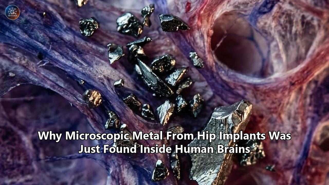
The beam of the field-emission scanning electron microscope focused on an ultra-thin slice of human temporal cortex, illuminating an anomaly that orthopedists and toxicologists had argued for decades could not exist.
Resting amid the delicate, web-like arborizations of dead neurons in a Chicago laboratory were distinct, jagged shards of inorganic matter. They measured mere tens of nanometers across—far smaller than a red blood cell, smaller even than most bacteria. When energy-dispersive X-ray spectroscopy bombarded the specks with electrons to read their elemental signatures, the spectrum produced sharp, undeniable peaks: cobalt, chromium, and titanium.
These heavy metals were not environmental pollutants inhaled from industrial smog, nor were they biological trace elements required for metabolic enzymes. They were the mechanical detritus of manufactured hip prostheses. Scoured from artificial ball-and-socket joints by millions of repetitive steps, the particles had entered the bloodstream, slipped through the body’s most heavily guarded anatomical firewall, and settled deep within the tissues of the human brain.
The study, led by materials scientist Robin Pourzal and a joint team of orthopedic pathologists and neuroscientists at Rush University Medical Center in Chicago alongside researchers from the Florey Institute of Neuroscience and Mental Health in Melbourne, was published in Acta Biomaterialia. By examining the autopsied brain tissue of 701 elderly participants enrolled in the long-running Rush Memory and Aging Project, Pourzal’s group confirmed what a handful of dismissed whistleblowers had suspected for over a decade: the degradation products of orthopedic joint replacements do not stay in the hip.
The spectrometric evaluation revealed that postmortem brain samples from patients who had received a total hip arthroplasty contained cobalt concentrations 8.9% higher across multiple cerebral regions than those of age- and sex-matched controls who had never received an artificial joint. In the inferior temporal cortex—a cerebral waypoint critical for visual processing, semantic memory, and one of the earliest staging grounds for neurodegenerative disease—elevated cobalt levels correlated with a statistically significant increase in amyloid-beta burden.
The finding delivers definitive physical evidence to a medical controversy that has simmered for twenty years. For generations of surgeons, total joint replacement was hailed as the undisputed triumph of twentieth-century biomechanics, an operation that restored pain-free mobility to tens of millions. The metallurgical consensus insisted that modern medical-grade alloys—principally cobalt-chromium-molybdenum and titanium-aluminum-vanadium—were bio-inert, walled off by scar tissue, and permanently isolated from systemic physiology.
The microscopic shards discovered inside human cerebral tissue prove that model obsolete.
The Autopsy Vault: Tracing Metal Through Chicago’s Cold Storage
The journey to this discovery began not in an orthopedic operating theater, but inside the brain bank of the Rush Alzheimer’s Disease Center on Chicago’s West Side.
Since 1997, the Rush Memory and Aging Project has tracked thousands of older adults across northeastern Illinois. Participants enroll with intact cognition and sign away their final earthly possession: upon their death, their brains are extracted within hours, methodically cataloged, flash-frozen or fixed in formalin, and subjected to microscopic autopsy. Every donor undergoes exhaustive annual evaluations while alive—a grueling 19-test cognitive battery assessing episodic memory, perceptual speed, and executive function.
Because the project correlates decades of meticulous behavioral data with the physical reality of the postmortem brain, it has long served as a premier engine for dementia research. But inside the Rush Department of Orthopedic Surgery, materials scientist Robin Pourzal and his colleagues saw the cohort through an entirely different lens.
"For years, we've known that particles can deposit in tissues surrounding the joint," Pourzal observed when summarizing the findings. "Total joint arthroplasty remains one of the most successful interventions in modern medicine, but wear particles from the implant, particularly in replacements involving accelerated wear, travel beyond the joint".
The orthopedic community had long known that prosthetic joints wear down. An active person takes between two and three million strides every year. Over a decade or two, the perpetual articulation of metal against polyethylene, ceramic against plastic, or metal against metal inevitably abrades the implant’s bearing surfaces. Histopathologists had spent decades documenting "metallosis"—the black, sludgy staining of synovial capsules caused by billions of sub-micron particles shed directly into the hip socket. When those joints failed, surgeons cut away liters of dead, necrotized muscle tissue stained the color of coal dust.
Yet standard dogma maintained that once metal left the joint cavity, it either remained sequestered within local drainage lymph nodes or filtered out through the renal tubules into the urine.
To test whether this was an illusion born of never having looked, Pourzal’s team drew upon 885 brains in the Rush depository. Cross-referencing Medicare Fee-For-Service claims data stretching from 1991 to 2016, they isolated 229 individuals who had undergone total joint replacements—146 hip arthroplasties and 83 knee or shoulder procedures—and placed them alongside 472 control subjects who had carried no metal hardware in their skeletons.
The researchers harvested tissue from four distinct anatomical zones: the midfrontal cortex, the anterior cingulate cortex, the cerebellum, and the inferior temporal cortex. They vaporized minute samples in high-temperature argon plasma, measuring elemental mass down to parts per billion using inductively coupled plasma mass spectrometry (ICP-MS).
The numbers registered clear disparities:
- Cobalt content across the cerebral regions of hip replacement recipients was 8.9% higher than in the control population.
- High-resolution scanning electron microscopy backed by elemental mapping spotted physical nanoparticles of cobalt directly embedded in the cortical matrix.
- In the inferior temporal cortex, the presence of cobalt was accompanied by a measurable elevation in amyloid-beta plaque load, the pathological hallmark of Alzheimer’s disease.
- Titanium, while also present in the brains of joint replacement patients, showed an inverse correlation with amyloid-beta.
Crucially, when the investigators combed through the donors’ decades of annual cognitive testing, they found no evidence that the presence of cobalt or titanium had hastened cognitive decline or increased the rate of clinical dementia diagnoses in the broader group.
The dichotomy was jarring. The metal was physically inside the brain. It was altering the local biochemical environment and driving up classic pathological proteins. Yet it had not produced overt dementia across the general cohort.
To understand how the metal breached the neural threshold in the first place, and why some individuals suffer catastrophic breakdown while others carry the debris invisibly, the evidence trail leads away from the pathology suite and into the biomechanics of modern hip design.
Mechanically Assisted Corrosion: How Hips Shed Atoms
The human hip is an unforgiving mechanical environment. A healthy hip joint experiences mechanical loads equivalent to three to five times a person’s total body weight during ordinary walking, and up to eight to ten times body weight during an accidental stumble or descent down a flight of stairs.
To withstand these staggering cyclical stresses, biomedical engineers turned to high-performance metallurgy. By the early 2000s, the alloy of choice for hip stems and femoral heads had become ASTM F75 or ASTM F1537: a mix of roughly 60% cobalt, 28% chromium, 6% molybdenum, and trace quantities of nickel, manganese, and carbon. The alloy's strength depends on its passivation layer—a microscopic skin of chromium oxide ($Cr_2O_3$), mere nanometers thick, that forms spontaneously when the metal contacts oxygen, shielding the underlying substrate from bodily fluids.
Corrosion should theoretically be impossible. But human biology is an aggressively corrosive chemical bath: warm, salty, oxygenated, and perpetually fluctuating in pH.
In a total hip arthroplasty, the natural ball and socket are excised. A titanium or cobalt-chromium stem is hammered into the hollowed marrow canal of the femur. Onto the tapered neck of this stem, surgeons seat a spherical metal or ceramic ball—the femoral head—locking it in place with a tap of a mallet. This junction is known in engineering as the trunnion. The ball then swivels inside an acetabular cup secured into the pelvic bone.
The source of systemic metal shedding is not solely the friction where ball meets socket. Even in modern "metal-on-polyethylene" joints—where a metal ball glides against a plastic cup—frictional wear is eclipsed by a insidious phenomenon: mechanically assisted crevice corrosion, or trunnionosis.
Every time an individual takes a step, asymmetrical bending forces torque the spherical head against the tapered neck of the femoral stem. This generates infinitesimal micro-motions measured in micrometers. At the microscopic contact ridges of the taper junction, this micro-motion mechanically scratches away the protective chromium oxide passivation layer.
Deprived of its passive shield, the raw alloy underneath is exposed to interstitial fluid. A microscopic crevice forms between the metal components, depleting local oxygen and allowing the fluid inside the crevice to turn acidic, with pH levels plummeting from a neutral 7.4 down to 1.5—comparable to stomach acid.
Under these conditions, galvanic and crevice corrosion dissolve the solid metal:
- Metallic cobalt ($Co^0$) oxidizes, shedding electrons to form toxic divalent cobalt ions ($Co^{2+}$).
- Chromium dissolves into trivalent ions ($Cr^{3+}$), which can re-precipitate into abrasive chromium phosphate or chromium oxide nanoparticles. Under inflammatory conditions, it can even oxidize into hexavalent chromium ($Cr^{6+}$), an aggressive carcinogen.
- Mechanical abrasion shears off chunks of alloy, generating millions of sub-micron wear particles ranging from 10 to 100 nanometers in diameter.
       Mechanical Stress & Cyclical Loading
                        │
                        ▼
       Micro-Motion at the Modular Neck Taper (Trunnionosis)
                        │
                        ▼
    Passive Chromium Oxide ($Cr_2O_3$) Layer Wiped Away
                        │
                        ▼
   Acidic Crevice Chemistry (pH drops to ~1.5) & Galvanic Corrosion
       ┌────────────────┴────────────────┐
       ▼                                 ▼
Soluble Heavy Metal Ions       Nano-Scale Alloy Particulates
 ($Co^{2+}$, $Cr^{3+}$, $Ti^{4+}$)       (10–100 nm Cr-Co debris)
       │                                 │
       ▼                                 ▼
 Bind to Serum Albumin/Transferrin   Phagocytosed by Macrophages
       └────────────────┬────────────────┘
                        │
                        ▼
         Dissemination into Systemic Circulation
                        │
                        ▼
  Breaching the Blood-Brain Barrier (BBB) & Infiltration into CNS
Once shed into the periprosthetic tissue, the immune system reacts. Macrophages engulf the metallic nanoparticles, attempting to digest them with intracellular acid and enzymes. The metal resists digestion. Overwhelmed, the macrophages rupture, spilling inflammatory cytokines and lysosomal enzymes into the joint capsule, inciting localized tissue necrosis and adverse local tissue reactions (ALTR).
Surviving macrophages and free-floating nanoparticles migrate into local lymph channels. From there, they drain into the thoracic duct and enter the venous bloodstream. At the same time, unbound cobalt and chromium ions bind to systemic transport proteins, predominantly albumin and transferrin, hitching a free ride through the cardiovascular circuit.
From that moment, the hip is no longer a localized mechanical problem. It has become a systemic chemical distributor.
Patient Zero: The Surgeon Who Poisoned Himself
Long before scanning electron microscopes identified these particles in Chicago donor brains, the real-world consequences of systemic metallosis emerged through a medical mystery in Anchorage, Alaska.
In 2006, Dr. Stephen Tower was a 51-year-old orthopedic surgeon with a thriving practice. An ultra-distance athlete who routinely rode hundreds of miles on a bicycle, Tower was suffering from worsening degenerative osteoarthritis in his right hip. Wanting to maintain his strenuous athletic regimen without the risk of dislocating a standard hip replacement, he opted for the newest technological marvel: a DePuy ASR metal-on-metal hip system.
The device, manufactured by Johnson & Johnson’s orthopedic subsidiary, substituted the traditional plastic liner with an all-metal cobalt-chromium cup and head. The pitch was seductively simple: metal-on-metal eliminated the plastic that historically wore out, promising a joint that would last a lifetime. Tower was such a believer that he implanted the same device in several of his own patients.
His recovery was fast. He was back on his bicycle within weeks, logging thousands of miles. But within eighteen months, Tower’s life began to unravel.
The initial symptoms were physical: an odd squeaking sound whenever he rotated his leg, followed by a deep, agonizing ache in his groin. Then the neurological decay took over. Tower noticed a tremor developing in his surgical hand. He suffered from ringing tinnitus, low-frequency hearing loss, and unexplained cardiac arrhythmias.
Soon, the breakdown penetrated his mind. The surgeon began experiencing cognitive fragmentation, erratic mood swings, and terrifying bouts of clinical mania. During a medical conference, overcome by compulsive cognitive hyperactivity, Tower paced his hotel room for hours before using a bar of soap as a stylus to scrawl complex geometric equations, flowcharts, and diagrams across every wall and mirror.
His partners and family feared he had developed early-onset Alzheimer’s, atypical Parkinson’s, or a psychotic illness. He was forced to stop operating.
Tower began suspecting his own artificial hip. As an orthopedist, he knew that metal hips could produce localized metallosis. But had anyone looked at what cobalt did when it entered the brain?
He sent his blood and urine to an environmental toxicology laboratory. The baseline reference level for cobalt in human blood is less than 0.1 micrograms per liter (parts per billion). Tower’s lab work returned with a blood cobalt level of 120 micrograms per liter—more than 1,000 times normal. His urine was dark, saturated with metal.
In 2009, a surgical colleague reopened Tower’s hip. When the scalpel punctured the joint capsule, gray-black fluid under pressure geysered into the surgical field, described in clinical notes as resembling "a crankcase full of dirty oil". The cobalt-chromium acetabular cup had worn through the passivation layer; the hip was grinding metal against metal at an angle that accelerated wear. The abraded metal had eaten through his rectus femoris muscle, turning healthy muscle tissue into grayish necrotic sludge.
The surgeon removed the metal monstrosity and replaced it with a conventional ceramic-on-polyethylene joint.
Within two weeks of the revision surgery, Tower’s blood cobalt levels plunged. His tremors vanished. The brain fog lifted. Within months, his executive function, memory, and spatial reasoning returned entirely to baseline.
Convinced that his case was not unique, Tower published his personal case report in the Journal of Bone and Joint Surgery in 2010. He coined a new medical phrase: Arthroplasty Cobalt Encephalopathy (ACE). Over the subsequent decade, he transformed into an industry whistleblower, tracking down scores of hip replacement recipients who had been placed in memory care facilities, locked wards, or placed on antipsychotic pharmaceuticals, their profound psychiatric degeneration misdiagnosed when they were suffering from an escalating reservoir of heavy metal in their blood.
"Orthopedic surgeons were trained to look at the X-ray," Tower argued in testimonies before regulators and in documentary appearances. "If the implant looks stable on the bone, they tell the patient the hip is fine. They never consider that the joint is poisoning the central nervous system."
For years, Tower’s warnings were handled by device manufacturers as eccentric edge cases—the result of extreme, anomalous implant malposition.
Then came the spinal fluid evidence from Berlin.
The Spinal Barrier Breach: The NeuroWear Trial
If the Chicago autopsy study confirmed that metal from hip implants was arriving in the brain, it was a 2025 study from Berlin that caught the metal in transit through the living central nervous system.
At the Charité-Universitätsmedizin Berlin, one of Europe’s premier medical centers, a team led by orthopedic researcher Dr. Anastasia Rakow launched the NeuroWear study. While Pourzal had examined postmortem brains, Rakow sought to look inside the living nervous system by examining cerebrospinal fluid (CSF)—the clear, sterile liquid that cushions the brain and spinal cord, produced by the choroid plexus and sealed off from systemic blood circulation by the blood-cerebrospinal fluid barrier.
Published in JAMA Network Open in March 2025, the cross-sectional study evaluated 204 adult patients:
- 103 individuals had at least one large joint replacement (hip or knee) in situ, with a median implant age of roughly 10 years.
- 101 age- and sex-matched controls had no artificial joint hardware.
The participants were undergoing elective procedures requiring spinal anesthesia or had been scheduled for diagnostic lumbar punctures, allowing clinicians to ethically harvest matched pairs of blood serum and CSF under pristine, metal-free analytical protocols.
The team analyzed the fluid for a wide spectrum of arthroplasty-associated elements: aluminum, cobalt, chromium, molybdenum, nickel, niobium, tantalum, titanium, vanadium, and zirconium.
The findings confirmed that systemic exposure was translating directly into central nervous system penetration:
- Cobalt Concentration Spikes: Patients with metal-containing joint replacements exhibited a significantly greater concentration of cobalt in their cerebrospinal fluid compared to arthroplasty-naive controls (median of 0.03 µg/L versus 0.02 µg/L, with peaks reaching 0.64 µg/L in implant patients).
- Chromium Accumulation: In patients carrying cobalt-chromium-molybdenum hardware, chromium levels in CSF were similarly elevated (0.31 µg/L versus 0.23 µg/L for controls), matching parallel elevations in their blood.
- Multi-Metal Permeability: Titanium, niobium, and zirconium—metals widely considered insoluble and biologically inert—were detected traversing into the spinal fluid of patients with titanium-alloy stems and modular revision prostheses.
"Notably, in the CSF of patients with at least one implant component made of cobalt-chromium-molybdenum, cobalt and chromium were found to be significantly higher than in matched controls," Rakow’s team reported. "This finding emphasizes that the use of cobalt-chromium-molybdenum alloys in particular bears the risk of exposure to arthroprosthetic metals in the central nervous system".
The NeuroWear data revealed an unsettling pharmacokinetic reality: the concentration ratio of cobalt between blood and cerebrospinal fluid suggested that cobalt was not merely leaking passively through holes in damaged blood vessels. Instead, correlation analysis suggested cobalt-specific transport mechanisms across neural barriers.
The brain was actively pulling the metal in.
Biochemical Sabotage: How Heavy Metals Cross the Blood-Brain Barrier
The central nervous system is insulated from the systemic circulatory system by the blood-brain barrier (BBB).
Unlike capillaries throughout the rest of the body, which have small fenestrations (pores) that let fluid and dissolved compounds pass freely, cerebral capillaries are forged from continuous endothelial cells welded together by high-resistance tight junctions: complex protein networks composed of claudin-3, claudin-5, occludin, and zonula occludens. These capillaries are wrapped in a basal lamina, cloaked by the contractile feet of pericytes, and enclosed in an outer sheath of astrocyte end-foot processes.
This neurovascular unit denies passage to roughly 98% of small-molecule drugs and virtually 100% of large biological macromolecules.
How, then, do industrial metals cast in a foundry end up across this defense system?
      BLOODSTREAM (Lumen)
   [ Co2+ ] [ Cr3+ ] [ Ti-Nanoparticles (15-30 nm) ]
       │        │                   │
       ▼        ▼                   ▼
   Transferrin / Albumin      Macropinocytosis / Transcytosis
       │        │                   │
   ═════════════════════════════════════════════════════════
      CEREBROVASCULAR ENDOTHELIUM (Tight Junctions: Claudin-5)
      * Metal ions induce ROS -> Calpain activation -> Tight junction breakdown
   ═════════════════════════════════════════════════════════
       │        │                   │
       ▼        ▼                   ▼
   Divalent Metal-Ion         Exocytosis across Basolateral
   Transporter 1 (DMT1)       Endothelial Membrane
       │        │                   │
       └────────┼───────────────────┘
                │
                ▼
      BRAIN PARENCHYMA (Interstitial Fluid & Neurons)
       ├── HIF-1α Pseudohypoxia Signaling
       ├── Mitochondrial Voltage-Dependent Anion Channel Blockade
       ├── Reactive Oxygen Species (ROS) & Microglial Neuroinflammation
       └── Amyloid-Beta Aggregation (Inferior Temporal Cortex)
The invasion relies on two mechanisms: molecular mimicry and particulate transcytosis.
1. Molecular Trojan Horses: Divalent Mimicry
The brain requires essential trace elements to survive—primarily iron, zinc, copper, and manganese. Because these metals are electrically charged ions, they cannot diffuse through the lipid membranes of endothelial cells. The BBB has evolved dedicated import systems to shuttle them across.
Cobalt ($Co^{2+}$) shares an ionic radius and coordination chemistry nearly identical to iron ($Fe^{2+}$) and calcium ($Ca^{2+}$). When cobalt enters the bloodstream from a corroding hip trunnion, it binds to transferrin—the primary plasma vehicle for systemic iron distribution.
When transferrin docks at the Transferrin Receptor 1 (TfR1) densely populated on the luminal surface of brain capillary endothelial cells, the receptor internalizes the protein via endocytosis. Once inside the endosome, the metal is released and transported into the endothelial cytoplasm by Divalent Metal-Ion Transporter 1 (DMT1).
DMT1 cannot distinguish divalent iron from divalent cobalt. The transporter moves the cobalt across the endothelial cell and discharges it into the cerebral parenchyma.
2. Endothelial Tight Junction Destruction
The metals do not merely ride existing systems; they break the machinery.
Free cobalt and chromium ions trigger oxidative stress in endothelial cells by uncoupling electron transfer chains, unleashing an overproduction of reactive oxygen species (ROS), including the hydroxyl radical ($\cdot OH$) and superoxide anion ($O_2^{\cdot-}$).
This oxidative burst triggers calcium influx into the endothelial cytoplasm, activating calpains—calcium-dependent proteases. These calpains cleave the cytosolic anchoring proteins of occludin and claudin-5. Within hours of prolonged exposure to micromolar levels of cobalt, the tight junctions loosen. The barrier turns leaky, allowing sub-micron nanoparticles (10 to 50 nm in size) to pass paracellularly through the intercellular gaps.
3. Particulate Transcytosis
Nanoparticles of chromium oxide and titanium dioxide are taken up by endothelial cells via non-specific macropinocytosis. Packaged inside vesicles, the particles bypass intracellular lysosomes and are expelled out the basolateral membrane into the brain’s extracellular space—a process known as adsorptive transcytosis.
Once on the neural side of the barrier, the metal interacts directly with astrocytes, microglia, and neurons.
Cobalt’s primary intracellular mechanism is the induction of "pseudohypoxia." Under normal physiological conditions, an enzyme called prolyl hydroxylase uses molecular oxygen and iron as cofactors to tag Hypoxia-Inducible Factor 1-alpha (HIF-1α) for rapid destruction. Cobalt directly displaces the iron atom in prolyl hydroxylase, rendering the enzyme inactive.
The cell is tricked into believing it is asphyxiating. HIF-1α accumulates, shuttles into the nucleus, and activates hundreds of genes that trigger inflammatory cascades, switch metabolism to anaerobic glycolysis, and trigger apoptotic cell death pathways.
At the same time, cobalt enters the mitochondria, where it binds to the voltage-dependent anion channel (VDAC) and compromises the inner mitochondrial membrane potential. Cytochrome c leaks into the cytoplasm, igniting caspase-9 and caspase-3: the definitive biochemical trigger for cellular suicide.
The Inferior Temporal Cortex and the Amyloid Mystery
The most provocative detail to emerge from Pourzal’s study in Acta Biomaterialia is the selective behavior of the metals inside the inferior temporal cortex.
Why did cobalt specifically track with an elevation of amyloid-beta in this specific anatomical region? And why did titanium show the opposite effect?
The inferior temporal cortex sits along the ventral visual pathway, responsible for semantic processing, high-level object representation, and face recognition. It is also an area that shows heightened vulnerability to the early stages of tau pathology and amyloid accumulation in Alzheimer’s disease.
Pathologists tracking neurodegeneration have noted for years that amyloid-beta—the peptide that aggregates into the plaques characteristic of Alzheimer’s disease—is not merely a toxic waste product. In molecular biology, amyloid-beta acts as an endogenous metalloprotein. Its primary amino acid sequence possesses a high-affinity binding domain for divalent metal cations, specifically copper, zinc, and iron.
When Pourzal’s co-authors, including prominent neurochemists Scott Ayton and Ashley Bush from the Florey Institute, analyzed the correlations, a biochemical hypothesis crystallized:
Amyloid-beta is acting as the brain’s emergency bio-scavenger.
When toxic, reactive divalent cobalt ($Co^{2+}$) crosses into the inferior temporal cortex, it generates an intensely pro-inflammatory, oxidatively stressed microenvironment. Neurons respond by upregulating amyloid precursor protein (APP) and cleaving it into amyloid-beta monomers. These amyloid-beta peptides contain histidine residues (His6, His13, His14) that act as natural chelators, binding to the free metal ions in an attempt to neutralize their reactivity.
The tragic consequence is that the binding of heavy metal ions to amyloid-beta alters the peptide's electrostatic charge, collapsing its tertiary shape. Instead of remaining soluble, the metal-bound amyloid peptides aggregate into insoluble, neurotoxic beta-sheet oligomers and fibrils.
The metal is safely trapped, but the resulting plaque clusters trigger chronic activation of microglia, surrounding the deposits and pumping out inflammatory cytokines that degrade local synapses.
       Soluble [ Co2+ ] Ions in Parenchyma
                    │
                    ▼
       Local Oxidative Stress & HIF-1α Activation
                    │
                    ▼
       Upregulation of Amyloid Precursor Protein (APP)
                    │
                    ▼
       Amyloid-Beta Secreted as an Emergency Chelator
                    │
                    ▼
     Histidine Residues Bind Co2+ -> Conformational Collapse
                    │
                    ▼
         Cross-Beta Sheet Oligomerization
                    │
                    ▼
       Insoluble Neurotoxic Plaque Aggregates
                    │
                    ▼
Synaptic Degradation & Microglial Neuroinflammation
Conversely, the study found that titanium was negatively correlated with amyloid-beta load. Unlike cobalt, titanium is tetravalent ($Ti^{4+}$) or present as inert titanium dioxide ($TiO_2$) particles, which do not participate in the same Fenton-type redox chemistry that destabilizes amyloid-beta folding.
Some researchers speculate that titanium wear particles, rather than stimulating amyloid generation, may trigger a foreign-body macrophagic response from microglia that upregulates general endocytic clearance, clearing extracellular amyloid debris as collateral cleanup.
Yet this leads to the central paradox of the Chicago findings: if cobalt drives amyloid accumulation in the brain, why didn’t the hip replacement recipients demonstrate higher rates of clinical dementia?
The answer exposes the profound difference between neuropathology and cognitive reserve.
Amyloid accumulation is a slow, multi-decade process. In Alzheimer’s disease, amyloid plaques often accumulate silently for 15 to 25 years before the brain's compensatory mechanisms buckle and symptomatic cognitive decline appears. In the Rush Memory and Aging cohort, the median age at death was over 85 years, and the duration of implant exposure averaged between 8 and 14 years.
Cobalt’s subtle 8.9% acceleration of amyloid burden was detectable with instruments measuring parts per billion, but for many participants, the clock simply ran out before that biological insult transformed into symptomatic dementia.
For a subset of patients—those with impaired kidney clearance, defective blood-brain barriers, or implants experiencing accelerated mechanical failure—that clock ticks at a catastrophic, compressed velocity.
The Regulatory Loophole: How Untested Metallurgy Entered Millions
The discovery of metal particles inside human brains is not merely a biological phenomenon. It is the end result of a regulatory system that let medical devices bypass clinical scrutiny for forty years.
In the United States, pharmaceuticals must endure a multi-phase gauntlet of animal models, human toxicity trials, and randomized placebo-controlled studies before reaching the public. Medical devices face no such requirement.
Under Section 510(k) of the 1976 Medical Device Amendments to the Federal Food, Drug, and Cosmetic Act, a manufacturer can bring a high-risk (Class III) medical implant to market without human clinical trials if the company demonstrates that the new device is "substantially equivalent" to a product already legally marketed prior to May 28, 1976—the date the law was passed.
This provision was intended as a temporary grandfather clause. Instead, it became the primary conduit for the global orthopedic industry.
Throughout the late 1990s and early 2000s, multinational device giants—including DePuy (Johnson & Johnson), Stryker, Zimmer, and Biomet—sought to capture an expanding demographic: younger, more active osteoarthritis patients who were wearing out their conventional plastic joint liners.
The manufacturers revived a concept from the 1960s: metal-on-metal (MoM) articulation. By utilizing polished cobalt-chromium-molybdenum for both the femoral head and the acetabular cup, the joint operated with virtually no plastic. In laboratory simulator machines, where robotic arms cycled joints through clean, fluid-lubricated arcs, the wear rates of metal-on-metal looked remarkably low.
Because cobalt-chromium and total hip systems had technically existed before 1976, device companies asserted "substantial equivalence". The FDA cleared devices like the DePuy ASR XL Acetabular System, the Stryker Rejuvenate modular stem, and the Zimmer Durom Cup through the 510(k) process with zero human clinical trial data.
Nobody had tested what happened when these alloys endured the micro-motions of a living body over decades:
- No animal studies investigated whether wear particles crossed the blood-brain barrier.
- No neurotoxicity profiling was conducted on chronic, low-dose exposure to cobalt and chromium.
- No testing accounted for mechanically assisted crevice corrosion in modular junctions.
Between 2003 and 2011, more than 31,000 patients in the United Kingdom and over 1,000,000 patients worldwide received metal-on-metal hip replacements. Millions more received modular "metal-on-polyethylene" hips featuring cobalt-chromium heads fitted to titanium stems.
By 2008, disaster struck. Rather than lasting a lifetime, metal-on-metal hips were failing at rates never before seen in modern orthopedics. Within five years of implantation, revision rates for some models soared beyond 12% to 20%.
Surgeons opening the hips of failing patients were horrified. Instead of stable implants, they encountered sprawling inflammatory pseudotumors, detached tendons, and melted bone. The regulatory framework, built to evaluate the device strictly as a mechanical wedge inside bone, had overlooked its role as a chronic heavy-metal delivery vehicle.
       1976 Medical Device Amendments Passed
                        │
                        ▼
       Section 510(k) Clearance Loophole Established
                        │
                        ▼
   2000s: Revival of Metal-on-Metal (MoM) Hip Designs
   * Bypassed human clinical trials via "substantial equivalence"
                        │
                        ▼
   Over 1 Million Patients Globally Receive High-Risk Implants
                        │
                        ▼
   2010–2012: The Collapse
   ├── DePuy ASR Global Recall (2010)
   ├── Stryker Rejuvenate / ABG II Recall (2012)
   └── MHRA & FDA Issue Safety Alerts for Metallosis
                        │
                        ▼
   Orthopedic Orthodoxy Minimizes Findings to "Local Reaction"
                        │
                        ▼
   2024–2026: Definitive Evidence of CNS Penetration
   ├── CSF Metal Translocation Confirmed (Berlin, 2025)
   └── Intracerebral Cobalt Debris Identified in Postmortem Brains (Rush, 2026)
In August 2010, DePuy recalled 93,000 ASR hip systems worldwide. In 2012, Stryker recalled its Rejuvenate and ABG II modular stems.
Yet when regulatory bodies such as the FDA and the UK Medicines and Healthcare products Regulatory Agency (MHRA) issued guidance, their warnings were confined almost entirely to orthopedic metrics: soft tissue destruction, pain, radiographic loosening, and pseudotumor formation.
The broader clinical manifestation—systemic poisoning that could reach beyond the pelvis—was minimized. Medical device companies paid out billions of dollars in multidistrict litigation settlements, sealing discovery documents and proprietary testing data behind non-disclosure agreements.
As a direct consequence of this compartmentalization, the full spectrum of metal hip implant side effects remained obscured from frontline clinical medicine.
The Clinical Silo: Why Primary Care and Neurology Miss the Signs
The discovery that metal from hip implants penetrates human brain tissue highlights an institutional blind spot in modern medicine: clinical hyper-specialization.
When an older adult experiences cognitive fog, uncharacteristic bouts of depression, memory loss, hand tremors, or visual disturbances, they do not consult their orthopedic surgeon. They consult a primary care physician, a geriatrician, or a neurologist.
These physicians look through the diagnostic lens of their own disciplines:
- A 72-year-old with tremors, slowed movement, and postural instability is diagnosed with Parkinson’s disease.
- A 68-year-old suffering from short-term memory lapses and spatial confusion is diagnosed with Mild Cognitive Impairment or early-stage Alzheimer’s disease.
- An adult presenting with sudden emotional volatility, severe insomnia, or mania is diagnosed with late-onset psychiatric disorder and prescribed selective serotonin reuptake inhibitors or antipsychotics.
Rarely does a neurologist ask an aging patient to drop their trousers to inspect for a surgical scar over their greater trochanter.
Even if a clinician suspects an environmental toxin, heavy metals like cobalt and chromium are absent from a comprehensive metabolic panel. Standard blood work measures sodium, potassium, liver enzymes, and creatinine. Specialized heavy-metal profiling requires inductively coupled plasma mass spectrometry conducted in trace-metal-free tubes—a test rarely ordered unless there is known occupational exposure in a foundry or battery factory.
"The patient is trapped between two worlds," explains Dr. David Bennett, Director of the Rush Alzheimer’s Disease Center and co-author of the Acta Biomaterialia study. "The orthopedic surgeon looks exclusively at the mechanics of the joint and the bone interface. If the prosthesis isn't loose on the X-ray, they dismiss the hip as the cause of distress. Meanwhile, the neurologist has no training in biomaterials or mechanically assisted crevice corrosion. They see neurodegeneration and assume it is idiopathy, genetics, or ordinary aging".
Compounding the dilemma is that a deteriorating implant does not always hurt.
Numerous clinical case studies of systemic cobaltism have emerged where the artificial joint was entirely asymptomatic: no pain, no clicking, no squeaking, and pristine X-rays showing stable bone ingrowth. Inside the modular taper junction, fretting corrosion had turned the joint into a silent chemical factory, pouring toxic ions into the bloodstream while the joint itself functioned with mechanical perfection.
The clinical manifestation of this unrecognized toxicity spans multiple systemic axes:
- Neurocognitive: Executive dysfunction, short-term memory deficits, processing speed reduction, severe brain fog.
- Neuropsychiatric: Rapid-onset depression, emotional blunting, uncharacteristic panic disorders, psychosis, and mania.
- Neurosensory: Tinnitus, high- and low-frequency neurosensory hearing loss, optic nerve atrophy, visual field constriction.
- Neuromuscular: Peripheral neuropathy, loss of vibratory sense, resting and action tremors, profound muscle weakness.
- Cardiovascular & Endocrine: Dilated cobalt cardiomyopathy, thyroid hormone disruption, secondary polycythemia.
These multisystem symptoms have long been documented as severe metal hip implant side effects in specialized toxicology literature, yet they rarely cross into mainstream clinical diagnostic practice.
When a patient is misdiagnosed with a neurodegenerative condition, they are managed with supportive therapies while the unrecognized toxin continues to leach into their nervous system. For those fortunate enough to identify the connection early, revision surgery removing the corroded metal alloy consistently halts the neurological decline, often triggering dramatic clinical recovery.
For those who remain undiagnosed, the microscopic metal remains embedded in their cortex, altering brain architecture for the rest of their lives.
Engineering the Post-Metal Joint
Over 1.5 million Americans undergo total joint replacements every year—a volume projected to surpass three million annually by 2030 as life expectancies rise and arthritis spreads across an aging global population.
Joint replacement surgery is indispensable. Without it, millions of individuals would spend their final decades crippled by agonizing osteoarthritic degradation, trapped in wheelchairs, and suffering the cascading cardiovascular and metabolic illnesses that accompany forced immobility. The total abandonment of joint replacement is neither possible nor desirable.
The imperative facing biomedical engineering is the elimination of vulnerable cobalt-chromium alloys and the redesign of modular junctions to prevent the shedding of neurotoxic debris.
| Implant Technology Generation | Articulation Materials | Primary Wear Debris | CNS Penetration Risk | Clinical Status |
|---|---|---|---|---|
| Generation 1: Classic Metal-on-Metal (1960s–1970s) | Cobalt-Chromium head on Cobalt-Chromium cup | Large-scale particulate cobalt and chromium | High (Severe systemic metallosis risk) | Obsolete; abandoned due to early catastrophic wear |
| Generation 2: Metal-on-Polyethylene (1980s–1990s) | Cobalt-Chromium head on conventional Polyethylene | Polyethylene sub-micron flakes; low metal release | Low to Moderate (Trunnion corrosion dependent) | Phased out; high rate of polyethylene-induced osteolysis |
| Generation 3: "Modern" Metal-on-Metal (2000s–2010s) | High-carbon Co-Cr-Mo alloy head on matching cup | Billions of nano-scale (10–50 nm) Co & Cr particles | Severe (Widespread documented CSF and organ translocation) | Global recalls (e.g., DePuy ASR); largely abandoned |
| Generation 4: Modern Modular Hybrid (2010s–Present) | Co-Cr or Ceramic head on cross-linked Polyethylene, Ti stem | Titanium particles; localized Co-Cr trunnion debris | Moderate (Trunnionosis remains active chemical vector) | Most widely implanted joint design globally today |
| Generation 5: Bio-Inert Ceramic & Surface Engineered | Ceramic-on-Ceramic or Oxidized Zirconium on XLPE | Biologically inert ceramic fragments ($Al_2O_3$, $ZrO_2$) | Negligible to Zero (Absence of toxic divalent metal ions) | Rapidly ascending standard of care for younger/active patients |
To address the mechanical and biological liabilities of earlier designs, modern orthopedic manufacturing is undergoing a transformation in biomaterials:
1. Ceramic-on-Polyethylene and Ceramic-on-Ceramic Bearings
The most effective barrier against cobalt release is eliminating cobalt from the articulating surface entirely. Ceramic femoral heads—forged from high-purity alumina ($Al_2O_3$), zirconia-toughened alumina (ZTA), or silicon nitride ($Si_3N_4$)—are chemically inert. They do not possess a metal lattice, cannot dissolve into divalent metal ions, and do not corrode in bodily fluids.
When paired with modern, highly cross-linked polyethylene (HXLPE)—plastic that has been irradiated to link its molecular chains, dramatically lowering wear rates—the amount of particulate debris generated by the joint plummets by more than 80%.
2. Oxidized Zirconium (Oxinium)
Another alternative is oxidized zirconium, a proprietary alloy comprising 97.5% zirconium and 2.5% niobium. The metal component is heated in air to over 500°C, causing oxygen to diffuse into the surface and transforming the outer five microns of the metal into a smooth, black ceramic monoclinic oxide layer.
The resulting surface provides the fracture resistance of solid metal with the low-friction wear properties of a ceramic, completely devoid of toxic cobalt or chromium.
3. Monolithic and Coated Tapers
Even when a ceramic ball is used, many designs still rely on a cobalt-chromium adapter sleeve to seat the ceramic onto a titanium femoral stem. If this sleeve experiences micro-motion, mechanically assisted crevice corrosion can still occur.
Engineers are designing monolithic, unibody stems or applying atomic layer deposition (ALD) and diamond-like carbon (DLC) coatings onto the male taper itself. These ceramicized coatings act as electrical insulators, interrupting the galvanic circuit between dissimilar metals and blocking corrosion.
4. 3D-Printed Trabecular Titanium
To fix the stem firmly within bone without cement, manufacturers are utilizing selective laser melting (SLM) 3D printing to fabricate porous titanium structures that mimic the spongy architecture of human trabecular bone.
By inducing immediate, deep bone ingrowth, these porous titanium surfaces eliminate the microscopic shifting and toggling of the stem that flexes the neck and drives trunnionosis.
While these advances safeguard patients undergoing joint replacement today, they do nothing for the millions of people who already carry older alloys inside their bodies.
The Looming Public Health Reckoning
The physical confirmation of microscopic hip implant metal inside human brain tissue shifts this issue from a localized surgical complication to an expansive public health challenge.
There are currently an estimated four to seven million individuals living in the United States alone who carry at least one artificial joint replacement. Hundreds of thousands of these individuals carry metal-on-metal systems implanted during the peak boom of the 2000s. Millions more carry modular systems that utilize a cobalt-chromium head anchored to a titanium stem.
The critical imperative is surveillance.
For over a decade, professional orthopedic associations advised that patients with metal implants who were pain-free required no regular blood testing. The findings of Pourzal’s group in Chicago and Rakow’s team in Berlin prove that biological containment cannot be assumed simply because an X-ray appears stable.
"What this data demands is a shift in clinical surveillance protocols," argues Dr. Stephen Tower. "We cannot wait for an artificial hip to fall apart or for a patient to develop psychiatric collapse or motor tremors before we check trace metal levels in their blood".
Medical researchers and epidemiological watchdogs are calling for concrete changes to standard care:
- Mandatory Baseline and Longitudinal Trace-Metal Blood Monitoring: Patients with cobalt-containing joint implants—particularly metal-on-metal or large-diameter modular designs—should receive annual testing for whole blood cobalt and chromium levels using inductively coupled plasma mass spectrometry. Establishing a patient's personal trajectory can identify runaway mechanically assisted crevice corrosion years before tissue necrosis or neurological translocation reaches critical thresholds.
- Interdisciplinary Diagnostic Cross-Talk: Health systems must dismantle the institutional silos separating orthopedics from neurology and psychiatry. Any patient with a history of joint replacement who presents with new-onset, unexplained tremors, severe depressive episodes, atypical cognitive decline, or sudden neurosensory loss (such as ringing in the ears or failing sight) should immediately have a blood heavy-metal panel included in their initial diagnostic workup.
- Establishing Neurological Safety Thresholds: Current regulatory guidelines for blood cobalt are based largely on the risk of local joint loosening and soft tissue destruction (typically 7 micrograms per liter in the UK and US). The NeuroWear trial and the Rush Memory autopsies demonstrate that metal accumulates in spinal fluid and brain tissue at concentrations well below these regulatory thresholds. New, physiologically grounded exposure limits are needed to protect sensitive neural structures.
- Targeted Neuroimaging in At-Risk Populations: For patients demonstrating elevated circulating cobalt and chromium, advanced neuroimaging—such as quantitative susceptibility mapping (QSM) MRI, which can quantify magnetic metal deposition in deep brain structures, and fluorodeoxyglucose (FDG) PET scans to evaluate regional cerebral metabolic dysfunction—should be deployed to track subclinical neural injury.
The most pressing, unresolved scientific question remains: what happens to the metal already inside the brain?
When a corroded implant is surgically removed and revised to ceramic, circulating blood levels of cobalt and chromium plunge within days. But heavy metals that have crossed the blood-brain barrier and bound to parenchymal matrices, or incorporated into insoluble amyloid plaques, do not have a simple clearance pathway. The brain possesses no lymphatic system equivalent to the rest of the body; its only clearance mechanism is the glymphatic system—a slow, fluid-exchange network active primarily during deep sleep.
Can the brain clear these nanoparticles once the source is severed? Or do these microscopic metallic specks remain embedded in the cerebral architecture for life, acting as permanent inflammatory focal points that subtly accelerate the trajectory of neurodegenerative aging?
These questions remain open. In laboratories across North America and Europe, researchers are tracking the surviving participants of the Rush Memory and Aging Project and launching multi-center clinical trials to analyze the long-term cognitive and neurological outcomes of joint replacement recipients.
The era of regarding the human body as a collection of isolated, independent systems is over. The discovery of hip implant debris lodged within human cerebral tissue confirms that whatever we implant into our bones will ultimately be reckoned with by the brain. Modern medicine has bridged the gap between mechanics and human flesh, but as the evidence on the pathology slides confirms, the border between the machine and the mind was far more porous than anyone imagined.
Reference:
- https://findanexpert.unimelb.edu.au/scholarlywork/2305224-cobalt-and-titanium-levels-in-the-brain-are-associated-with-alzheimer%E2%80%99s-disease-pathology-but-not-cognition--a-study-of-older-adults-with-and-without-total-joint-replacement
- https://newatlas.com/brain/alzheimers-dementia/particles-joint-replacement-enter-brain/
- https://medtruth.com/articles/patient-stories/cobalt-toxicity-the-poison-in-her-hips/
- https://pmc.ncbi.nlm.nih.gov/articles/PMC13242630/
- https://www.neurology.org/doi/10.1212/WNL.0000000000001935
- https://newatlas.com/brain/alzheimers-dementia/particles-joint-replacement-enter-brain/
- https://www.diamond.ac.uk/Science/Research/Highlights/2018/metal-deposition-in-organs.html
- https://www.nationalinjuryhelp.com/cobalt-chromium-poisoning-hip-implants/
- https://inkorr.com/en/u-mozku-ludej-z-endoprotezami-suglobiv-viavili-metalevi-mikrocastinki-345382
- https://www.researchgate.net/publication/396968961_Knee_arthroplasty_failure_is_associated_with_significant_systemic_multimetal_exposure
- https://bonezonepub.com/2023/05/22/concerns-about-cobalt-chrome-spur-innovation-in-implant-design/
- https://www.theguardian.com/film/2018/jul/25/the-bleeding-edge-netflix-documentary-medical-devices
- https://www.cbsnews.com/news/investigation-most-medical-devices-implanted-in-patients-without-testing/
- https://www.medpagetoday.com/orthopedics/orthopedics/114877
- https://medicaldialogues.in/orthopaedics/news/patients-with-metal-containing-joint-replacement-have-increased-metal-concentrations-in-csf-jama-145721
- https://sciprofiles.com/profile/2739448
- https://www.medpagetoday.com/orthopedics/orthopedics/114877
- https://www.aboutlawsuits.com/joint-implants-metal-patient-bloodstream-spinal-fluid-study/
- https://www.igb.illinois.edu/article/nanoparticle-transport-across-blood-brain-barrier-increases-alzheimer%E2%80%99s-and-age-study-finds
- https://www.diamond.ac.uk/Science/Research/Highlights/2021/Metallic-nanoparticles-can-cross-the-blood-brain-barrier--BBB--.html
- https://premierscience.com/pjn-25-981/
- https://pmc.ncbi.nlm.nih.gov/articles/PMC7966284/
- https://pmc.ncbi.nlm.nih.gov/articles/PMC5259873/
- https://pmc.ncbi.nlm.nih.gov/articles/PMC11115421/
- http://www.healthwatchusa.org/HWUSA-Initiatives/PDF-Downloads/20170606-NewsRelease-Cobaltism-14385830.pdf
- https://www.medicalrepublic.com.au/metal-hip-implants-dont-affect-your-brain-integrity/105182