In an operating theater at Royal Prince Alfred Hospital in Sydney, a surgical team led by cardiothoracic surgeon Paul Bannon and cardiologist Sean Lal paused during an open-chest coronary artery bypass procedure. With the patient supported on a heart-lung bypass machine, the surgeons excised millimeter-sized wedges of myocardial tissue from two distinct regions of the exposed left ventricle: the scarred, oxygen-starved edge of an acute infarction and a remote, well-perfused zone of healthy muscle.
Rather than depositing the tissue into formalin—a routine step that cross-links proteins and immobilizes cellular structures in a permanent chemical stasis—the researchers immediately transferred the live biopsies into oxygen-saturated, temperature-controlled cardioplegic transport media. Within minutes, the living samples were delivered to an adjacent laboratory equipped for single-nucleus RNA sequencing, high-resolution confocal microscopy, and spatial proteomics.
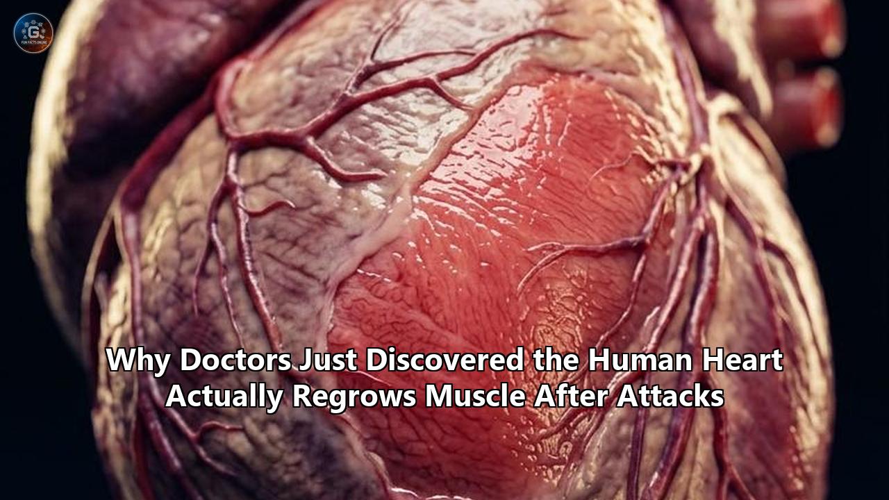
The data extracted from those live human tissue samples, published in Circulation Research by a multi-institutional team led by first author Robert Hume and senior author Sean Lal, revealed something medical textbooks insisted did not happen: adult human heart muscle cells, known as cardiomyocytes, were actively dividing in response to the injury.
       ISCHEMIC CASSETTE: THE ADULT CARDIOMYOCYTE PARADOX

   Remote Healthy Zone                 Peri-Infarct Border Zone
  ┌──────────────────────┐            ┌──────────────────────────┐
  │ • Mature binucleated │            │ • Reversible dediffer-   │
  │   cardiomyocytes     │            │   entiation              │
  │ • 90% Fatty Acid     │   Infarct  │ • Glycolytic switch      │
  │   Oxidation          │   Stress   │ • Aurora B midbodies     │
  │ • Strict cell-cycle  │ ─────────> │ • MKLP1+ cleavage furrow │
  │   quiescence         │            │ • Mononuclear cytokinesis│
  │ • Intact sarcomeres  │            │ • Partial mass renewal   │
  └──────────────────────┘            └──────────────────────────┘
For more than a hundred years, cardiology operated on the immovable assumption that the adult human heart is a terminally differentiated organ incapable of generating new muscle cells after damage. When a heart attack strikes, cutting off blood supply and suffocating billions of contractile cells, conventional medicine has treated the resulting dead tissue as an irreversible wound that can heal only through the deposition of an inert collagen scar.
The Sydney study proved that the human heart does mount an intrinsic, self-directed proliferative response. In the ischemic border zone—the knife-edge territory between living muscle and dead tissue—adult cardiomyocytes re-enter the cell cycle, copy their genetic code, and undergo both mitosis and complete cytokinesis, splitting into brand-new daughter cells.
This is not a cure for heart attacks, nor does the natural level of division suffice to replace the billion cells lost during a massive coronary occlusion. Yet the identification of this latent biological machinery alters the foundational principles of cardiovascular biology. It provides direct, physical evidence that heart muscle regeneration is an existing human program that can be studied, manipulated, and potentially amplified through targeted therapeutics.
Understanding why this mechanism stayed hidden for over a century—and why scientists missed it until now—requires an examination of scientific errors, forensic nuclear physics, deep-seated medical politics, and the extreme technical challenges of measuring division inside the body's most hard-working muscle.
The Century-Old Doctrine of the Permanent Heart
The belief that the heart cannot grow new muscle traces back to 19th-century cellular pathology. In 1894, the Italian pathologist Giulio Bizzozero classified mammalian tissues based on their regenerative turnover:
- Labile tissues, such as the skin epidermis and intestinal epithelium, which proliferate continuously throughout life.
- Stable tissues, including the liver and kidney parenchyma, which maintain low turnover but retain the ability to divide rapidly following injury.
- Permanent tissues, composed of cells that drop out of the cell cycle shortly after birth, never to divide again.
Bizzozero placed the adult central nervous system and striated heart muscle in the permanent category. His reasoning was sound for his era: when clinicians examined hearts under light microscopes weeks, months, or years after a myocardial infarction, they saw a dense, fibrous collagen scar devoid of newly formed muscle cells.
               HISTORICAL TISSUE TAXONOMY (BIZZOZERO, 1894)

      ┌─────────────────────────────────────────────────────────┐
      │ Labile: Epithelium, Bone Marrow (Continuous renewal)     │
      ├─────────────────────────────────────────────────────────┤
      │ Stable: Liver, Kidney (Dormant; divide upon injury)     │
      ├─────────────────────────────────────────────────────────┤
      │ Permanent: Neurons, Cardiomyocytes (Never divide)       │
      └─────────────────────────────────────────────────────────┘
Over the following decades, physiological logic reinforced this anatomical dogma. The adult human heart beats roughly 100,000 times every day, pumping more than 2,000 gallons of blood through miles of vascular network. To accomplish this, individual cardiomyocytes are packed with an intricate, highly organized lattice of contractile proteins known as myofibrils—repeating units of actin and myosin organized into sarcomeres. These internal cables are physically anchored to the cell's outer membrane by the dystrophin-glycoprotein complex and joined end-to-end to neighboring cells by intercalated discs loaded with mechanical junctions (fascia adherens, desmosomes) and electrical portals (gap junctions made of connexin 43).
For an animal cell to undergo mitosis, it must undergo radical structural changes. It typically rounds up, breaks down its nuclear envelope, disassembles its internal architecture, and builds an elaborate mitotic spindle made of microtubules to separate duplicate chromosomes.
Biologists believed that if an adult cardiomyocyte were to take apart its sarcomeres to form a mitotic spindle, the cell would lose its ability to generate force. If millions of cells did so at once, the ventricular wall would weaken, stretch, and rupture under intracardiac pressures. Worse, dismantling intercalated discs and gap junctions would interrupt electrical conduction, inviting catastrophic, fatal ventricular arrhythmias.
Evolution, scientists argued, made an absolute trade-off: in exchange for relentless mechanical power and electrical stability, mammals surrendered the ability to divide their heart cells once they reached adulthood. When a coronary artery occluded, the remaining muscle cells were thought to have only one defensive option: hypertrophy. They could swell in size, stretching their remaining fibers to bear the hemodynamic load, but they could never increase in number.
For almost a hundred years, this hypothesis went unchallenged. Textbooks, cardiology training fellowships, and pharmaceutical pipelines treated the myocardium as a fixed biological engine. Anyone claiming otherwise was dismissed as misinformed.
The Specter of Piero Anversa: How Fabricated Science Paralyzed a Field
The scientific establishment's refusal to accept cardiac turnover was further solidified by one of the most damaging scientific scandals in modern medicine.
In 2001, Dr. Piero Anversa, then a researcher at New York Medical College, published a paper in Nature that sent shockwaves through the scientific world. Anversa and his colleagues claimed that bone marrow cells expressing the surface receptor c-kit could, when injected into the damaged hearts of mice, transdifferentiate into brand-new cardiomyocytes, repairing more than 60% of the dead tissue within days. Two years later, Anversa went a step further in a paper published in Cell: he declared that the adult mammalian heart contained its own population of endogenous cardiac stem cells—also identified by the c-kit marker—that constantly regenerated heart muscle and could be harnessed to cure heart failure.
The medical establishment, eager for clinical interventions, embraced the findings. Anversa moved to Harvard Medical School and Brigham and Women’s Hospital, drawing tens of millions of dollars in National Institutes of Health (NIH) grant funding. Biotech startups formed around c-kit cardiac stem cells.
Clinical trials, including the NIH-sponsored SCIPIO trial and private commercial studies, quickly pushed the concept into human patients. Patients undergoing cardiac bypass or catheterization were infused with their own isolated, expanded c-kit cells in hopes of rebuilding their infarcted ventricles.
       CHRONOLOGY: THE RISE AND COLLAPSE OF THE C-KIT HYPOTHESIS

  2001: Anversa claims bone marrow c-kit+ cells regenerate dead myocardium
  2003: Anversa reports endogenous c-kit+ cardiac stem cells in the adult heart
  2004: Independent groups (Murry, Molkentin) demonstrate failure to replicate
  2014: Genetic fate-mapping confirms c-kit+ cells produce only blood vessels
  2015: Harvard and Brigham & Women's open internal investigative inquiry
  2017: Partners HealthCare pays $10M civil settlement to U.S. government
  2018: Harvard recommends retraction of 31 papers; field enters deep chill
The story soon began to fall apart. Independent laboratories led by researchers including Charles Murry at the University of Washington, Loren Field at Indiana University, and Jeffery Molkentin at Cincinnati Children’s Hospital tried to replicate the experiments. They consistently failed.
Using rigorous genetic fate-mapping techniques—in which c-kit-expressing cells were permanently tagged with fluorescent reporter proteins—Molkentin’s group demonstrated in 2014 that endogenous c-kit cells in the heart produce blood vessels, but their contribution to cardiomyocytes is functionally negligible, occurring at a rate of less than 0.005%. The cells Anversa claimed were rebuilding hearts were not generating new muscle cells at all.
An internal investigation at Harvard Medical School and Brigham and Women’s Hospital confirmed extensive data manipulation, improper image alterations, and outright fabrication across dozens of papers. In 2017, Partners HealthCare (the parent company of Brigham and Women’s) paid a $10 million settlement to the federal government to resolve civil allegations that Anversa’s laboratory had used falsified data to obtain NIH research grants. In 2018, Harvard recommended the retraction of 31 papers authored by Anversa's group.
The fallout was devastating. It poisoned the well of cardiovascular regenerative medicine for over a decade. Researchers working in the field faced deep skepticism from grant review committees, scientific journals, and private investors.
Many researchers swung to the opposite extreme: if the c-kit adult stem cell was a mirage, then the heart must be utterly, completely incapable of any new muscle formation. The dogma of the permanent, non-regenerative heart was rebuilt, stronger than before. For years, any investigator presenting evidence that heart cells were dividing risked professional isolation.
The field did not need more stem cell promises. It needed an objective, indisputable physical measurement of whether human heart muscle cells ever divided under real-world clinical conditions.
Atomic Clocks in the Nucleus: The Cold War Physics Behind Cellular Age
The first real cracks in the non-regenerative consensus did not come from stem cell biology, but from an unconventional application of Cold War atmospheric nuclear weapons data.
During the late 1950s and early 1960s, above-ground nuclear detonations conducted by the United States and the Soviet Union doubled the atmospheric concentration of Carbon-14 ($^{14}\text{C}$), a heavy radioactive isotope of carbon. Following the Limited Test Ban Treaty of 1963, atmospheric nuclear testing ceased, and the concentration of $^{14}\text{C}$ in atmospheric carbon dioxide began a steady, predictable exponential decline, diluted by the global carbon cycle and the burning of fossil fuels.
       ATMOSPHERIC CARBON-14 SPIKE AND GENOMIC STABILIZATION

  Atmospheric
     14C
      ▲         [1963 Test Ban Treaty]
      │                 * *
      │             *         *
      │           *             *
      │          *               *
      │         *                 *   Atmospheric Decay Curve
      │        *                   * * * * * * * * * * *
      │       *                                         *
      │  * * *                                           *
      └─────────────────────────────────────────────────────► Time
         1950    1960    1970    1980    1990    2000    2010

      [ Genomic DNA fixes atmospheric 14C level precisely at division ]
Every living organism incorporates atmospheric carbon into its biomolecules through the food chain. When a human cell divides, it synthesizes a new copy of its genomic DNA, pulling carbon atoms from the blood pool and locking them into the phosphodiester backbone of the double helix.
Once that cell undergoes its final division and exits the cell cycle, its genomic DNA is chemically stable; unlike proteins or lipids, genomic DNA does not turn over. The amount of $^{14}\text{C}$ integrated into that cell's genome serves as an immutable time stamp, recording the exact atmospheric concentration of $^{14}\text{C}$ present at the moment the cell was born.
In 2009, a team led by Jonas Frisén and Olaf Bergmann at the Karolinska Institute in Sweden realized they could use this atmospheric decay curve to solve the question of human cardiomyocyte turnover.
The logistical hurdles were daunting. Bergmann and Frisén had to collect post-mortem hearts from individuals who lived through the nuclear testing era, isolate intact cardiomyocyte nuclei using fluorescence-activated cell sorting (FACS) to separate them from non-muscle cells, extract the purified genomic DNA, convert it into pure graphite pellets, and measure the tiny isotopic ratios using accelerator mass spectrometers.
Their results, published in Science, provided the first undeniable physical proof: adult human cardiomyocytes do, in fact, renew.
The turnover was modest:
- At age 20, a healthy human heart replaces approximately 1% of its cardiomyocytes per year.
- By age 75, that rate slows to roughly 0.45% per year.
- Over a normal human lifespan, an individual exchanges approximately 40% of their total cardiomyocytes.
The heart was not a biological brick frozen in amber at birth. It possessed an ongoing, baseline turnover driven by the division of pre-existing cardiomyocytes.
Then came a critical follow-up. In late 2024, Bergmann and his team published a study in Circulation looking at 52 patients with advanced heart failure, 28 of whom had received a mechanical pump known as a Left Ventricular Assist Device (LVAD). An LVAD takes over the pumping workload of the failing left ventricle, dropping mechanical wall stress to near-zero levels.
The Swedish team discovered that in end-stage, unassisted failing hearts, cardiomyocyte renewal was suppressed to minimal levels—18 to 50 times lower than in healthy individuals. But in patients who received an LVAD and showed clinical cardiac recovery, the rate of cardiomyocyte renewal jumped more than sixfold compared to healthy hearts, reaching an annual turnover of 3.1%.
┌──────────────────────────────────────────────────────────────────────────┐
│      MEASURED ANNUAL CARDIOMYOCYTE RENEWAL RATES (CARBON-14 DATING)      │
├──────────────────────────────────────────────────────────────────────────┤
│ Healthy Young Adult (Age ~20)                    │ 1.0% per year        │
│ Healthy Older Adult (Age ~75)                    │ 0.45% per year       │
│ End-Stage Heart Failure (Untreated)              │ 0.02%–0.05% per year │
│ Failing Heart Unloaded via Mechanical LVAD Support│ 3.1% per year        │
└──────────────────────────────────────────────────────────────────────────┘
The Karolinska findings revealed two points. First, chronic heart failure shuts down the heart's natural regenerative ability. Second, when mechanical stress is removed, human cardiomyocytes retain an intrinsic capacity to wake up and divide at elevated rates.
Yet the Carbon-14 method had a major blind spot: it could calculate mathematical averages over years of survival, but it could not visualize the living, real-time mechanics of cell division in the immediate aftermath of an acute heart attack. It could not show where the cells divided, what molecular triggers kicked off the process, or whether adult cardiomyocytes could execute full, physical cell division inside the ischemic human border zone.
The Pre-Mortem Breakthrough: How Sydney Clinicians Captured Living Mitosis
To understand why the Sydney study succeeded where decades of prior investigations failed, one must look at the mechanics of biological sample collection.
Nearly everything scientists knew about human heart pathology came from two tissue sources:
- Autopsy specimens taken hours or days after death.
- Explanted hearts removed from end-stage heart failure patients during organ transplantation.
Both sources introduce serious technical artifacts. When a patient dies, blood flow stops, systemic oxygen vanishes, and autolysis begins within minutes. Cellular enzymes leak, mRNA degrades, and phosphorylation marks—essential for tracking active cell signaling—are rapidly stripped away by unchecked phosphatases.
Critically, the delicate protein structures required for cellular division, such as the mitotic spindle and the cytokinetic midbody, are extremely unstable. If a cardiomyocyte were in the middle of dividing when the patient died, that cellular apparatus would break down long before an autopsy pathologist placed the heart on a cutting board.
Explanted transplant hearts carry the opposite bias: they represent burned-out, end-stage fibrotic tissue that has suffered years of metabolic exhaustion, chronic neurohormonal overactivation, and inflammatory scarring—conditions that, as the Karolinska group proved, suppress cell turnover down to almost nothing.
The team at Royal Prince Alfred Hospital bypassed both problems by building a "pre-mortem" tissue collection protocol. Working under institutional human research ethics protocols, they identified patients undergoing open-chest Coronary Artery Bypass Grafting (CABG) who had recently suffered an acute myocardial infarction.
               PRE-MORTEM LIVING BIOPSY PROTOCOL

  Live Patient During CABG Surgery
                │
                ├───────► Ischemic Border Zone (BZ LV) Biopsy
                │
                └───────► Remote Healthy Myocardium (RZ LV) Biopsy
                                │
                                ▼
          Immediate Immersion in Ice-Cold Cardioplegia
                                │
        ┌───────────────────────┴───────────────────────┐
        ▼                                               ▼
  Flash Fixation / Cryopreservation        Single-Nucleus Suspension
        │                                               │
        ├─ High-Resolution 3D Confocal                  ├─ snRNA-Seq
        ├─ Aurora B / MKLP1 Cleavage Furrows            ├─ Epigenetic Profiling
        └─ Alpha-Actinin Structural Integrity           └─ Spatial Proteomics
During the bypass surgery, while the patient’s circulation was maintained via extracorporeal membrane oxygenation or cardiopulmonary bypass, the surgical team harvested living tissue samples from two distinct locations:
- The peri-ischemic border zone (BZ LV), the viable margin directly adjacent to the dead infarct.
- The remote non-ischemic zone (RZ LV), an uninjured area of the same patient's heart.
The tissue was placed into an ice-cold, hyperkalemic cardioplegic buffer, arresting all electrical and metabolic consumption within seconds to keep the cells intact. Transferred immediately to the laboratory, portions of the live human tissue were fixed in ultra-pure paraformaldehyde for structural imaging, while companion pieces were rapidly dissociated for single-nucleus RNA sequencing (snRNA-seq), mass-spectrometry-based proteomics, and untargeted metabolomics.
When Hume and his colleagues examined the living border-zone samples, the data were unmistakable. The tissue showed a striking enrichment of cardiomyocytes expressing markers of active cell division. Adult human heart muscle cells were not sitting inertly next to the scar. They were attempting to regrow.
The Polyploidy Trap: Why Science Kept Confusing Nuclear Swelling with Division
To understand why the findings sparked debate across the global cardiology community, one must grasp a biological technicality that has trapped cardiovascular researchers for thirty years: the difference between karyokinesis, polyploidization, and true cytokinesis.
A cell cycle is divided into several discrete phases:
- $G_1$ Phase: The cell grows and checks its surroundings.
- $S$ Phase: The cell replicates its nuclear DNA, turning a normal diploid set ($2n$) into a tetraploid set ($4n$).
- $G_2$ Phase: The cell double-checks the replicated DNA and prepares for division.
- $M$ Phase (Mitosis/Karyokinesis): The cell condenses its chromosomes, breaks down its nuclear envelope, lines up its genetic material on a mitotic spindle, and separates the chromosomes into two distinct nuclei.
- Cytokinesis: The final physical event. An actin-myosin contractile ring pinches the cell’s cytoplasm down the middle, creating a midbody cleavage furrow that physically cuts the original cell into two separate, independent daughter cells, each with its own membrane, its own nucleus, and its own set of organelles.
                  THE CARDIOMYOCYTE CELL-CYCLE SPLIT

                               ┌──────── G1 Phase ────────┐
                               │     (Cellular Growth)    │
                               └────────────┬─────────────┘
                                            ▼
                               ┌──────── S Phase ─────────┐
                               │     (DNA Replication)    │
                               └────────────┬─────────────┘
                                            ▼
                               ┌──────── G2 Phase ────────┐
                               │   (Division Checkpoint)  │
                               └────────────┬─────────────┘
                                            ▼
                               ┌──────── M Phase ─────────┐
                               │  (Mitosis/Karyokinesis)  │
                               └────────────┬─────────────┘
                                            │
                    ┌───────────────────────┴───────────────────────┐
                    ▼                                               ▼
         ABORTIVE TERMINATION                              TRUE CYTOKINESIS
     ┌─────────────────────────────┐                 ┌─────────────────────────────┐
     │ • Nuclear division without  │                 │ • Assembly of actin-myosin  │
     │   cellular cleavage         │                 │   contractile ring          │
     │ • Result: Binucleated or    │                 │ • Localization of Aurora B  │
     │   Polyploid (4n/8n) Cell    │                 │   kinase and MKLP1 at mid-  │
     │ • NO NEW CELLS CREATED      │                 │   body                      │
     │                             │                 │ • Complete cleavage furrow  │
     │                             │                 │ • Two functional daughter   │
     │                             │                 │   cells created             │
     └─────────────────────────────┘                 └─────────────────────────────┘
In human infants, cardiomyocytes divide rapidly via this full, canonical pathway. But within the first few months of postnatal life, an abrupt transition occurs.
As systemic blood pressures jump and oxygen levels spike, human cardiomyocytes begin skipping cytokinesis. They progress through $S$ phase and copy their DNA; they may even progress through $M$ phase and divide their nucleus; but they abort the cycle before pinching into two cells.
The result is that by childhood, more than 60% of human cardiomyocytes are binucleated (possessing two distinct nuclei), and a huge fraction of those nuclei are polyploid—carrying four, eight, or even sixteen copies of the human genome.
Polyploidy is an effective physiological strategy for stress management: it allows an enlarged cell to produce vast quantities of mRNA to service its massive volume without needing to disassemble its mechanical contractile machinery.
This characteristic caused decades of confusion in cardiac research. In previous studies, researchers would stain heart tissue with classic markers of cell-cycle entry, such as Ki-67, PCNA, or thymidine analogs like BrdU and EdU. When they saw these markers light up inside a cardiomyocyte nucleus, they often proclaimed that the cells were dividing.
Critics repeatedly proved them wrong. Staining positive for Ki-67 or incorporating BrdU simply meant the cell had copied its DNA. It meant the cell was undergoing endoreplication, becoming polyploid, or turning into a binucleated cell. It did not mean the cell was undergoing cytokinesis.
Because polyploid cells do not create new functional units, mistaking endoreplication for cell division was a recurring error. To prove that the adult human heart actually regrows muscle cells, a study had to produce visual and biochemical proof of cytokinesis.
┌────────────────────────────────────────────────────────────────────────┐
│                   CELL-CYCLE MARKERS VS. ACTUAL MITOSIS                │
├────────────────────────────────────────────────────────────────────────┤
│ Marker                      │ What It Actually Proves                  │
├─────────────────────────────┼──────────────────────────────────────────┤
│ Ki-67 / PCNA                │ Cell has exited G0; S-phase entry        │
│ BrdU / EdU Incorporation    │ DNA synthesis / Endoreplication          │
│ Phospho-Histone H3 (pH3)    │ Chromosome condensation (Karyokinesis)   │
│ Aurora Kinase B             │ Cytokinetic midbody localization         │
│ MKLP1 (Mitotic Kinesin)     │ Functional contractile cleavage furrow   │
│ Caveolin-3 Boundary Tracing │ Complete physical, dual-membrane abscission│
└────────────────────────────────────────────────────────────────────────┘
This is where the Sydney findings changed the scientific equation. In the live human border-zone biopsies, Hume and Lal did not rely solely on Ki-67 or simple DNA markers. They deployed high-resolution multi-channel immunofluorescence targeting:
- Phospho-Histone H3 (pH3): Staining condensed chromosomes in early and late metaphase.
- Aurora Kinase B: A catalytic component of the chromosomal passenger complex that localizes precisely to the central spindle and the midbody during cytokinesis.
- MKLP1 (Mitotic Kinesin-Like Protein 1): An essential motor protein that cross-links antiparallel microtubules in the intercellular bridge to drive the final physical cut between daughter cells.
- Caveolin-3 and Dystrophin: Structural membrane markers, combined with three-dimensional confocal z-stacks, used to prove that the two emerging daughter cells were enclosed within completely distinct, newly formed sarcolemmal membranes.
The team identified clear instances of cardiomyocytes caught in late-stage cytokinesis, complete with active MKLP1-positive cleavage furrows pinching between two separating nuclei.
Crucially, the cells performing this division were not large, multinucleated cells. They were a distinct, localized subpopulation of small, mononuclear cardiomyocytes nestled within the ischemic border zone. The adult human heart was running an active, structurally complete cytokinetic program.
The Border Zone Cauldron: Hypoxia, Mechanical Strain, and Metabolic Reversion
Cardiomyocytes do not divide everywhere in the injured heart. In their tissue surveys, the researchers found that mitotic activity in the remote, well-perfused myocardium was vanishingly low, matching the baseline levels established by the Karolinska Institute. The surge in cell division was concentrated within the peri-infarct border zone.
Why does this narrow rim of tissue permit something that the rest of the heart prohibits? The answer lies in the microenvironment that develops along the edge of an ischemic injury.
       THE BORDER-ZONE TRANSCRIPTIONAL & METABOLIC TRANSITION

                 Normoxic Remote Zone               Ischemic Border Zone
           ┌───────────────────────────────┐  ┌───────────────────────────────┐
  Energy   │ • 90% Fatty Acid β-Oxidation  │  │ • Glycolytic switch           │
  Flow     │ • Massive mitochondrial ROS   │  │ • Mitochondrial clearance     │
           │ • High ATP per oxygen ratio   │  │ • Low ROS production          │
           └──────────────┬────────────────┘  └──────────────┬────────────────┘
                          ▼                                  ▼
  DNA      │ • Permanent oxidative DNA     │  │ • Downregulated DNA damage    │
  State    │   damage response (DDR)       │  │   checkpoint signaling        │
           │ • Persistent ATM/Chk2 block   │  │ • Epigenetic remodeling       │
           └──────────────┬────────────────┘  └──────────────┬────────────────┘
                          ▼                                  ▼
  Pheno-   │ • Rigid sarcomeres (Titin)    │  │ • Partial dedifferentiation   │
  type     │ • Large binucleated cells     │  │ • Small mononuclear phenotype │
           │ • COMPLETE CELL-CYCLE ARREST  │  │ • CYTOKINESIS & MITOSIS       │
           └───────────────────────────────┘  └───────────────────────────────┘
In healthy adult myocardium, heart cells run on a hyper-efficient metabolic engine. To generate the continuous energy required to beat without pause, adult cardiomyocytes derive 70% to 90% of their ATP from mitochondrial fatty acid $\beta$-oxidation (FAO), with the remaining fraction coming from the oxidation of glucose, lactate, and ketone bodies.
While fatty acid oxidation generates large quantities of ATP, it carries a heavy biological cost: high rates of electron leakage in the mitochondrial respiratory chain, producing continuous clouds of reactive oxygen species (ROS) such as superoxide radicals and hydrogen peroxide.
Over the past decade, work by Hesham Sadek at the University of Texas Southwestern Medical Center helped explain why this metabolic profile blocks heart muscle regeneration.
When juvenile mammals are exposed to atmospheric oxygen after birth, their cardiomyocytes transition from fetal glycolysis to mitochondrial fatty acid oxidation. The resulting burst of ROS inflicts oxidative damage on cardiomyocyte genomic DNA.
This oxidative stress activates the cell's internal DNA damage response (DDR), governed by the kinases ATM, ATR, and Chk1/2. The DDR enforces a permanent checkpoint arrest by stabilizing the cyclin-dependent kinase inhibitors p21 (CIP1) and p27 (KIP1). The cell cycle slams shut.
In the border zone of an acute myocardial infarction, this metabolic landscape is upended:
- Severe localized hypoxia reduces the oxygen available to feed the mitochondrial respiratory chain.
- The snRNA-seq and metabolomic data from Hume and Lal confirmed that border-zone cardiomyocytes undergo an immediate, adaptive metabolic switch: they downregulate fatty acid oxidation enzymes and reactivate the fetal gene program, relying on anaerobic glycolysis to survive.
- By dimming their mitochondrial furnaces, these hypoxic cells see a drop in intracellular ROS production.
- The oxidative DNA damage response eases, removing the persistent molecular brake that keeps p21 and p27 tethered to cell-cycle complexes.
At the same time, these cells undergo partial dedifferentiation. Confocal imaging of border-zone cardiomyocytes showed that they downregulate several structural proteins, including cardiac troponin T and heavy-chain myosins, and loosen their rigid sarcomeric lattices. They shrink in volume, disassemble their intercalated discs, and revert to a plastic, immature phenotype that closely mimics embryonic and neonatal heart cells.
This altered state, however, balances on a mechanical knife-edge. As Kevin King and colleagues at the University of California San Diego reported in Nature, surviving cardiomyocytes trapped within the border zone are subjected to extreme biomechanical shear forces. With their dead neighbors unable to contract, the remaining cells must stretch to hold the ventricular wall together.
King’s team showed that this mechanical stress frequently causes the cell's nuclear envelope to rupture, spilling genomic DNA directly into the cytoplasm. This triggers the intracellular cGAS-STING pathway, igniting a powerful type I interferon (IFN) inflammatory response that can induce cellular senescence, wall thinning, and rupture.
The border zone is therefore caught in a biological tug-of-war. The unique mix of low oxygen and metabolic remodeling primes adult cardiomyocytes to wake up and divide. But if the mechanical strain and resulting sterile inflammation become too intense, the cells are driven into cell death or irreversible scarring.
Hume and Lal captured living human cells that managed to navigate that balance, successfully completing cell division before destructive inflammatory cues took over.
Deciphering the Circuitry: Hippo-YAP, Cyclin A2, and Epigenetic Brakes
The demonstration of active cytokinesis in post-infarction human hearts answers the anatomical question: can adult human heart cells divide? Yes.
The next question is mechanistic: what molecular pathways control this process, and why does this endogenous response usually fall short of fully healing the heart?
Three primary biochemical systems govern this checkpoint: the Hippo-YAP pathway, cyclin-dependent kinase complexes, and chromatin accessibility architecture.
          BIOCHEMICAL CONTROL AXIS OF CARDIOMYOCYTE PROLIFERATION

                 [Extracellular Mechanical & Contact Signals]
                                      │
                                      ▼
                        MST1/2 Kinase Activation
                                      │
                                      ▼
                        LATS1/2 Kinase Activation
                                      │
                     ┌────────────────┴────────────────┐
                     ▼                                 ▼
             Hippo Pathway ON                  Hippo Pathway OFF
      ┌─────────────────────────────┐   ┌─────────────────────────────┐
      │ • LATS1/2 phosphorylates    │   │ • Unphosphorylated YAP/TAZ  │
      │   YAP at Serine 127         │   │   translocates to nucleus   │
      │ • 14-3-3 protein binds YAP  │   │ • YAP binds TEAD factors    │
      │ • YAP trapped in cytoplasm  │   │ • Transcriptional activation│
      │   or degraded by proteasome │   │   of Cyclins, Aurora B,     │
      │ • MITOTIC GENES REPRESSED   │   │   and cytokinesis motors    │
      └─────────────────────────────┘   └──────────────┬──────────────┘
                                                       │
                                                       ▼
                                            [ Cyclin A2 Re-expression ]
                                                       │
                                                       ▼
                                            Overcomes p21/p27 block;
                                            Drives S-Phase & Cytokinesis
The Hippo-YAP Signaling Cascade
The evolutionary master switch controlling mammalian organ size is the Hippo signaling pathway. When the Hippo pathway is active (switched "ON"), a kinase cascade consisting of MST1/2 and LATS1/2 phosphorylates two key transcriptional co-activators: YAP (Yes-associated protein) and TAZ.
Phosphorylation of YAP at Serine-127 creates a docking site for 14-3-3 proteins, trapping YAP inside the cytoplasm or sending it to the ubiquitin-proteasome pathway for destruction. In healthy adult cardiomyocytes, continuous mechanical tension and cell-to-cell contacts keep the Hippo pathway active, locking YAP out of the cell nucleus.
When the Hippo pathway is conditionally deleted or genetically knocked out in animal models—work pioneered by James Martin at Baylor College of Medicine—unphosphorylated YAP flows freely into the cardiomyocyte nucleus.
Once inside, YAP binds to TEAD family transcription factors, turning on a large transcriptional cassette that drives cell-cycle re-entry, chromatin remodeling, sarcomere disassembly, and mitotic entry.
The snRNA-seq datasets from the Sydney study revealed that in the human border zone, surviving mononuclear cardiomyocytes exhibit transient, localized downregulation of Hippo signaling, with clear signatures of YAP nuclear target gene activation.
The Cyclin A2 Gateway
Even if YAP slips into the nucleus, it encounters a second molecular barrier: cell-cycle regulatory arrest. Mammalian cell division requires an ordered sequence of cyclin proteins binding to cyclin-dependent kinases (CDKs) to phosphorylate downstream targets, including the retinoblastoma protein (Rb).
During fetal development, human heart cells express a full suite of cyclins. Shortly after birth, however, the gene encoding Cyclin A2 (CCNA2) is permanently turned off.
Cyclin A2 is unique because it governs both transitions of the cell cycle: it pairs with CDK2 to drive the cell through $S$ phase, and then pairs with CDK1 to push the cell through the $G_2/M$ checkpoint into active mitosis.
┌─────────────────────────────────────────────────────────────────────────┐
│               CYCLIN-CDK COMPLEXES IN CARDIOMYOCYTE CYCLING             │
├─────────────────────────────────────────────────────────────────────────┤
│ Complex               │ Normal Adult Status │ Induced Regeneration Status│
├───────────────────────┼─────────────────────┼───────────────────────────┤
│ Cyclin D1 / CDK4/6    │ Low / Basal         │ Initiates G1 progression  │
│ Cyclin E / CDK2       │ Suppressed by p27   │ G1 to S phase transition  │
│ Cyclin A2 / CDK2      │ SILENCED AT BIRTH   │ DNA synthesis execution   │
│ Cyclin A2 / CDK1      │ SILENCED AT BIRTH   │ Entry into mitosis (G2/M) │
│ Cyclin B1 / CDK1      │ Low / Ineffective   │ Spindle / Cytokinesis drive│
└─────────────────────────────────────────────────────────────────────────┘
Without Cyclin A2, adult cardiomyocytes that get coaxed into the cell cycle by stress signals stall during DNA replication or get stuck in $G_2$ phase, resulting in polyploidy rather than cell division.
In research published in late 2025 in NPJ Regenerative Medicine, Hina Chaudhry and her team at the Icahn School of Medicine at Mount Sinai showed that delivering a functional CCNA2 gene back into middle-aged, non-human primate and adult human heart cells using viral vectors breaks this roadblock, safely releasing cardiomyocytes from their cell-cycle arrest and driving complete, functional cell division without inducing cellular instability.
The Sydney post-MI biopsy data revealed that in patients surviving acute myocardial infarction, a small number of border-zone cardiomyocytes naturally manage to transiently reactivate endogenous Cyclin A2 transcription, providing a biochemical explanation for how these cells slip past the $G_2/M$ checkpoint to complete cytokinesis.
The Epigenetic Architecture
The final, and perhaps most resilient, barrier is the physical organization of the cardiomyocyte genome. Over years of life, the chromatin of an adult cardiomyocyte is shaped by epigenetic marks—such as dense DNA methylation and histone modifications like H3K9me3 and H3K27me3—that pack the promoter regions of proliferative genes into tightly coiled, inaccessible heterochromatin.
Simultaneously, the genes required for contractile sarcomeres, calcium handling, and fatty acid transport are kept accessible as open euchromatin.
To divide, an adult cardiomyocyte must loosen this epigenetic architecture. It has to temporarily unpack its embryonic cell-cycle genes while repressing its mature structural programs.
In most adult cardiomyocytes, this epigenetic lock is too rigid to open; the cells simply cannot remodel their chromatin fast enough to divide before apoptotic or fibrotic signals trigger cell death.
Only a tiny subpopulation of cardiomyocytes—primarily smaller, mononuclear cells that maintain a less restrictive epigenetic profile—can navigate this chromatin remodeling process in the border zone.
The Engineering Chasm: Turning Endogenous Division into Functional Myocardium
The Sydney study establishes an empirical fact: adult human hearts are biologically capable of regrowing muscle cells after an infarction. But the clinical reality of cardiovascular medicine is shaped by numbers, and the arithmetic of a heart attack remains unforgiving.
                THE CARDIOMYOCYTE ARITHMETIC GAP

  Total Left Ventricle Pool:  ~4,000,000,000 cells
  Lost in Anterior STEMI:     ~1,000,000,000 cells (25%)

  Natural Border Zone Renewal:
  [▒] < 0.5% - 2.0% of border zone pool (~10-20 million cells)

  Deficit Remaining:
  [███████████████████████████████████████████████████████████]
  > 980,000,000 cells short of structural reconstitution
A healthy adult human left ventricle contains roughly 4 billion cardiomyocytes. A standard transmural ST-elevation myocardial infarction (STEMI) involving the proximal left anterior descending (LAD) coronary artery kills between 800 million and 1 billion cardiomyocytes in a few hours.
The natural, endogenous proliferative burst detected by Hume and Lal affects a small fraction of the surviving cells in the immediate border zone—amounting to tens of millions of new cells at best.
While that is biologically meaningful, it is not enough to rebuild the wall. The heart is trapped in a race against physics:
- If a 1-billion-cell defect is not rapidly stabilized, mechanical wall stress skyrockets according to the Law of Laplace:
$$\text{Wall Stress} = \frac{P \times r}{2h}$$
where $P$ is ventricular pressure, $r$ is the chamber radius, and $h$ is wall thickness.
- As wall thickness ($h$) drops, the wall stress on the surviving muscle spikes, stretching the ventricle out of shape.
- To prevent the ventricle from rupturing, the immune system and cardiac fibroblasts lay down a dense collagen patch.
- Collagen cannot contract, nor can it conduct electricity.
- Over months and years, the remaining muscle faces progressive mechanical overload, eventually leading to congestive heart failure.
To turn this biological capacity into a meaningful medical therapy, clinicians cannot simply let nature take its course. They must find safe ways to amplify this natural heart muscle regeneration by an order of magnitude.
Doing so requires overcoming three engineering hurdles:
1. The Sarcomeric Breakdown Problem
If a therapy floods the heart with signals telling cardiomyocytes to divide, hundreds of millions of cells will simultaneously disassemble their sarcomeres, detach their intercalated discs, and enter mitosis.
While those cells are dividing, they cannot contract. If a significant percentage of the left ventricle stops beating at the same moment, the patient’s cardiac output will plummet, sending them into cardiogenic shock.
Any viable regenerative therapy must deliver a controlled, wave-like, or strictly localized proliferative stimulus, nudging a small fraction of cells into division at any one time while leaving surrounding muscle intact to maintain pump function.
2. The Electrophysiological Arrhythmia Risk
Newborn cardiomyocytes do not emerge with their mature electrical circuitry fully formed. They start small, with high electrical resistance, low expression of inward rectifier potassium channels ($I_{\text{K1}}$), and fragmented distribution of the gap junction protein connexin 43.
If millions of newly divided cells are peppered throughout an ischemic ventricle, they risk forming electrically unstable zones of slow, disorganized conduction.
In animal models, crude attempts to drive cardiac proliferation—such as overexpressing microRNA-199a or uncontrolled YAP activation—consistently improved contractile numbers, but often triggered lethal ventricular tachycardia and fibrillation.
Newly created cardiomyocytes must integrate electrically into the surrounding muscle network, establishing synchronous electromechanical coupling with every heartbeat.
3. The Neoplasia Threat
The signaling pathways that instruct a cardiomyocyte to divide—Hippo-YAP, Wnt/$\beta$-catenin, Cyclin A2, and CDK networks—are the same pathways mutated in human cancers.
Cardiomyocytes rarely form primary cancers precisely because they are so locked down in cell-cycle arrest. If doctors administer systemic drugs or permanent gene therapies that shut off the Hippo pathway or lock cell-cycle regulators in the "ON" position, they risk inducing cardiac tumors or driving malignancies in other organ systems.
Any translational drug must have a built-in off-switch, providing a transient signal that prompts the cell to divide once or twice before the natural brakes re-engage.
The Emerging Pipeline: From Biological Mechanism to Clinical Protocol
Now that researchers have proved adult human heart muscle cells can divide, the focus of cardiovascular science is shifting. The era of harvesting random stem cells and injecting them blindly into scarred hearts is winding down, replaced by targeted strategies designed to amplify the heart's own cellular machinery.
              NEXT-GENERATION CARDIAC REGENERATION MATRIX

   Delivery Modality           Molecular Target               Clinical Status
  ┌──────────────────────┐    ┌──────────────────────┐    ┌──────────────────────┐
  │ Cardiac-Targeted     │    │ Modified mRNA        │    │ Pre-clinical large   │
  │ Lipid Nanoparticles  │───>│ (Transient Cyclin A2 │───>│ animal trials        │
  │ (LNPs)               │    │ or active YAP5SA)    │    │ (Swine models)       │
  └──────────────────────┘    └──────────────────────┘    └──────────────────────┘
  ┌──────────────────────┐    ┌──────────────────────┐    ┌──────────────────────┐
  │ Electromechanical    │    │ Small-Molecule       │    │ Early Phase I/II     │
  │ Guided Catheter      │───>│ LATS1/2 Inhibitors   │───>│ trials (Ischemic     │
  │ Injections (NOGA)    │    │ or p27 degraders     │    │ cardiomyopathy)      │
  └──────────────────────┘    └──────────────────────┘    └──────────────────────┘
  ┌──────────────────────┐    ┌──────────────────────┐    ┌──────────────────────┐
  │ Temporary Mechanical │    │ Hemodynamic Wall     │    │ Active clinical      │
  │ Unloading (Impella   │───>│ Stress Relievers     │───>│ protocol design      │
  │ / Microaxial Pumps)  │    │ + Epigenetic primers │    │ (Post-STEMI patients)│
  └──────────────────────┘    └──────────────────────┘    └──────────────────────┘
Several translational approaches are advancing through pre-clinical and early clinical development:
Transient mRNA Delivery via Targeted LNPs
Instead of using permanent viral vectors like adeno-associated viruses (AAV), which integrate or persist for years, labs are using synthetic modified messenger RNA (modRNA) packaged inside heart-homing lipid nanoparticles (LNPs).
These LNPs are surface-engineered with peptides that target receptors upregulated on ischemic cardiomyocytes, such as angiotensin II type 1 receptors or specific integrins.
Once inside the cell, the modRNA translates a burst of a pro-mitotic factor—such as Cyclin A2 or an active YAP variant—for 48 to 72 hours before the RNA degrades naturally.
This transient window is long enough to push border-zone cardiomyocytes through cytokinesis, but short enough to avoid neoplastic transformation or sustained structural disarray.
Small-Molecule Inhibitors of Hippo Kinases
Medicinal chemists have synthesized potent, reversible small-molecule inhibitors of LATS1 and LATS2, the gatekeeper kinases of the Hippo cascade.
By temporarily blocking LATS1/2 activity for a few days after an acute heart attack, these drugs allow endogenous YAP to transit to the nucleus, unlocking the downstream genes that drive border-zone division.
Because small molecules can be cleared by the liver and kidneys, their dosing can be titrated or halted if side effects or arrhythmias appear.
Catheter-Guided Border Zone Microinjections
Injecting regenerative agents into a vein dilutes them throughout the body, raising the risk of off-target effects.
Cardiologists are pairing regenerative biologics with high-resolution electromechanical mapping systems, such as the NOGA catheter system.
By running an electrode-tipped sensor across the interior surface of the left ventricle, clinicians can measure the local electrical voltage and mechanical motion of every square millimeter of the heart wall.
They can map the non-contractile, electrically silent scar, the normally functioning remote muscle, and the low-voltage, surviving peri-infarct border zone.
The catheter can then deploy a micro-needle to inject pro-regenerative drugs directly into that border zone, delivering the therapy straight to the cells that have already initiated their own regenerative programs.
Mechanical Unloading Combinations
Building on the Karolinska Institute’s LVAD findings, researchers are designing clinical trials that pair mechanical unloading with regenerative pharmacology.
By inserting temporary, catheter-based microaxial pumps—such as the Impella device—into the left ventricle of a patient immediately after opening a blocked artery, interventional cardiologists can reduce chamber pressures and mechanical wall stress.
This mechanical rest prevents nuclear envelope ruptures, calms destructive type I interferon signaling, and lowers oxygen consumption, providing an ideal microenvironment for pro-regenerative drugs to coax surviving cells into productive division.
The Road Ahead: Overcoming the Remaining Questions
The work by Hume, Lal, and their colleagues has shifted how science views the human heart. The idea that the adult heart is biologically incapable of generating new muscle has been replaced by a more nuanced picture: it is an organ with a real, but throttled, intrinsic capacity for self-repair.
               THE EVOLVING VIEW OF THE ADULT HUMAN HEART

   Old Scientific Consensus                    New Empirical Reality
  ┌───────────────────────────────┐           ┌───────────────────────────────┐
  │ • Permanently post-mitotic    │           │ • Retains latent cell-cycle   │
  │ • Zero capacity for division  │           │   machinery                   │
  │ • Purely fibrotic scar repair │  ──────>  │ • Measurable cytokinesis in   │
  │ • Hypertrophy is only defense │           │   the ischemic border zone    │
  │ • Stem-cell injection models  │           │ • Metabolic switch triggers   │
  │   (largely discredited)       │           │   partial dedifferentiation   │
  │                               │           │ • Endogenous programs can be  │
  │                               │           │   pharmacologically boosted   │
  └───────────────────────────────┘           └───────────────────────────────┘
The task now is working out the details. Researchers still need to determine:
- The precise therapeutic window for intervening after an infarction. Does pro-mitotic signaling need to be initiated within the first 48 hours, or can dormant border-zone cells be coaxed into dividing months after a scar has matured?
- Which patients have the highest numbers of mononuclear, division-ready cardiomyocytes? Does age, sex, diabetes, or long-term medication use change an individual's intrinsic regenerative capacity?
- How to guarantee that newly divided cardiomyocytes mature quickly enough to avoid triggering lethal electrical rhythms while still contributing to contractile force.
Answers to these questions will emerge over the coming years as data from living human tissue slices, spatial transcriptomics, and large-animal preclinical trials converge.
What is settled is the foundational biology: when an attack strikes the human heart, the muscle does not simply submit to scarring. Deep within the oxygen-starved margins of the injury, human cardiomyocytes do something science insisted was impossible: they start to divide. Medicine's challenge is learning how to help them finish the job.
Reference:
- https://scitechdaily.com/world-first-study-reveals-human-hearts-can-regenerate-after-a-heart-attack/
- https://www.findarticles.com/human-heart-tissue-cardiomyocyte-division-heart-attack/
- https://www.ahajournals.org/doi/10.1161/CIRCRESAHA.125.327486
- https://www.ahajournals.org/doi/abs/10.1161/CIRCRESAHA.125.327486
- https://scitechdaily.com/world-first-study-reveals-human-hearts-can-regenerate-after-a-heart-attack/
- https://www.sciencedaily.com/releases/2024/08/240829132407.htm
- https://www.researchgate.net/publication/398330200_Human_Hearts_Intrinsically_Increase_Cardiomyocyte_Mitosis_After_Myocardial_Infarction
- https://www.ahajournals.org/doi/10.1161/CIRCULATIONAHA.123.067156
- https://www.sydney.edu.au/news-opinion/news/2026/01/20/human-heart-regrows-muscle-cells-after-heart-attack-world-first-study-shows.html
- https://www.ahajournals.org/doi/10.1161/CIRCRESAHA.125.327486
- https://www.researchgate.net/scientific-contributions/Robert-D-Hume-2168700441
- https://academic.oup.com/eurheartj/article/38/30/2333/3977787
- https://pmc.ncbi.nlm.nih.gov/articles/PMC8345975/
- https://www.mountsinai.org/about/newsroom/2025/a-specific-human-gene-can-help-the-heart-repair-itself-from-heart-attack-or-heart-failure
- https://www.sciencedaily.com/releases/2026/09/260909231754.htm