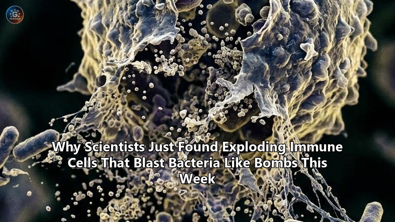
Researchers from Stanford University and Ben-Gurion University of the Negev (BGU) have uncovered a previously unknown class of immune cells that protect host tissues by literally erupting like microscopic grenades. Published in the journal Cell, the findings demonstrate that these specialized cells—named ruptoblasts—undergo an explosive form of programmed self-destruction termed ruptosis. When activated by biochemical distress signals, ruptoblasts flood with calcium, burst open within minutes, and release a potent chemical shockwave that shatters the membranes of nearby invading bacteria.
For decades, immunology textbooks have taught that animal immune systems rely almost exclusively on white blood cells that fight pathogens through direct contact, phagocytosis (engulfing microbes), or the gradual secretion of targeted antibodies and cytokines. Ruptoblasts break every rule in that classic framework. Instead of belonging to the blood-forming cell lineage, these exploding immune cells derive from an entirely separate family of glandular cells, proving that cellular defense mechanisms can evolve from outside traditional white blood cell pathways.
Traditional Immune Defense vs. Ruptosis
┌──────────────────────────────────────────────────────────┐
│ Traditional Phagocytosis / NETosis                       │
│ White Blood Cells ──► Engulf Microbes / Slow Pore Leak   │
│ (Takes hours; gradual inflammatory signaling)            │
└──────────────────────────────────────────────────────────┘
                            VS.
┌──────────────────────────────────────────────────────────┐
│ Ruptosis (Newly Discovered)                              │
│ Glandular Ruptoblast ──► Activin Trigger ──► Ca²⁺ Flood │
│ ──► Explosive Rupture (Minutes) ──► Membrane Destruction  │
└──────────────────────────────────────────────────────────┘
The discovery, led by Prof. Bo Wang of Stanford’s Department of Bioengineering and Prof. Benyamin Rosental of BGU’s Shraga Segal Department of Microbiology, Immunology, and Genetics, provides a radically new model for cellular suicide as a frontline defense. Because the chemical blast delivered by ruptoblasts physically destroys cell membranes regardless of species, the finding holds immediate implications for engineering targeted cell therapies capable of eliminating drug-resistant bacteria and solid cancerous tumors.
The Anomaly in the Lab: Tracking the Instantaneous Suicide
The breakthrough emerged during high-resolution live-cell imaging of Planaria—aquatic flatworms widely studied for their extraordinary regenerative capabilities. While examining how primitive organisms mobilize defenses against tissue injury and bacterial infection, lead researchers Dr. Chew Chai and Dr. Eliya Sultan noticed a distinct cellular anomaly that defied established paradigms.
Using a combination of fluorescent dye markers, high-speed live microscopy, and flow cytometry (laser-assisted cell sorting), the research team observed that exposing the flatworms to pathogenic Escherichia coli bacteria triggered a sudden, localized wave of cellular destruction. A tiny population of glandular cells did not migrate toward the bacteria to engulf them slowly. Instead, these cells began swelling rapidly, erupted violently in a matter of seconds, and completely vanished within five minutes, leaving no trace behind other than a zone of dead bacteria.
Ruptoblast Detonation Timeline
[0:00 Min] Activin Flare Detected by Ruptoblast Receptor
   │
[1:00 Min] Rapid Cytosolic Calcium (Ca²⁺) Influx Begins
   │
[2:30 Min] Extreme Internal Osmotic Pressure & Membrane Ballooning
   │
[3:00 Min] Violent Cell Membrane Eruption (Ruptosis)
   │
[3:15 Min] Broad-Spectrum Antimicrobial Payload Ejected
   │
[5:00 Min] Target Bacteria Destroyed; Ruptoblast Vanishes Completely
"Some mammalian cells and bacteria may also do an explosive sort of cell death, but the timescale is really long," explained Dr. Chew Chai in detailing the observations. "They are exploding, but it's more like pores that slowly leak things out over the course of several hours. Ruptosis happens within seconds to minutes."
To confirm whether this rapid destruction was an active biological defense or a passive response to stress, the team isolated ruptoblasts and tested them against multiple cellular targets in vitro. When exposed to E. coli bacteria, mammalian kidney cells, and murine blood cells, the ruptoblasts consistently executed their explosive routine, delivering a localized killing wave that shattered every foreign cell type within the immediate blast zone.
Anatomy of an Eruption: The Biochemical Detonation Chain
To understand how a single cell transforms itself into a precision bomb, the research team mapped the molecular cascade that controls ruptosis. Unlike unprogrammed necrosis, which occurs randomly when a cell suffers physical trauma, ruptosis is governed by a strict, multi-step biochemical detonation sequence.
Step 1: Receiving the Distress Flare (Activin Signaling)
Ruptoblasts do not detonate spontaneously. They remain dormant until they detect a sharp increase in a signaling hormone called activin. In animal physiology, activin acts as a molecular alarm flare released by surrounding tissue cells when they experience bacterial invasion or severe physical damage.
Step 2: The Calcium Influx
When activin binds to specialized receptors on the surface of a ruptoblast, it sets off an intracellular chain reaction. Within seconds, channels across the cell membrane open, driving a massive flood of calcium ions ($Ca^{2+}$) into the cytosol. This sudden alteration in intracellular ionic concentration causes rapid osmotic swelling.
Step 3: Membrane Rupture and Payload Ejection
As internal pressure mounts, the ruptoblast's outer membrane ballooning reaches a critical threshold. The cell violently bursts, discharging its entire internal cocktail of broad-spectrum toxic agents into the surrounding extracellular space.
Step 4: Physical Membrane Destruction
The chemical cocktail released during ruptosis acts directly on nearby cellular structures. Rather than interfering with complex metabolic pathways inside the target pathogen, the toxins physically disrupt lipid membranes, causing invading bacterial walls to fragment instantly.
Comparative Cell Death Mechanisms
─────────────────────────────────────────────────────────────────────────
Mechanism    Time to Execution   Primary Trigger       Targeting Strategy
─────────────────────────────────────────────────────────────────────────
Apoptosis    6 to 24 Hours       Caspase Cascade       Silent / Non-inflammatory
Pyroptosis   1 to 6 Hours        Gasdermin Pores       Cytokine Leakage / Inflammation
NETosis      2 to 4 Hours        ROS / Chromatin Net   Sticky Extracellular Traps
Ruptosis     1 to 5 Minutes      Activin / Ca²⁺ Flood  Explosive Physical Rupture
─────────────────────────────────────────────────────────────────────────
This rapid execution distinguishes ruptosis from other known forms of programmed cell death. In classical pyroptosis, mammalian cells use gasdermin proteins to punch small pores into their own membranes, allowing water to trickle in and inflammatory cytokines like interleukin-1β (IL-1β) to leak out over several hours. In NETosis, neutrophils undergo a controlled death that releases sticky webs of DNA studded with antimicrobial peptides to entangle microbes. Ruptosis skips the slow leakage and net-building entirely, opting instead for a instantaneous physical detonation.
Nature's Failsafes: Preventing Runaway Tissue Destruction
Deploying self-destructing cells within living tissue carries immense risk. If exploding immune cells were to detonate indiscriminately or set off chain reactions among neighboring cells, an infection could quickly transform into widespread tissue necrosis.
The Stanford-BGU study revealed that nature implemented strict safety features to keep ruptoblasts under control:
- Specific Chemical Lock: Mechanically crushing or physically tearing a ruptoblast does not release its lethal cocktail. The killing agents require the precise biochemical activation of the activin pathway to become lethal. If a ruptoblast dies from physical trauma, its contents remain completely harmless to neighboring tissue.
- Short Toxin Half-Life: The chemical payload released during an eruption degrades within roughly 15 minutes. This short active lifespan ensures that the blast radius remains confined to a perimeter of just a few micrometers around the detonating cell.
- No Chain Detonation: Neighboring ruptoblasts caught within the shockwave of an exploding cell do not detonate in response. Because they require a sustained activin hormone signal rather than a physical shockwave to trigger calcium flooding, the explosion cannot cascade out of control.
These spatial and temporal controls turn the ruptoblast into a surgical strike weapon. It unleashes total physical destruction inside a tiny, localized zone while leaving surrounding host structures intact.
Outside the White Blood Lineage: An Evolutionary Shift
Beyond its tactical mechanics, the discovery of ruptoblasts fundamentally alters basic assumptions about how immunity evolved across the animal kingdom.
In mammals and other vertebrates, cellular immunity is anchored in hematopoiesis—the process by which multipotent stem cells in the bone marrow differentiate into specialized white blood cells, such as macrophages, neutrophils, T cells, and B cells. Biologists have long treated hematopoiesis as the universal foundation of complex cellular defense.
The Evolutionary Lineages of Immunity
┌────────────────────────────────────────────────────────┐
│ Classical Immune Lineage (Hematopoietic)               │
│ Bone Marrow Stem Cells ──► White Blood Cells           │
│ (Neutrophils, Macrophages, T Cells, B Cells)           │
└────────────────────────────────────────────────────────┘
                           AND
┌────────────────────────────────────────────────────────┐
│ Newly Identified Lineage (Glandular / Non-Hematopoietic)│
│ Epithelial/Glandular Lineage ──► Ruptoblasts           │
│ (Executes Ruptosis via Activin-Triggered Detonation)   │
└────────────────────────────────────────────────────────┘
Single-cell RNA sequencing and genetic lineage tracing revealed that ruptoblasts do not originate from hematopoietic stem cells. Instead, they belong to a specialized lineage of glandular cells—a lineage historically associated with secreting mucus, enzymes, or structural proteins rather than conducting immune surveillance.
Genetic mapping across primitive marine and freshwater organisms suggests that this glandular defense tactic is deeply ancient. Long before complex vertebrates evolved circulating white blood cells and bone marrow, primitive organisms repurposed glandular secretion pathways into explosive, suicidal landmines to defend their epithelial surfaces.
"These findings reveal a completely new strategy that directly links hormonal signals with explosive immune defense," noted the study's senior authors. "It shows how diverse nature can be when inventing ways to fight off infection."
The Dark Side: How Ruptoblasts Drive Graft Rejection
While ruptoblasts serve as a broad-spectrum shield against pathogenic bacteria, the researchers discovered that this explosive defense mechanism comes with a severe trade-off: it acts as a primary driver of tissue graft rejection.
To study how ruptoblasts interact with foreign host tissue, the team performed surgical tissue fusion experiments, combining body segments from two genetically distinct planarian flatworms. The biological incompatibility between the two tissue types triggered an immediate hyper-inflammatory response.
Tissue Fusion Experiment & Rejection Pathway
[Genetically Distinct Flatworm Tissues Fused]
                       │
                       ▼
    [Incompatibility Triggers Mass Activin Surge]
                       │
                       ▼
   [Widespread Synchronized Ruptoblast Detonations]
                       │
                       ▼
  [Massive Localized Lesions & Total Graft Rejection]
                       │
                       ▼
[Genetically Knocking Out Ruptoblasts Stops Rejection Entirely]- Activin Buildup: Tissue incompatibility caused neighboring cells to release massive, sustained quantities of activin.
- Mass Detonation: The local surge in activin detonated hundreds of ruptoblasts simultaneously across the graft boundary.
- Lesion Formation: The combined chemical blast shattered host and donor cells alike, creating deep tissue lesions and resulting in total rejection of the transplanted tissue.
To verify that ruptoblasts were responsible for the rejection, the researchers used genetic editing to knock out ruptoblasts in test flatworms. When tissues from two distinct worms were fused in the absence of ruptoblasts, the destructive rejection response vanished completely, allowing the transplanted tissue to integrate smoothly.
This finding provides organ transplant researchers with a new biological framework. In human organ transplantation, graft-versus-host disease (GVHD) and acute rejection are driven by complex T-cell signaling. Discovering that primitive organisms use non-hematopoietic, explosive immune cells to execute organ rejection opens new avenues for investigating whether localized, non-white-blood-cell pathways contribute to human transplant failure.
Therapeutic Horizons: Weaponizing Ruptosis Against Superbugs and Cancer
The discovery of exploding immune cells arrives at a critical juncture in global medicine. As global rates of antimicrobial resistance (AMR) rise, common bacterial pathogens are increasingly defying conventional antibiotic treatments.
Most clinical antibiotics function by inhibiting specific bacterial enzymes or disrupting metabolic synthesis pathways—biochemical mechanisms that bacteria can mutate around over successive generations. Ruptosis bypasses biochemical pathways entirely, using localized physical destruction to shatter bacterial cell membranes. Mutating to resist a physical shockwave is infinitely more difficult for a microbe than mutating a single target enzyme.
Translational Medical Applications of Ruptosis Research
┌──────────────────────────────────────────────────────────┐
│ Antibiotic-Resistant "Superbug" Therapy                  │
│ Synthetic antimicrobial cocktails mimicking ruptoblast   │
│ payloads to physically shatter bacterial membranes.      │
└──────────────────────────────────────────────────────────┘
                            │
                            ▼
┌──────────────────────────────────────────────────────────┐
│ Precision Cancer Immunotherapy                           │
│ CAR-T / CAR-NK cells engineered with activin-sensitive   │
│ "ruptosis switches" to detonate inside solid tumors.     │
└──────────────────────────────────────────────────────────┘
                            │
                            ▼
┌──────────────────────────────────────────────────────────┐
│ Anti-Rejection & Autoimmune Therapeutics                 │
│ Activin-blockers and ruptosis inhibitors to prevent      │
│ tissue lesion formation during organ transplantation.    │
└──────────────────────────────────────────────────────────┘
Beyond infectious disease, the broad-spectrum potency of ruptoblast payloads offers a template for oncology. In laboratory trials conducted by the Stanford-BGU team, the chemical cocktail harvested from detonated ruptoblasts successfully destroyed human renal cell carcinoma (kidney cancer) cells in culture.
By analyzing the structure of these exploding immune cells, bioengineers hope to develop synthetic cell therapies:
- Engineered Oncolytic Cells: Synthetic immune cells could be programmed with activin-sensitive "detonation switches." When injected into a solid cancer tumor—where activin levels are naturally elevated—these cells would migrate into the tumor core and execute ruptosis, shattering tumor cells and opening up dense tumor microenvironments without causing systemic toxicity.
- Targeted Antimicrobial Payloads: Isolating the active proteins responsible for ruptoblast membrane shattering could lead to a new class of topical or localized biopharmaceuticals designed to clear multi-drug resistant wound infections.
What Lies Ahead: Key Questions for Immunologists
While the identification of ruptoblasts solves a long-standing mystery regarding invertebrate survival, it opens several new research frontiers for cell biologists and medical researchers:
1. Identifying the Active Payload Molecules
Scientists have not yet cataloged every protein and peptide contained within the ruptoblast chemical payload. High-throughput mass spectrometry is currently underway at Stanford to isolate the exact molecular compounds that execute physical membrane shattering within seconds.
2. Searching for Mammalian Equivalents
While ruptoblasts were identified in planarian flatworms, genetic mapping shows similar glandular defense pathways across diverse invertebrate species. Researchers are now searching human tissue databases to determine whether hidden subsets of mammalian glandular or epithelial cells possess latent ruptosis-like pathways.
3. Fine-Tuning Synthetic Activation Triggers
For clinical applications, scientists must ensure that engineered ruptosis mechanisms can be triggered by custom, synthetic biomarkers rather than relying solely on endogenous activin. Establishing synthetic control over cellular detonation will be crucial before testing ruptosis-based cell therapies in animal models.
As research into these exploding immune cells progresses from basic invertebrate biology into translational biomedical engineering, the discovery promises to reshape how medicine approaches cellular defense, tissue rejection, and the continuous fight against drug-resistant pathogens.
Reference:
- https://www.bgu.ac.il/en/news-and-articles/researchers-discover-explosive-immune-cells/
- https://www.biocompare.com/Life-Science-News/626312-A-Newly-Discovered-Immune-Cell-Defends-the-Body-by-Exploding/
- https://indianexpress.com/article/technology/science/scientists-immune-response-flatworms-cell-grenade-discovery-stanford-study-10742893/
- https://news.illinois.edu/new-study-overturns-orthodoxy-on-how-macrophages-kill-bacteria/
- https://www.thehealthsite.com/news/scientists-discover-immune-cells-that-explode-like-tiny-bombs-1336822/
- https://www.sciencedaily.com/releases/2026/08/260803080914.htm
- https://www.eurekalert.org/news-releases/1132729
- https://pmc.ncbi.nlm.nih.gov/articles/PMC12994850/
- https://pmc.ncbi.nlm.nih.gov/articles/PMC12304206/
- https://www.news-medical.net/news/20210912/Neutrophil-extracellular-traps-boost-the-bacterial-killing-power-of-macrophages.aspx