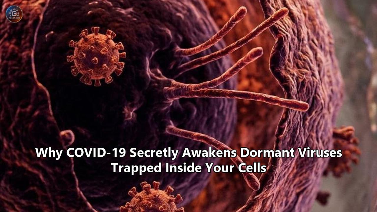
When multi-omics data from international post-viral research consortia converged earlier this year, they confirmed what high-containment laboratories and neuro-immunology clinics had suspected for months: the long-tail pathology of SARS-CoV-2 is not driven solely by the coronavirus itself. Instead, high-throughput proteomic and transcriptomic mapping reveals that SARS-CoV-2 acts as a cellular disruptor, triggering a coordinated, multi-pathogen reactivation cascade across human tissue reservoirs.
For tens of millions of people suffering from debilitating post-acute sequelae, the root cause of prolonged neurological deficits, microvascular injury, and profound metabolic exhaustion often lies in the forced awakening of latent pathogens. The intricate interplay between covid-19 and dormant viruses represents a major shift in how clinicians understand acute respiratory infections, proving that a single viral challenge can destabilize lifelong immune equilibrium.
                     SARS-CoV-2 INVASION
                              │
             ┌────────────────┴────────────────┐
             ▼                                 ▼
   MITOCHONDRIAL SABOTAGE            CYTOKINE HYPERACTIVATION
   • OMM fragmentation               • IL-6, TNF-α, IL-1β surge
   • MAVS proteolysis                • Hyper-phosphorylated Sp1/c-Jun
   • ROS & mtDNA leakage             • Epigenetic locus remodeling
             │                                 │
             └────────────────┬────────────────┘
                              │
                              ▼
                T-CELL BLACKOUT & EXHAUSTION
                • CD28 loss / CD57 elevation
                • Overexpressed PD-1, TIM-3, LAG-3
                • Impaired cytotoxic surveillance
                              │
                              ▼
                THE LATENT-TO-LYTIC SWITCH
   ┌──────────────────────────┼──────────────────────────┐
   ▼                          ▼                          ▼
HERPESVIRIDAE            NEUROTROPIC AGENTS       ENDOGENOUS RETROVIRUSES
• EBV (Zta/Rta fire)     • HHV-6A/B microglial    • HERV-W Env expressed
• CMV endothelial flare    infection              • HERV-K transcription
• VZV ganglion escape    • HSV-1 neuroinflammation • Neurotoxic demyelination
Behind closed doors at academic medical centers, researchers are mapping the biochemical cascade through which SARS-CoV-2 disables epigenetic locks, breaks open cellular reservoirs, and exhausts cytotoxic T-cell defenses. What appears on the surface as lingering post-viral fatigue or autonomic dysfunction is, under the microscope, an active multi-front war between host tissues and long-dormant viral passengers that have lived silently inside human cells for decades.
The Sleeping Virome: What Lies Trapped Within Human Cells
The human body is not a sterile biological system. It is a dense, heavily negotiated ecosystem hosting trillions of microorganisms, including a permanent virome that colonizes host cells shortly after birth. Under normal physiological conditions, the adaptive immune system maintains a state of latency: an evolutionary detente where viral genomes remain quiescent within specific cellular niches, neither replicating aggressively nor causing overt disease.
┌─────────────────────────────────────────────────────────────────────────────┐
│                       HUMAN LATENT VIROME ATLAS                             │
├──────────────────────┬─────────────────────────────┬────────────────────────┤
│ Pathogen             │ Primary Cellular Reservoir  │ Population Seroprevalence│
├──────────────────────┼─────────────────────────────┼────────────────────────┤
│ Epstein-Barr (EBV)   │ Resting Memory B Cells      │ 90% – 95%              │
│ Cytomegalovirus (CMV)│ Myeloid Progenitors/Endoth. │ 60% – 85%              │
│ HHV-6A / HHV-6B      │ CD4+ T Cells, Astrocytes    │ >95% (HHV-6B)          │
│ Varicella-Zoster     │ Dorsal Root/Cranial Ganglia │ >90% (Adults >40)      │
│ HSV-1 / HSV-2        │ Trigeminal/Sensory Ganglia  │ 60% – 80% (HSV-1)      │
│ HERV Elements (W, K) │ Integrated Host Genome (~8%)│ 100% (Endogenous DNA)  │
└──────────────────────┴─────────────────────────────┴────────────────────────┘
The most ubiquitous members of this latent passenger community belong to the Herpesviridae family:
- Epstein-Barr Virus (EBV / HHV-4): Present in more than 90% to 95% of the global adult population, EBV resides inside resting memory B lymphocytes. In its latent state, it survives as a circular extrachromosomal episome tethered to host chromatin by EBV Nuclear Antigen 1 (EBNA-1), expressing minimal viral proteins to evade CD8+ cytotoxic T-cell recognition.
- Cytomegalovirus (CMV / HHV-5): Infecting between 60% and 85% of adults worldwide, CMV establishes long-term persistence in CD34+ hematopoietic progenitor cells, monocytes, and vascular endothelial beds. CMV devotes massive amounts of its large double-stranded DNA genome to subverting immune detection.
- Human Herpesviruses 6A and 6B (HHV-6A/B): HHV-6B is acquired by nearly all children within the first two years of life, remaining latent in peripheral blood mononuclear cells, salivary glands, and brain tissue. In about 1% of the population, HHV-6 integrates directly into the telomeres of human chromosomes, passing through the germline.
- Varicella-Zoster Virus (VZV / HHV-3) & Herpes Simplex Viruses (HSV-1, HSV-2): These neurotropic pathogens retreat into the dorsal root ganglia, cranial nerve ganglia, and trigeminal ganglia following primary infection, held in check by local populations of tissue-resident memory T (TRM) cells.
Beyond exogenous herpesviruses lies a vast, ancient genomic archive: Human Endogenous Retroviruses (HERVs). Comprising roughly 8% of the human genetic code, these sequences represent remnants of ancestral germline infections that occurred millions of years ago.
Normally, host cells silence HERVs using dense chromatin methylation, histone modifications (specifically H3K9me3 marks), and Kruppel-associated box zinc finger proteins (KRAB-ZFPs). When these epigenetic barriers fail, HERV retroelements transcribe pathogenic proteins, including the neurotoxic HERV-W envelope protein (W-ENV) and HERV-K reverse transcriptase, triggering autoimmune and neurodegenerative cascades.
The Molecular Ignition: How SARS-CoV-2 Breaches Viral Silencing
The awakening of latent viruses is neither an accident nor an unlinked secondary infection. It is a direct consequence of the unique biochemical environment created when SARS-CoV-2 infects host tissues.
The primary trigger is a molecular switch: the transition of latent herpesvirus episomes from transcriptional dormancy into the lytic replication cycle.
                     SARS-CoV-2 CELLULAR INGRESS
                                  │
      ┌───────────────────────────┴───────────────────────────┐
      ▼                                                       ▼
STRESS KINASE ACTIVATION                             CHROMATIN REMODELING
Spike/NSPs trigger hyper-phosphorylation            HDAC displacement &
of p38 MAPK, JNK, and PI3K/Akt                       H3K27me3 histone erasure
      │                                                       │
      └───────────────────────────┬───────────────────────────┘
                                  │
                                  ▼
                 EPIGENETIC DEREPRESSION OF BZLF1 (Zp)
               Host Sp1, MEF2D, ATF-2, & c-Jun bind to Zp
                                  │
                                  ▼
                     EXPRESSION OF Zta (ZEBRA)
                 Pioneer transcription factor binds
                     methylated viral promoters
                                  │
                                  ▼
                 LYTIC CASCADE: EARLY & LATE GENES
               BALF2, BALF5, BMRF1 (DNA Polymerase)
                                  │
                                  ▼
                  DE NOVO VIRION SYNTHESIS & BUDDING
1. Epigenetic Derepression of the BZLF1 and BRLF1 Promoters
In latent EBV infections, the switch to the lytic cycle depends on the activation of two immediate-early viral genes: BZLF1 (which encodes the protein Zta, also known as ZEBRA) and BRLF1 (encoding Rta).
Under normal immune surveillance, the promoter controlling BZLF1 (termed the Zp promoter) is kept silent by repressive histone marks (H3K27me3 and H4K20me3) and dense DNA methylation.
When SARS-CoV-2 infects cells, its non-structural proteins (NSPs) and spike glycoprotein initiate hyper-activation of intracellular stress-signaling cascades, specifically the mitogen-activated protein kinase (MAPK)/p38, c-Jun N-terminal kinase (JNK), and phosphoinositide 3-kinase (PI3K)/Akt pathways. Phosphorylation events driven by these kinases activate host transcription factors, including Sp1, MEF2D, ATF-2, and c-Jun.
These host transcription factors translocate into the nucleus and bind with high affinity to the regulatory elements of the Zp and Rp promoters. This binding recruits histone acetyltransferases, displacing histone deacetylases (HDACs) and erasing repressive chromatin marks.
Once Zta is expressed, it functions as a pioneer transcription factor. Unlike most human transcription factors, Zta preferentially binds to methylated CpG dinucleotides within viral promoters that are normally silenced.
Zta then drives the transcription of BRLF1, and together they initiate the early lytic phase: transcribing the viral DNA polymerase processivity factor (BMRF1), the single-stranded DNA-binding protein (BALF2), and the catalytic DNA polymerase subunit (BALF5). The dormant virus begins replicating its genome millions of times over.
┌─────────────────────────────────────────────────────────────────────────────┐
│                   THE THREE STAGES OF EBV LYTIC ACTIVATION                  │
├─────────┬──────────────────────────────┬────────────────────────────────────┤
│ Stage   │ Transcriptional Drivers      │ Downstream Functional Impact       │
├─────────┼──────────────────────────────┼────────────────────────────────────┤
│ Stage 1 │ BZLF1 (Zta), BRLF1 (Rta)     │ Immediate-early transactivation;   │
│         │ Activated via MAPK/p38 & Sp1 │ pioneer chromatin remodeling       │
├─────────┼──────────────────────────────┼────────────────────────────────────┤
│ Stage 2 │ BALF2, BALF5, BMRF1, BALF1   │ Viral DNA polymerase assembly;     │
│         │ Activated by Zta/Rta binding │ viral genome replication begins    │
├─────────┼──────────────────────────────┼────────────────────────────────────┤
│ Stage 3 │ VCA (gp125), MA (gp350),     │ Capsid and envelope assembly;      │
│         │ Viral Glycoproteins          │ Virion maturation, lysis & egress  │
└─────────────────────────┴──────────────┴────────────────────────────────────┘
2. Mitochondrial Sabotage and Innate Immune Collapse
SARS-CoV-2 attacks cellular mitochondria directly. The viral accessory proteins ORF9b, ORF3a, and NSP8 localize to the outer mitochondrial membrane (OMM).
ORF9b binds the translocase of outer mitochondrial membrane 70 (TOM70), suppressing downstream signaling, while ORF3a forms ion-permeable channels that disrupt mitochondrial membrane potential ($\Delta\Psi_m$).
         SARS-CoV-2 ORF9b / ORF3a / NSP8
                       │
                       ▼
       ┌───────────────────────────────┐
       │ OUTER MITOCHONDRIAL MEMBRANE  │
       │         DISRUPTION            │
       └───────────────┬───────────────┘
                       │
       ┌───────────────┴───────────────┐
       ▼                               ▼
OMM TOM70/MAVS DEGRADATION     MEMBRANE COLLAPSE & ROS
• MAVS cleared via autophagy   • Superoxide radical burst
• Type-I IFN synthesis halted  • mtDNA fragments into cytosol
       │                               │
       ▼                               ▼
BLOCKADE OF HOST INTERFERONS   cGAS-STING ABNORMAL SIGNALING
Epigenetic latency breaks down  Persistent neuroinflammatory loop
This mitochondrial destabilization has severe consequences:
- MAVS Proteolysis: Mitochondrial Antiviral Signaling protein (MAVS), anchored to the OMM, forms the central hub for RIG-I and MDA5 sensing of RNA viruses. SARS-CoV-2 induces the ubiquitination and autophagic degradation of MAVS, shutting down the downstream activation of Interferon Regulatory Factors 3 and 7 (IRF3/IRF7) and preventing type-I and type-III interferon expression. Without baseline interferon, the epigenetic suppression of latent herpesviruses breaks down.
- Mitochondrial DNA (mtDNA) Leakage: As the mitochondrial network fragments, fragments of double-stranded mtDNA leak into the host cytoplasm. Cytosolic mtDNA activates the cGAS-STING (cyclic GMP-AMP synthase - stimulator of interferon genes) pathway. However, because SARS-CoV-2 simultaneously inhibits STING phosphorylation, the cell fails to clear the virus, locking it into a state of chronic, unresolved reactive oxygen species (ROS) generation.
- Redox-Driven Viral Induction: The resulting intracellular oxidative stress produces hydrogen peroxide and superoxide radicals that oxidize critical cysteine residues on host repressive complexes, directly driving transcription from the latent viral promoters of EBV, CMV, and HHV-6.
Immunological Exhaustion: The Crippling of CD8+ Cytotoxic T Cells
While intracellular signaling triggers viral transcription inside infected cells, the immune system would ordinarily dispatch CD8+ cytotoxic T lymphocytes (CTLs) and Natural Killer (NK) cells to eliminate those cells before infectious virions escape. In SARS-CoV-2 infection, however, the cytotoxic defense system is compromised.
Flow cytometry profiling of patients with acute and post-acute COVID-19 reveals profound dysfunction within the T-cell compartment:
┌─────────────────────────────────────────────────────────────────────────────┐
│                    CYTOTOXIC T-CELL PHENOTYPIC DRIFT                        │
├──────────────────────┬─────────────────────────────┬────────────────────────┤
│ Surface Marker       │ Healthy / Latent Control    │ Post-COVID Reactivation│
├──────────────────────┼─────────────────────────────┼────────────────────────┤
│ CD28 (Co-stimulatory)│ High Expression (>80%)      │ Markedly Diminished    │
│ CD11a (Integrin)     │ Normal Co-expression        │ Downregulated          │
│ CD57 (Senescence)    │ Low Baseline (<15%)         │ Markedly Elevated      │
│ PD-1 (Inhibitory)    │ Low/Transient Expression    │ Sustained High Levels  │
│ TIM-3 / LAG-3        │ Absent/Basal                │ Strongly Co-expressed  │
└──────────────────────┴─────────────────────────────┴────────────────────────┘
The immunological drivers behind this surveillance blackout include:
CD28 Downregulation and Replicative Senescence
CD8+ T cells specific for EBV and CMV show significant loss of the co-stimulatory receptor CD28 and the adhesion integrin CD11a. Without CD28, T cells cannot receive the "Signal 2" needed for effective activation upon encountering antigen-presenting cells.
Concurrently, these cells upregulate CD57, a terminal marker indicating severe replicative senescence and shortened telomeres. The T cells are locked in an exhausted, non-proliferative state, unable to divide when challenged by reactivating herpesviruses.
                     ANTIGEN RECOGNITION
                              │
               ┌──────────────┴──────────────┐
               ▼                             ▼
       HEALTHY SURVEILLANCE          POST-COVID T-CELL
     • Intact CD28 co-stimulation   • Loss of CD28/CD11a (Signal 2 loss)
     • Perforin/Granzyme release    • High CD57 replicative senescence
     • Rapid clearance of lytic cell • Sustained PD-1/TIM-3 checkpoints
               │                             │
               ▼                             ▼
       VIRAL RE-SILENCING            IMMUNOLOGICAL BLACKOUT
       (Episome stays dormant)       (Unchecked lytic replication)
Exhaustion Checkpoint Overexpression
High-throughput immune profiling reveals sustained upregulation of programmed cell death protein 1 (PD-1), T-cell immunoglobulin and mucin-domain containing-3 (TIM-3), and lymphocyte activation gene 3 (LAG-3) on CMV- and EBV-specific CD8+ T cells.
This multi-checkpoint exhaustion dampens production of interferon-gamma (IFN-$\gamma$), tumor necrosis factor-alpha (TNF-$\alpha$), and cytotoxic granzyme B, allowing reactivating cells to shed intact virions.
Autoantibodies Against Type-I Interferons
Research led by the COVID Human Genetic Effort has uncovered another major mechanism: between 10% and 20% of patients with life-threatening acute COVID-19—and a significant subpopulation of Long COVID patients—harbor circulating autoantibodies that neutralize their own type-I interferons, specifically IFN-$\alpha2$ and IFN-$\omega$.
These autoantibodies blind the immune system to early viral replication, creating a permissive environment for dormant viruses to replicate unchecked.
The Pathogen Spectrum: Clinical Anatomy of Reactivation
The medical consequences of covid-19 and dormant viruses are not uniform. They vary depending on which viral reservoirs are breached, the tissue distribution of those reservoirs, and the host's underlying genetic vulnerabilities.
┌─────────────────────────────────────────────────────────────────────────────┐
│                   SPECTRUM OF REACTIVATED PATHOGENS                         │
├──────────────┬───────────────────────────────┬──────────────────────────────┤
│ Pathogen     │ Primary Tissue / Organ Targets│ Dominant Clinical Pathology  │
├──────────────┼───────────────────────────────┼──────────────────────────────┤
│ EBV          │ B Cells, Epithelium, Endoth.  │ Extreme Fatigue, PEM, Brain  │
│ (HHV-4)      │ Upregulates host ACE2         │ Fog, New Autoantibody Panels │
├──────────────┼───────────────────────────────┼──────────────────────────────┤
│ CMV          │ Endothelium, Gut Mucosa,      │ GI Mucosal Ulceration,       │
│ (HHV-5)      │ Myeloid Progenitor Cells      │ Microvascular Thrombosis     │
├──────────────┼───────────────────────────────┼──────────────────────────────┤
│ HHV-6A /     │ Astrocytes, Oligodendrocytes, │ Neuroinflammation, Severe    │
│ HHV-6B       │ Hippocampus, Glial Networks   │ Cognitive Deficits, POTS     │
├──────────────┼───────────────────────────────┼──────────────────────────────┤
│ VZV          │ Sensory/Cranial Ganglia,      │ Shingles, Ramsay Hunt,       │
│ (HHV-3)      │ Peripheral Nerve Axons        │ Intractable Neuropathic Pain │
├──────────────┼───────────────────────────────┼──────────────────────────────┤
│ HERV-W /     │ Neuronal Glia, Cerebral White │ Neurodegeneration, White     │
│ HERV-K       │ Matter, Perivascular Cells    │ Matter Loss, Demyelination   │
└──────────────┴───────────────────────────────┴──────────────────────────────┘
1. Epstein-Barr Virus (EBV): The Multi-System Driver
EBV reactivation is observed in up to 65% to 70% of hospitalized COVID-19 patients and in a substantial subset of non-hospitalized Long COVID cohorts.
When BZLF1 expression initiates the lytic cycle, EBV does not just replicate quietly:
- ACE2 Receptor Upregulation: EBV lytic replication upregulates the expression of Angiotensin-Converting Enzyme 2 (ACE2) on mucosal epithelial cells and vascular endothelium. This creates a damaging positive feedback loop: SARS-CoV-2 reactivates EBV, and active EBV increases the surface receptor density required for SARS-CoV-2 entry, driving tissue injury.
- Vascular Endothelial Activation: Lytic EBV stimulates human monocytes and macrophages to secrete high levels of TNF-$\alpha$ and IL-6. These cytokines upregulate vascular cell adhesion molecule-1 (VCAM-1) and intercellular adhesion molecule-1 (ICAM-1) on endothelial cells, promoting leukocyte adhesion, microvascular inflammation, and the formation of fibrin amyloid microclots.
- Clinical Presentation: Patients present with the classic symptom cluster of post-viral syndromes: extreme post-exertional malaise (PEM), unrefreshing sleep, temperature dysregulation, and neurocognitive impairment.
                     SARS-CoV-2 CELL ENTRY
                               │
                               ▼
                    EBV LYTIC REACTIVATION
                               │
             ┌─────────────────┴─────────────────┐
             ▼                                   ▼
   UPREGULATED ACE2 DENSITY             CYTOKINE SURGE: TNF-α & IL-6
   • Increased viral entry              • Monocyte hyperactivation
   • Higher SARS-CoV-2 viral load       • Endothelial VCAM-1 / ICAM-1
             │                                   │
             └─────────────────┬─────────────────┘
                               │
                               ▼
               MICROVASCULAR INJURY & CLOTTING
            Endothelial activation, fibrin microclots,
                   and chronic tissue hypoxia
2. Cytomegalovirus (CMV): Endothelial Damage and Tissue Inflammation
Reactivation of CMV during COVID-19 leads to immune remodeling. The virus leaves latency within myeloid cells and infects vascular endothelial beds, causing vascular permeability and microvascular thrombosis.
CMV-driven pathology in this setting includes:
- Expansion of Late-Stage CD8+ EMRA T Cells: CMV reactivation drives the expansion of terminally differentiated effector memory CD8+ T cells expressing CD45RA (termed TEMRA cells). These cells secrete high levels of inflammatory cytokines, accelerating immunosenescence.
- Gastrointestinal Reservoir Seeding: In patients with persistent gastrointestinal Long COVID symptoms, active CMV replication has been identified in gut mucosal tissue biopsies even when CMV DNA remains undetectable in peripheral blood plasma. This localized reactivation damages the intestinal epithelial barrier, allowing microbial lipopolysaccharide (LPS) to enter the systemic circulation and sustain systemic inflammation.
3. Human Herpesvirus 6 (HHV-6A and 6B): Central Nervous System Disruption
HHV-6 is uniquely neurotropic, with a strong affinity for olfactory pathways, glial cells, and hippocampal structures.
- Glial Tropism and Neuroinflammation: Reactivated HHV-6 infects human astrocytes and microglia, inducing cellular swelling and suppressing glutamate transporters (specifically EAAT2/GLT-1). This leads to excitotoxic extracellular glutamate levels in the synaptic cleft, damaging surrounding neurons.
- Hippocampal and Autonomic Dysfunction: Clinical studies link HHV-6 reactivation directly to memory consolidation deficits, spatial disorientation, and dysautonomia, including Postural Orthostatic Tachycardia Syndrome (POTS). Cerebrospinal fluid (CSF) testing in severe post-COVID neurocognitive cases shows elevated levels of HHV-6 DNA accompanied by high concentrations of IL-6, IL-1$\beta$, and neurofilament light chain (NfL), a sensitive biomarker of axonal injury.
                  HHV-6 ASTROCYTE INVASION
                              │
             ┌────────────────┴────────────────┐
             ▼                                 ▼
   GLUTAMATE CLEARANCE LOSS           PRO-INFLAMMATORY CYTOKINES
   • Downregulation of GLT-1/EAAT2    • Elevation of IL-6 & IL-1β in CSF
   • Synaptic glutamate accumulation  • Microglial activation & priming
             │                                 │
             └────────────────┬────────────────┘
                              │
                              ▼
                NEURONAL EXCITOTOXICITY & DAMAGE
              Axonal shedding (elevated NfL in CSF),
             hippocampal dysfunction, and severe POTS
4. Varicella-Zoster Virus (VZV) and HSV-1: Ganglionic Escape
The clinical manifestations of VZV and HSV-1 reactivation are direct and visible:
- VZV Ganglion Egress: Reactivation presents as herpes zoster (shingles) along defined dermatomes, frequently involving cranial nerves. Severe cases lead to Ramsay Hunt syndrome (herpes zoster oticus), causing facial nerve paralysis, intractable vestibular vertigo, and hearing loss.
- Trigeminal Ganglion Reactivation: HSV-1 flares manifest as recurrent oral-facial ulcerations, herpes keratitis, and in rare, severe cases, subacute focal encephalitis localized to the temporal and frontal lobes.
5. Human Endogenous Retroviruses (HERV-W and HERV-K): The Genomic Awakenings
The reactivation of endogenous retroviral sequences integrated into the human genome represents a deeply concerning consequence of severe viral infection.
                     SARS-CoV-2 SPIKE PROTEIN
                                  │
                                  ▼
                     TOLL-LIKE RECEPTOR 4 (TLR4)
                                  │
                                  ▼
                     NF-κB TRANSLOCATION TO NUCLEUS
                                  │
                                  ▼
               EPIGENETIC DEMETHYLATION OF HERV LTRs
                                  │
                                  ▼
              TRANSCRIPTION OF PATHOGENIC HERV-W ENV
                                  │
             ┌────────────────────┴────────────────────┐
             ▼                                         ▼
   NEUROTOXIC TLR4 ENGAGEMENT              MYELIN REPAIR SUPPRESSION
   • Microglial hyper-activation           • Oligodendrocyte Precursor Cell
   • Neurofilament degradation               maturation arrest
• Blood-Brain Barrier breakdown • Progressive white matter loss- Spike-Mediated HERV Induction: SARS-CoV-2 spike protein binds to host Toll-like Receptor 4 (TLR4) on monocytes and glial cells, triggering an NF-$\kappa$B transcriptional wave that demethylates silenced Long Terminal Repeats (LTRs) flanking HERV genes.
- Pathogenicity of HERV-W Envelope Protein (W-ENV): Once transcribed and translated, the W-ENV protein acts as a potent agonist on TLR4 receptors throughout the central nervous system. W-ENV triggers microglial activation, damages the blood-brain barrier, and blocks the maturation of Oligodendrocyte Precursor Cells (OPCs).
- Structural Neurodegeneration: By halting OPC differentiation, W-ENV prevents the normal remyelination of axons, providing a molecular explanation for the white matter loss, cognitive deficits, and neurodegenerative profiles observed in post-COVID neuroimaging studies.
De Novo Autoimmunity: Molecular Mimicry and Epitope Spreading
The forced reactivation of dormant viruses inside human tissues alters how the immune system perceives self-antigens. Prolonged exposure to dual viral antigens—SARS-CoV-2 structural proteins alongside reactivated herpesvirus and HERV proteins—breaks peripheral immune tolerance through two key mechanisms:
                       DUAL PATHOGEN PRESENCE
                     (SARS-CoV-2 + Reactivated EBV)
                                  │
      ┌───────────────────────────┴───────────────────────────┐
      ▼                                                       ▼
MOLECULAR MIMICRY                                     EPITOPE SPREADING
Cross-reactive T-cell receptor /                      Lytic viral lysis releases
antibody engagement with host                         intracellular host autoantigens
      │                                                       │
      ▼                                                       ▼
AUTOANTIBODY GENERATION                               NOVEL AUTOIMMUNE TARGETS
• Anti-GlialCAM (Demyelination)                       • Anti-Ro/SSA & Anti-La/SSB
• Anti-G-Protein Coupled Receptors (POTS)             • Anti-Nuclear Antibodies (ANA)
• Anti-Phospholipid Autoantibodies (Thrombosis)       • Anti-Mitochondrial Abs
      │                                                       │
      └───────────────────────────┬───────────────────────────┘
                                  │
                                  ▼
                     SYSTEMIC AUTOIMMUNE DISEASE
Molecular Mimicry
The clearest example involves Epstein-Barr Virus Nuclear Antigen 1 (EBNA-1). Structural biology and high-throughput peptide mapping demonstrate high sequence and structural homology between EBNA-1 and host GlialCAM, an adhesion molecule expressed on oligodendrocytes and astrocytes.
When SARS-CoV-2 triggers EBV lytic bursts, the subsequent expansion of high-avidity anti-EBNA-1 IgG antibodies leads to cross-reactivity with host GlialCAM, triggering demyelinating auto-inflammatory responses that mirror multiple sclerosis pathology.
Similarly, viral cross-reactivity triggers autoantibodies directed against G-protein coupled receptors (GPCRs)—specifically $\beta2$-adrenergic receptors and M2 muscarinic acetylcholine receptors—directly contributing to the severe dysautonomia, resting tachycardia, and orthostatic intolerance seen in POTS.
Epitope Spreading and Bystander Activation
As lytic herpesviruses destroy infected host cells, they release internal cellular antigens into an inflamed microenvironment filled with activated antigen-presenting cells.
This leads to epitope spreading: the immune system begins generating de novo autoantibodies against nuclear antigens (ANA), ribonucleoproteins (anti-Ro/SSA, anti-La/SSB), and mitochondrial enzymes, locking the patient into a state of chronic systemic autoimmunity long after acute SARS-CoV-2 has been cleared.
The Diagnostic Dilemma: Why Standard Blood Tests Fail
One of the greatest clinical challenges regarding covid-19 and dormant viruses is the widespread failure of standard diagnostic assays to detect tissue-level viral reactivation.
┌─────────────────────────────────────────────────────────────────────────────┐
│                    DIAGNOSTIC RESOLUTION MATRIX                             │
├──────────────────────┬─────────────────────────────┬────────────────────────┤
│ Diagnostic Modality  │ Clinical Limitations        │ Diagnostic Sensitivity │
├──────────────────────┼─────────────────────────────┼────────────────────────┤
│ Plasma qPCR (EBV)    │ Detects only free viremia;  │ Low in tissue-confined │
│                      │ misses lymph/tissue latency │ flares (<25%)          │
├──────────────────────┼─────────────────────────────┼────────────────────────┤
│ Standard IgG Panel   │ Distinguishes only past     │ Inadequate; cannot     │
│ (EBNA-1, VCA IgG)    │ exposure; misses flares     │ assess active lysis    │
├──────────────────────┼─────────────────────────────┼────────────────────────┤
│ Early Antigen (EA-D) │ Sensitive marker of active  │ High for active EBV    │
│ IgG Titers           │ lytic cycle transcription   │ lytic replication      │
├──────────────────────┼─────────────────────────────┼────────────────────────┤
│ Digital Droplet PCR  │ Absolute quantification of  │ Very High for low-copy │
│ (ddPCR)              │ low-copy viral DNA          │ tissue-level shedding  │
├──────────────────────┼─────────────────────────────┼────────────────────────┤
│ PhIP-Seq / VirScan   │ Comprehensive whole-virome  │ Gold Standard for      │
│ Profiling            │ epitope reactivity mapping  │ deep immune profiling  │
└──────────────────────┴─────────────────────────────┴────────────────────────┘
The primary diagnostic blind spots stem from:
- Viremia vs. Tissue-Level Compartmentalization: Routine hospital and commercial laboratory testing relies almost exclusively on quantitative PCR (qPCR) conducted on circulating blood plasma or serum. However, herpesviruses such as EBV, CMV, and HHV-6 can undergo active lytic replication inside lymph nodes, deep bone marrow niches, mucosal lamina propria, and central nervous system parenchyma without shedding detectable viral DNA into peripheral blood. A negative plasma PCR test frequently yields a false-negative assessment of deep-tissue viral reactivation.
- Misinterpretation of Standard Serology: Most standard clinical blood panels test for anti-EBNA-1 IgG (a marker of past infection) and viral capsid antigen (VCA) IgM. In chronic reactivation, VCA IgM often fails to re-emerge. Instead, the gold-standard serological marker for active EBV lytic cycle replication is anti-Early Antigen D (anti-EA-D) IgG, combined with elevated VCA IgG titers. Yet EA-D IgG testing is rarely included in standard clinical evaluations.
- High-Resolution Immunoprofiling Technologies: Next-generation diagnostic pipelines are moving away from single-analyte PCR assays toward systems virology tools:
Phage Immunoprecipitation Sequencing (PhIP-Seq / VirScan): Employs synthetic oligonucleotide libraries encoding complete proteomes of all known human viruses, allowing simultaneous, unbiased measurement of antibody responses across thousands of viral epitopes.
Digital Droplet PCR (ddPCR): Offers absolute quantification of viral DNA target molecules with single-copy sensitivity, detecting minimal viral reactivation in peripheral blood mononuclear cell (PBMC) fractions that conventional qPCR misses.
Soluble Biomarker Panels: Combining anti-EA-D IgG titers with serum neurofilament light chain (NfL), complement activation markers (C4d, sC5b-9), and microclot imaging provides a clearer picture of underlying viral reactivation.
Therapeutic Strategies: Disrupting the Multi-Viral Cycle
Addressing the complex biology of covid-19 and dormant viruses requires a shift away from single-target interventions toward combination therapies designed to silence lytic transcription, rebuild cytotoxic surveillance, and restore metabolic balance.
                   COMPREHENSIVE THERAPEUTIC REGIMEN
                                   │
      ┌────────────────────────────┼────────────────────────────┐
      ▼                            ▼                            ▼
ANTIVIRAL BLOCKADE         IMMUNE RECONSTITUTION        METABOLIC RESTORATION
• Valacyclovir /           • Anti-PD-1/LAG-3            • CoQ10 & NAD+ precursors
  Valganciclovir             checkpoint modulation      • Quench ROS & seal OMM
• Nirmatrelvir/Ritonavir   • Target IL-6/JAK-STAT       • Restore mitochondrial
• Monoclonal anti-W-ENV      (Baricitinib) to close       membrane potential
  (Temelimab)                Zp promoter access         • Inhibit cGAS-STING leak
┌─────────────────────────────────────────────────────────────────────────────┐
│ THERAPEUTIC PIPELINE & TARGETS │
├──────────────────────┬───────────────────────────────┬──────────────────────┤
│ Therapeutic Class │ Molecular Target │ Clinical Objective │
├──────────────────────┼───────────────────────────────┼──────────────────────┤
│ Nucleoside Analogues │ Herpesvirus DNA Polymerase │ Halt EBV, CMV, VZV │
│ (Valacyclovir, etc.) │ (BALF5, UL54) │ viral DNA elongation │
├──────────────────────┼───────────────────────────────┼──────────────────────┤
│ Anti-HERV Monoclonals│ HERV-W Envelope Protein │ Neutralize neurotoxin;│
│ (Temelimab / GNbAC1) │ (W-ENV) / TLR4 signaling │ allow OPC remyelination│
├──────────────────────┼───────────────────────────────┼──────────────────────┤
│ JAK-STAT Inhibitors │ JAK1 / JAK2 Signaling │ Block IL-6 feedback │
│ (Baricitinib) │ Downstream of IL-6/IFN-γ │ loop to close Zp │
├──────────────────────┼───────────────────────────────┼──────────────────────┤
│ Mitochondrial Redox │ CoQ10, Nicotinamide │ Restore OMM integrity;│
│ Modulators │ Riboside, Alpha-Lipoic Acid │ quench lytic-driver ROS│
└──────────────────────┴─────────────────────────────┴────────────────────────┘1. Targeted Antiviral Regimens
Standard nucleoside analogues—including valacyclovir, ganciclovir, valganciclovir, and famciclovir—specifically inhibit viral DNA polymerases (such as EBV BALF5 or CMV UL54) without interfering with host DNA replication.
In clinical cohorts where early lytic reactivation is detected, prolonged courses of high-dose bioavailable antivirals show efficacy in reducing viral loads, lowering systemic inflammation, and easing cognitive and post-exertional symptoms.
Researchers are now evaluating combination trials pairing anti-herpesvirus drugs with oral SARS-CoV-2 proteases (such as nirmatrelvir/ritonavir) to clear persistent coronavirus reservoirs and latent flares simultaneously.
2. Neutralizing Retroviral Proteins: The Temelimab Trials
The therapeutic potential of targeting endogenous retroviruses was tested directly in the Phase 2 clinical trial of temelimab (GNbAC1), a specialized monoclonal antibody engineered to bind and neutralize the pathogenic HERV-W envelope protein (W-ENV).
                     TEMELIMAB MECHANISM OF ACTION
                                   │
                                   ▼
                   MONOCLONAL ANTIBODY BINDING (IgG4)
                     Binds surface epitope of W-ENV
                                   │
                                   ▼
                   BLOCKADE OF TLR4 ENGAGEMENT ON GLIA
                     Stops downstream NF-κB inflammatory
                         and neurodegenerative firing
                                   │
                                   ▼
                   RESTORATION OF OLIGODENDROCYTES
                    OPCs resume differentiation and
                       remyelinate damaged axons
While top-line data from broad, unstratified Long COVID cohorts highlighted the difficulty of using single-target therapies in heterogeneous patient groups, sub-analyses demonstrated that in patients with verified, persistent W-ENV expression and shorter disease duration, neutralizing the retroviral protein led to measurable improvements in neurocognitive performance and markers of brain injury. The findings underscore the need for biomarker-driven patient selection in post-viral clinical trials.
3. Immunomodulation and Kinase Inhibition
To break the cycle of T-cell exhaustion and chronic cytokine signaling, clinical protocols are exploring repurposing Janus kinase (JAK) inhibitors, such as baricitinib and tofacitinib.
By inhibiting the JAK-STAT pathway, these agents suppress the excessive IL-6 and IFN signaling that keeps the Zp and Rp* promoters active. This approach deprives latent herpesviruses of the host transcription factors needed to sustain the lytic cascade, allowing T cells to recover functionality.
4. Mitochondrial Membrane Stabilization
Because mitochondrial breakdown is the foundational trigger linking SARS-CoV-2 entry to redox-driven viral awakening, metabolic and mitochondrial interventions are moving into clinical focus:
- Targeted Antioxidants and Cofactors: High-potency formulations of Coenzyme Q10 (ubiquinol), alpha-lipoic acid, and acetyl-L-carnitine are used to restore the mitochondrial electron transport chain and quench ROS.
- NAD+ Precursors: Supplementation with Nicotinamide Riboside (NR) or Nicotinamide Mononucleotide (NMN) replenishes depleted cellular NAD+ pools, activating mitochondrial sirtuins (SIRT1/SIRT3) that deacetylate histones and reinforce chromatin silencing over latent viral episomes.
What Comes Next: The Paradigm Shift in Post-Viral Medicine
The realization that an acute SARS-CoV-2 infection can destabilize the human virome is reshaping clinical immunology, neurology, and virology.
For decades, post-viral syndromes like Myalgic Encephalomyelitis/Chronic Fatigue Syndrome (ME/CFS) were poorly understood, often dismissed because single-pathogen blood tests came back inconclusive.
The molecular insights gained from studying covid-19 and dormant viruses demonstrate that post-viral illness is rarely driven by one pathogen acting in isolation. It is a systems-level breakdown in which an initial viral insult compromises host defenses, allowing an entire community of latent passengers to emerge and drive multi-organ pathology.
┌─────────────────────────────────────────────────────────────────────────────┐
│                    THE FUTURE POST-VIRAL PROTOCOL                           │
├─────────────────────────────────────────────────────────────────────────────┤
│ 1. ROUTINE VIROME PANELS: Move beyond single PCRs to broad multiplex panels │
│    measuring EA-D IgG, ddPCR viral copies, and HERV-W transcription.       │
├─────────────────────────────────────────────────────────────────────────────┤
│ 2. BIOMARKER STRATIFICATION: Match patients with targeted combinations      │
│    (e.g., Valganciclovir for CMV, Temelimab for HERV-W, Baricitinib for IL-6). │
├─────────────────────────────────────────────────────────────────────────────┤
│ 3. RESTORING METABOLIC BRAKES: Combine antivirals with mitochondrial        │
│    therapeutics (NAD+, CoQ10) to reinforce epigenetic chromatin locks.      │
├─────────────────────────────────────────────────────────────────────────────┤
│ 4. EARLY PROPHYLAXIS: Implement high-risk viral reactivation screenings     │
│    during acute COVID-19 to treat lytic flares before tissue damage occurs. │
└─────────────────────────────────────────────────────────────────────────────┘
The medical milestones to watch over the coming months are clear:
- The launch of larger, biomarker-stratified clinical trials testing combination antiviral therapies against both persistent SARS-CoV-2 and reactivated herpesviruses.
- The regulatory adoption of high-resolution diagnostic panels that evaluate the human virome comprehensively rather than relying on single-analyte plasma tests.
- A fundamental rethinking of acute viral clinical management: recognizing that preventing long-term disability requires monitoring and protecting the fragile epigenetic locks that keep dormant viruses safely trapped inside our cells.
Reference:
- https://insight.jci.org/articles/view/194314
- https://www.researchgate.net/publication/398627636_Long_COVID_involves_activation_of_proinflammatory_and_immune_exhaustion_pathways
- https://www.frontiersin.org/research-topics/79588/latent-virus-reactivation-in-ageing-chronic-disease-or-long-covid-mechanisms-immunity-and-pathogenic-synergy
- https://pmc.ncbi.nlm.nih.gov/articles/PMC13106337/
- https://www.mdpi.com/1422-0067/25/12/6389
- https://pmc.ncbi.nlm.nih.gov/articles/PMC9967513/
- https://pmc.ncbi.nlm.nih.gov/articles/PMC13092548/
- https://pmc.ncbi.nlm.nih.gov/articles/PMC12300560/
- https://www.mdpi.com/1999-4915/14/9/1872
- https://elifesciences.org/articles/86015
- https://tandf.figshare.com/articles/dataset/Reactivation_of_Human_herpesviruses_HHVs_following_SARS-CoV-2_infection_and_vaccination_a_systematic_review_of_clinical_studies/31287516
- https://www.swissbiotech.org/news/geneuro-announces-completion-of-recruitment-of-its-long-covid-phase-2-trial-and-confirms-top-line-results-for-june-2024/
- https://www.fiercebiotech.com/biotech/geneuros-long-covid-trial-fails-triggering-layoffs-and-search-strategic-alternatives
- https://www.frontiersin.org/journals/immunology/articles/10.3389/fimmu.2026.1909260/full
- https://academic.oup.com/femspd/article/doi/10.1093/femspd/ftae002/7590836
- https://www.springermedizin.de/sars-cov-2-and-ebv-the-cost-of-a-second-mitochondrial-whammy/19814132
- https://pmc.ncbi.nlm.nih.gov/articles/PMC8556816/