On a quiet Tuesday morning inside a specialized intensive care unit in Chicago, a 46-year-old patient lies in a medically induced coma. His skin and the whites of his eyes are a deep, luminous saffron—the telltale clinical signature of bilirubin saturating tissue. His kidneys have begun to shut down. His blood refuses to clot normally, its coagulation cascades starved of essential proteins synthesized by the liver.
To the casual observer, this appears to be the acute aftermath of a catastrophic toxic binge. But the clinical chart reveals a confounding reality: the patient has not consumed a drop of alcohol in over six weeks.
For decades, medicine treated this clinical trajectory as an exasperating mystery. The human liver possesses legendary regenerative power; surgical resections of up to 70% of the organ routinely regrow into full, functional tissue within weeks. Yet in severe alcohol-associated liver disease and severe alcohol-associated hepatitis, that miraculous biological engine suddenly seizes. Even when the toxic insult is entirely removed, the liver frequently refuses to heal. Instead, it enters a lethal downward spiral that kills up to 50% of severe patients within 90 days without an emergency organ transplant.
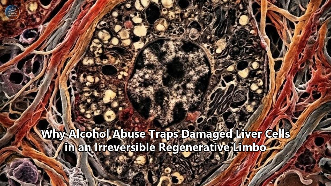
Now, a series of multi-institutional investigations has cracked open the molecular black box of this regenerative failure. Published across landmark papers in Nature Communications, Gut, and the Journal of Hepatology, researchers from the University of Illinois Urbana-Champaign, Duke University, the University of Edinburgh, and the Chan Zuckerberg Biohub have uncovered a striking biological phenomenon: alcohol abuse does not merely kill liver cells—it lures them into a developmental detour and traps them in a state of irreversible regenerative limbo.
The findings dismantle old assumptions that alcohol-associated liver collapse is simply the result of cell death outpacing cell division. Instead, the evidence reveals a far more insidious mechanism: damaged liver cells attempt to repair the organ by rewinding their genetic clocks toward an embryonic state, only to find the molecular bridge back to maturity permanently burned behind them.
The Autopsy of a Stalled Engine
To understand how the liver gets caught in this cellular trap, one must first look at the unique way it heals under normal physiological conditions.
Unlike the heart or the central nervous system, which rely on rare stem cell pools or scar formation to patch up damage, the liver is composed of workhorse cells known as hepatocytes. These mature, highly specialized cells carry out more than 500 vital metabolic tasks: detoxifying metabolic waste, manufacturing albumin, synthesizing blood-clotting factors, and managing lipid and glucose homeostasis.
When a healthy liver is cut or chemically injured, mature hepatocytes perform a controlled cellular maneuver. They temporarily downregulate their high-demand metabolic machinery and dedifferentiate—shifting into a flexible, proliferative, fetal-like state. In this quasi-progenitor state, they rapidly divide to produce new biomass. Once sufficient tissue mass is restored, an intricate cascade of genetic switches flips them back into mature, functioning adult cells.
Normal Liver Regeneration:
[Mature Hepatocyte] ──(Injury)──► [Proliferative Quasi-Progenitor] ──(Division)──► [Mature Hepatocyte Restored]

Alcohol-Trapped Regeneration:
[Mature Hepatocyte] ──(Alcohol/Inflammation)──► [Quasi-Progenitor Limbo] ──(Splicing & Epigenetic Arrest)──► ✖ PARALYSIS / SASP
In livers ravaged by chronic alcohol abuse, this cycle breaks down halfway through.
"We knew that the liver stops functioning and stops regenerating in patients with alcohol-related hepatitis and cirrhosis, even when a patient has discontinued consuming alcohol, but we didn't know why," explains Dr. Auinash Kalsotra, professor of biochemistry at the University of Illinois Urbana-Champaign, who co-led one of the primary investigations alongside Duke University School of Medicine professor Dr. Anna Mae Diehl. "The only real life-saving treatment option once a patient reaches the liver failure stage in those diseases is transplantation. But if we understood why these livers were failing, maybe we could intervene".
Analyzing explanted liver tissue from patients with severe alcohol-associated hepatitis alongside healthy donor livers from the National Institute on Alcohol Abuse and Alcoholism (NIAAA) repository at Johns Hopkins University, the research teams mapped out gene expression at single-cell resolution.
The forensic evidence pointed to an unexpected cellular demographic: the diseased livers were not empty wastelands stripped bare of hepatocytes. They were crowded with living cells that had successfully begun the journey backward into a regenerative state, but had become utterly unable to finish the transition.
"They are neither functional adult cells nor proliferative progenitor cells," explains Dr. Sushant Bangru and Dr. Ullas Chembazhi, co-first authors of the UIUC study. "Since they are not functioning, more pressure builds on the remaining cells. So they try to regenerate, and they're all ending up in this unproductive quasi-progenitor state, and that's what is causing liver failure".
The Splicing Trap: When RNA Processing Goes Rogue
Why do these cells get stuck? The investigative trail led Kalsotra, Diehl, and their collaborators straight to the cell's internal editing room: alternative RNA splicing.
When a gene is transcribed inside a cell, the raw precursor messenger RNA (pre-mRNA) must be spliced—cut and reassembled—to produce the final template for functional proteins. By including or excluding different sections of genetic material (exons), a single gene can produce distinct protein variations, known as isoforms, tailored for specific cellular tasks. A fetal liver cell, for example, relies on an entirely different suite of protein isoforms than a mature hepatocyte managing toxic bile acids.
Under ordinary circumstances, the master regulator of adult liver splicing is an RNA-binding protein called Epithelial Splicing Regulatory Protein 2 (ESRP2). During embryonic development, ESRP2 levels are low, allowing fetal liver cells to proliferate wildly without carrying the burden of full metabolic function. As an organism matures, ESRP2 expression surges, locking the splicing machinery into an adult configuration that produces mature metabolic enzymes, structural proteins, and cell-cycle checkpoints.
In livers suffering from advanced alcohol liver damage, the researchers uncovered a profound collapse of this splicing control system.
┌────────────────────────────────────────────────────────────────────────┐
│                        THE MOLECULAR SPLICING JAM                      │
├────────────────────────────────────────────────────────────────────────┤
│ Chronic Ethanol & Toxic Metabolites (Acetaldehyde, ROS)                 │
│                                │                                       │
│                                ▼                                       │
│ Massive Influx of Inflammatory Cytokines (TGF-β, TNF-α, IL-1β)         │
│                                │                                       │
│                                ▼                                       │
│ Severe Repression of Splicing Factors (ESRP2, ZFP36L1)                 │
│                                │                                       │
│                                ▼                                       │
│ Widespread Missplicing of Pre-mRNA (Exon Skipping, Aberrant Isoforms) │
│                                │                                       │
│                                ▼                                       │
│ Production of Malfunctioning Structural & Cell-Cycle Proteins          │
│                                │                                       │
│                                ▼                                       │
│ Hepatocytes Jammed in "Quasi-Progenitor" Limbo (Cannot Divide or Work) │
└────────────────────────────────────────────────────────────────────────┘
Chronic ethanol intake and its reactive breakdown products trigger a continuous flood of pro-inflammatory signaling molecules—chief among them Transforming Growth Factor-beta (TGF-β) and Tumor Necrosis Factor-alpha (TNF-α). This unrelenting inflammatory milieu directly suppresses ESRP2 and related RNA-binding proteins like ZFP36L1.
Deprived of their splicing conductors, the regenerating hepatocytes begin missplicing thousands of transcripts. Key proteins involved in the mitotic spindle, chromosomal segregation, and metabolic detoxification are produced in abnormal, defective, or embryonic isoforms.
The cell attempts to divide, but its internal structural machinery is deformed. It attempts to clear toxins, but its metabolic enzyme isoforms are non-functional. Caught in this mechanical jam, the hepatocyte triggers an emergency internal brake: it arrests its own cell cycle permanently to prevent catastrophic chromosomal damage, without executing programmed cell death (apoptosis).
It becomes a biological zombie—alive, consuming nutrients, occupying physical space, but functionally dead to the organ and incapable of completing regeneration.
The Histological Crime Scene: The Ductular Reaction Illusion
For over a century, pathologists looking down the barrels of microscopes at biopsies from patients with end-stage alcohol-related cirrhosis saw a hallmark pattern known as the "ductular reaction".
The periportal zones of the liver—the areas surrounding incoming blood supplies—are consistently choked with dense, web-like tangles of cells resembling primitive bile ducts. These cells express markers characteristic of both bile duct cells (cholangiocytes) and hepatocytes, displaying proteins such as Cytokeratin 7 (KRT7/CK7), Cytokeratin 19 (CK19), and Albumin (ALB).
HISTOLOGICAL MARKERS IN ALCOHOL-INDUCED DUCTULAR REACTION
┌──────────────────────┬────────────────────────┬───────────────────────────────────────────┐
│ Cellular Marker      │ Normal Cell Source     │ Pathological State in ALD                 │
├──────────────────────┼────────────────────────┼───────────────────────────────────────────┤
│ Albumin (ALB)        │ Mature Hepatocytes     │ Co-expressed in trapped progenitor cells   │
│ Cytokeratin 7 (KRT7) │ Biliary Cholangiocytes │ Aberrantly turned on in dediff. parench.  │
│ Cytokeratin 19 (CK19)│ Biliary Cholangiocytes │ Marks expansive, disorganized ductules    │
│ p21 (CDKN1A)         │ Transient arrest cells │ Staining saturation indicates senescence  │
│ SOX9                 │ Progenitor/Biliary     │ Locked on; prevents hepatocyte maturation │
└──────────────────────┴────────────────────────┴───────────────────────────────────────────┘
Historically, this ductular explosion was interpreted as a valiant, if desperate, emergency repair effort. Medical wisdom held that when mature hepatocytes were destroyed in massive numbers, dormant Hepatic Progenitor Cells (HPCs) located in the Canals of Hering stepped forward, dividing rapidly to generate new parenchyma to replace the lost liver volume.
Recent multiomic spatial profiling conducted at the University of Edinburgh's Institute for Regeneration and Repair tells a far darker story.
By tracking individual cellular lineages in human liver biopsies across time, researchers like Professor Stuart Forbes discovered that the ductular reaction in severe alcohol injury is not an organized rescue brigade. It is a biological mirage.
The cells comprising this ductular reaction are largely mature hepatocytes that have dedifferentiated in an effort to heal the tissue, alongside biphenotypic progenitor cells that have begun differentiating toward hepatocytes. But due to the disrupted signaling microenvironment of the alcoholic liver, they become stranded in this intermediate state.
"In a healthy liver subjected to acute injury, hepatocytes can transiently dedifferentiate, proliferate, and then redifferentiate into mature, functioning hepatocytes," says Dr. Jan Petrasek, an expert in transplant hepatology and alcohol-associated liver disease at the Texas Liver Institute and the University of Texas Health Science Center at San Antonio. "In severe alcohol-associated liver disease and severe alcohol-associated hepatitis, hepatocytes enter that initial transitional, fetal-like state but do not complete the maturation step. Instead of repopulating functional parenchyma, they remain metabolically inefficient and synthetically weak".
Dr. Petrasek emphasizes that this is what clinicians see at the bedside: "Functionally, in patients, that looks like persistent hyperbilirubinemia, severe coagulopathy, and plummeting albumin levels despite the complete removal of the inciting toxin".
Rather than forming functioning liver plates, these trapped ductular cells form primitive, blind-ended, lumenless micro-tubules. They cannot process bile, which backs up into the bloodstream, worsening cholestasis and poisoning other organ systems.
The Epigenetic Deadbolt: Silencing HNF4α
To understand why a cell cannot simply snap out of this limbo once the patient stops drinking, one must look at the chromatin architecture that defines cellular identity.
Every cell in the human body contains the exact same DNA sequence. What makes a hepatocyte distinct from a neuron or a white blood cell is its epigenetic programming—the specific patterns of chemical tags, histone modifications, and chromatin packing that determine which genes can be transcribed and which are locked away.
At the center of mature liver identity sits a master transcription factor called Hepatocyte Nuclear Factor 4-alpha (HNF4α). HNF4α functions like an operating system for liver parenchyma. It binds directly to the regulatory regions of thousands of genes responsible for:
- The urea cycle (converting neurotoxic ammonia into excretable urea)
- Gluconeogenesis and glycogen storage
- Bile acid synthesis and conjugation
- Coagulation factor production (Fibrinogen, Prothrombin, Factors VII, IX, X)
- Apolipoprotein production for cholesterol transport
                       ┌─────────────────────────────────────┐
                       │  HEALTHY ADULT LIVER HOMEOSTASIS    │
                       │  P1 Promoter Active ──► High HNF4α  │
                       └──────────────────┬──────────────────┘
                                          │
                                          ▼
     ┌────────────────────────────────────┴────────────────────────────────────┐
     │ Maintains: Urea Cycle, Coagulation Factors, Bile Conjugation, Glycogen  │
     └─────────────────────────────────────────────────────────────────────────┘
                                          │
                                          │ Chronic Ethanol Metabolism
                                          │ (Acetaldehyde, ROS, TGF-β, Histone Deacetylation)
                                          ▼
                       ┌─────────────────────────────────────┐
                       │      EPIGENETIC COLLAPSE IN ALD     │
                       │  P1 Silenced / P2 Switched / PRC2   │
                       └──────────────────┬──────────────────┘
                                          │
                                          ▼
     ┌────────────────────────────────────┴────────────────────────────────────┐
     │ Loss of HNF4α ──► Chromatin Compaction of Metabolic Genes               │
     │ Hyper-Activation of Biliary & Fetal Programs (SOX9, KRT7, Jagged1)      │
     │ RESULT: "Epigenetic Deadbolt" — Irreversible Metabolic Amnesia          │
     └─────────────────────────────────────────────────────────────────────────┘
In human liver tissue severely damaged by alcohol, investigators have discovered that HNF4α expression is crushed.
This is not a temporary dip in production; it is an epigenetic shutdown driven by an upstream promoter switch and aggressive chromatin remodeling. The HNF4α gene possesses two distinct promoters: the canonical P1 promoter, which drives expression in adult hepatocytes, and the P2 promoter, which is active during embryonic fetal development. Chronic alcohol-induced cellular stress, combined with the presence of TGF-β, prompts histone deacetylases (HDACs) and Polycomb Repressive Complex 2 (PRC2) to place repressive methylation marks (such as H3K27me3) across the P1 promoter.
The cell switches to the embryonic P2 promoter or stops expressing the protein altogether.
When HNF4α is lost, the mature hepatocyte suffers a form of cellular amnesia. The chromatin regions harboring adult metabolic genes physically condense into tightly wrapped, inaccessible heterochromatin. Meanwhile, genomic loci containing fetal and biliary markers—such as SOX9, KRT7, and Jagged1—are unwrapped and transcribed.
The cell loses its identity. It can no longer remember how to act as an adult metabolic engine, but it lacks the complete developmental roadmap required to function as an independent progenitor cell. The epigenetic deadbolt has been turned from the inside.
The Secretory Poison: Paracrine Senescence and SASP
If trapped quasi-progenitor cells simply sat silently in the liver, the remaining healthy tissue might eventually compensate. But senescent cells do not stay quiet.
When hepatocytes enter this stalled state, they trigger the high-level activation of the cyclin-dependent kinase inhibitors p21 (CDKN1A) and p16 (CDKN2A). This arrests their cell cycles permanently in the G1 phase, completely preventing DNA replication.
┌──────────────────────────────────────────────────────────────────────────────────┐
│               THE SENESCENCE-ASSOCIATED SECRETORY PHENOTYPE (SASP)                │
├──────────────────────────────────────────────────────────────────────────────────┤
│                                                                                  │
│   [ Trapped Quasi-Progenitor / Senescent Hepatocyte ]                             │
│                           │                                                      │
│       ┌───────────────────┼───────────────────┬───────────────────┐              │
│       │ (IL-6, IL-8)      │ (TGF-β, PDGF)     │ (MMP-2, MMP-9)    │ (CCL2, CXCL1)│
│       ▼                   ▼                   ▼                   ▼              │
│  [Paracrine Contagion] [Stellate Cells]   [Matrix Stiffness]  [Neutrophil Blast] │
│  Infects neighboring   Converts HSCs into Degrades normal      Massive influx of │
│  viable hepatocytes    hyperactive        scaffolding; lays   destructive       │
│  with p21-senescence   myofibroblasts     collagen cage       leukocytes        │
│                                                                                  │
└──────────────────────────────────────────────────────────────────────────────────┘
Once locked into this state, the cells develop a hyperactive secretory condition known as the Senescence-Associated Secretory Phenotype (SASP). Rather than performing metabolic work, the cell converts into a cytokine factory, pumping out toxic streams of:
- Pro-inflammatory Interleukins (IL-1β, IL-6, IL-8): Signaling molecules that act as chemical flares, drawing swarms of inflammatory cells directly into the liver parenchyma.
- Transforming Growth Factor-Beta (TGF-β): A master driver of tissue scarring and a potent inhibitor of healthy cell division.
- Chemokines (CCL2, CXCL1): Signals that recruit non-specific neutrophils and monocytes, which release reactive oxygen species and cytotoxic proteases, damaging adjacent healthy tissue.
- Matrix Metalloproteinases (MMPs): Enzymes that tear down the liver's delicate extracellular matrix, destroying the natural spatial scaffolding necessary for organized tissue architecture.
This process causes a dangerous chain reaction termed paracrine senescence.
In a landmark trial analysis published by an international consortium studying longitudinal transjugular liver biopsies from patients with severe alcohol-associated hepatitis participating in the ISAIAH clinical trial, researchers proved that SASP acts as a localized toxin.
A single hepatocyte trapped in senescence secretes enough TGF-β, IL-6, and reactive oxygen species to force adjacent, undamaged hepatocytes into senescence as well. The condition spreads through the liver parenchyma like a brushfire. Even if a patient achieves complete sobriety, the self-sustaining SASP microenvironment continues to infect healthy bystander cells, progressively shrinking the pool of functional liver tissue long after the last drink.
The Biomechanical Cage: How Stellate Cells Lock the Grid
As the SASP brew floods the liver microenvironment, it impacts another key cell type: the Hepatic Stellate Cell (HSC).
In a healthy liver, quiescent stellate cells sit quietly in the Space of Disse—the microscopic gap between hepatocytes and blood capillaries (sinusoids)—storing Vitamin A and maintaining baseline matrix chemistry.
When exposed to TGF-β and Platelet-Derived Growth Factor (PDGF) pouring out of senescent, trapped hepatocytes, stellate cells undergo transdifferentiation into highly contractile, proliferative myofibroblasts. These activated myofibroblasts begin churning out massive quantities of dense, cross-linked Type I and Type III collagen.
                      ┌────────────────────────────────────────┐
                      │    THE MECHANICAL-BIOLOGICAL MATRIX    │
                      └──────────────────┬─────────────────────┘
                                         │
                                         ▼
                 Trapped Hepatocytes Secrete Profuse TGF-β & PDGF
                                         │
                                         ▼
                 Hepatic Stellate Cells Transdifferentiate into Myofibroblasts
                                         │
                                         ▼
                 Massive Deposition of Cross-Linked Type I/III Collagen
                                         │
                                         ▼
                 Tissue Microenvironment Stiffens Dramatically (Fibrosis)
                                         │
                                         ▼
                 Hippo/YAP Mechanotransduction Distorted; Sinusoids Capillarize
                                         │
                                         ▼
                 Paracrine Factors Cannot Clear; Hepatocytes Starved of Oxygen
This collagen deposition creates physical barriers that encase trapped hepatocytes in rigid, fibrotic cages.
This physical stiffening fundamentally changes the biomechanics of the liver. Through mechanotransduction pathways governed by the Hippo signaling cascade and its transcriptional co-activators YAP/TAZ, extreme physical stiffness signals to hepatocytes that they should not divide as normal epithelial sheets. Instead, mechanical stress enforces the progenitor arrest.
At the same time, the specialized, fenestrated endothelial cells lining the liver's blood vessels undergo "capillarization". The microscopic pores that normally allow blood plasma, nutrients, and oxygen to flow freely over hepatocytes close up, replaced by a continuous basement membrane of scar tissue.
The trapped hepatocytes are physically sealed off from their blood supply. Starved of oxygen (hypoxia) and unable to exchange waste products, their intracellular metabolism breaks down entirely.
The Bioenergetic Blackout: Mitochondrial Collapse
A regenerating cell is an energy-hungry machine. To duplicate three billion base pairs of DNA, synthesize millions of structural proteins, and build new cellular membranes, a single hepatocyte requires vast quantities of Adenosine Triphosphate (ATP).
In patients with advanced alcohol liver damage, that bioenergetic currency is completely unavailable.
                          ┌────────────────────────┐
                          │   ETHANOL METABOLISM   │
                          └───────────┬────────────┘
                                      │
                         ADH / CYP2E1 │ Generation of Acetaldehyde
                                      ▼ & Superoxide (ROS)
                 ┌──────────────────────────────────────────┐
                 │       MITOCHONDRIAL DNA (mtDNA) ADDUCTS  │
                 └────────────────────┬─────────────────────┘
                                      │
                                      ▼
                 Breakdown of Cytochrome c Oxidase & Complexes I/III
                                      │
                                      ▼
                 Loss of Mitochondrial Membrane Potential (ΔΨm)
                                      │
                                      ▼
                 ATP Production Plummets by up to 80%
                                      │
                                      ▼
                 Metabolic Energy Insufficient for Mitosis & Cytokinesis
Ethanol metabolism occurs primarily via two enzymatic pathways: Alcohol Dehydrogenase (ADH) in the cytoplasm and Cytochrome P450 2E1 (CYP2E1) in the endoplasmic reticulum. Both pathways generate high levels of acetaldehyde—a reactive chemical that binds directly to proteins, lipids, and nucleic acids—along with massive surges of reactive oxygen species (ROS).
Mitochondria possess their own distinct circular genome (mtDNA), which lacks protective histones and sophisticated DNA repair mechanisms. Acetaldehyde and hydroxyl radicals cause extensive mtDNA deletions and form disruptive DNA adducts.
Multiomic investigations into human liver biopsies have pinpointed a breakdown of Cytochrome c Oxidase (Complex IV of the electron transport chain) and mitochondrial Complexes I and III in hepatocytes subjected to chronic alcohol exposure. The mitochondrial inner membrane potential ($\Delta\Psi_m$) collapses.
Under electron microscopy, the mitochondria of these trapped cells do not appear as healthy, elongated organelles. They are swollen, fractured, and disorganized—a pathological state known as "megamitochondria."
With cellular ATP production dropping by up to 80%, the hepatocyte cannot generate the energy needed to power the mitotic machinery. The cell enters metabolic exhaustion. It is caught between two worlds: it has initiated a resource-heavy dedifferentiation process, but lacks the basic ATP required to complete division or restore baseline mature function.
The Signaling Tug-of-War: Notch Versus Wnt
When a liver cell attempts to regenerate, its developmental direction is controlled by two opposing evolutionary signaling cascades: the Wnt/$\beta$-catenin pathway and the Notch pathway.
- Wnt/$\beta$-catenin is the pro-hepatocyte signal. When Wnt signaling is dominant, liver progenitor cells and dedifferentiated hepatocytes are guided toward maturing into adult, functional hepatocytes capable of clearing toxins and producing albumin.
- Notch is the pro-biliary signal. When Notch signaling is activated, it instructs cells to adopt a biliary phenotype, directing them to form bile ducts and cholangiocyte-like structures.
                     ┌─────────────────────────────────────────┐
                     │ THE REGENERATIVE SIGNALING CROSSROADS   │
                     └────────────────────┬────────────────────┘
                                          │
                  ┌───────────────────────┴───────────────────────┐
                  ▼                                               ▼
         [ WNT / β-CATENIN ]                                  [ NOTCH ]
     (Pro-Hepatocyte Pathway)                          (Pro-Biliary Pathway)
                  │                                               │
     Directs maturation into                          Directs branching into
     functional adult hepatocytes                     tubular bile ducts
                  │                                               │
                  │ Ethanol Exposure                              │ High Jagged1 &
                  │ Severely Inhibits                             │ TGF-β Induces
                  ▼                                               ▼
       ❌ REGENERATION CRUSHED                        ⚠️ RUNAWAY DUCTULAR CHAOS
   (Synthetic & metabolic arrest)                 (Non-functional, profibrotic nests)
In severe alcohol-induced disease, this signaling balance swings out of control.
Ethanol metabolism directly represses the canonical Wnt/$\beta$-catenin signaling pathway in parenchymal cells. At the same time, the surrounding inflamed immune cells and activated stellate cells overexpress Jagged1, the primary ligand that activates Notch receptors on neighboring hepatocytes and progenitors.
This creates an insurmountable directional conflict.
Cells that have dedifferentiated in an effort to regenerate receive zero Wnt cues telling them how to rebuild functional liver plates. Instead, they are bombarded by Notch and TGF-β signals forcing them down an incomplete biliary pathway.
They begin expressing bile duct markers like Cytokeratin 7 and Cytokeratin 19, yet they lack the proper architectural environment to assemble functional bile ducts.
A recent single-cell transcriptomic study evaluating epithelial populations across distinct stages of alcohol-associated liver disease identified these expanding ALB+/KRT7+ biphenotypic cells as an evolutionary dead-end.
Instead of rescuing hepatic mass, this trapped population acts as a nidus for chronic inflammation, secreting profibrotic growth factors that accelerate cirrhosis. In advanced stages, these stalled cells can even acquire secondary oncogenic mutations, transforming this regenerative limbo into a direct breeding ground for hepatocellular carcinoma (HCC).
The Clinical Crossroads: The Illusion of Abstinence
The discovery of this multi-tiered molecular arrest transforms how hepatologists interpret the clinical progression of severe alcohol-associated liver disease.
For decades, public health messaging and traditional clinical wisdom operated under a simple premise: alcohol is an external toxin; remove the toxin, and the liver will heal itself.
While this holds true in early stages of fatty liver (steatosis) and mild inflammation, it breaks down completely once hepatocytes cross the threshold into the quasi-progenitor, senescent state.
STAGE 1: Simple Steatosis (Fatty Liver)
  - Ethanol metabolism increases triglyceride storage.
  - Fully reversible within 2 to 4 weeks of complete abstinence.

STAGE 2: Steatohepatitis & Early Fibrosis
  - Infiltration of neutrophils and initial stellate cell activation.
  - High capacity for tissue repair; regenerative pathways intact.

STAGE 3: Severe Alcohol-Associated Hepatitis / Decompensated Cirrhosis
  - Alternative splicing machinery breaks down (ESRP2/ZFP36L1 lost).
  - Widespread hepatocyte senescence (p21 activation & SASP secretion).
  - Massive, non-functional ductular reaction (ALB+/KRT7+ expansion).
  - Epigenetic silencing of HNF4α.
  - BIOLOGICAL RESULT: Regenerative Limbo. Abstinence alone fails to restore
    parenchymal function; 90-day mortality climbs to 30–50%.
When a patient arrives in the hospital with severe alcohol-associated hepatitis—often marked by sudden-onset jaundice, ascites (fluid accumulation in the abdomen), hepatic encephalopathy (brain dysfunction caused by toxic ammonia build-up), and systemic inflammatory response syndrome (SIRS)—their liver is already caught in this cellular deadlock.
"This explains why patients with severe alcohol-related hepatitis frequently continue to deteriorate and die even after they have completely stopped drinking," says Dr. Kalsotra. "The insult started the fire, but the inflammatory and missplicing loops keep it burning indefinitely".
Because the cells are trapped in a non-functional state, the metabolic workload on the few remaining healthy hepatocytes multiplies exponentially.
These overworked survivor cells are forced to attempt their own dedifferentiation and division cycles to compensate, only to enter the same hostile, cytokine-rich microenvironment, where they promptly succumb to splicing failure, epigenetic silencing, and paracrine senescence.
The disease accelerates through an internal domino effect.
                     ┌─────────────────────────────────────────┐
                     │ THE INTERNAL DOMINO EFFECT IN ALD       │
                     └────────────────────┬────────────────────┘
                                          │
                                          ▼
                      Hepatocytes Enter Regenerative Limbo
                                          │
                                          ▼
                      Metabolic Capacity of the Liver Drops
                                          │
                                          ▼
                      Excess Workload Shifts to Surviving Cells
                                          │
                                          ▼
                      Surviving Cells Attempt Emergency Division
                                          │
                                          ▼
                      Hostile SASP Microenvironment Jams Them
                                          │
                                          ▼
                      Systemic Failure: Encephalopathy, Jaundice, Death
Mapping the Escape Routes: Next-Generation Therapies
Understanding that regenerative failure is an active, locked state—rather than a passive absence of living cells—has opened up unexpected avenues for therapeutic intervention.
Current pharmacological care for severe alcohol-associated hepatitis remains reliant on corticosteroids, such as prednisolone. Corticosteroids act as broad anti-inflammatory agents, but their real-world clinical benefits are modest: they improve short-term 28-day survival by only a small margin and do nothing to reverse long-term mortality, while significantly increasing the risk of life-threatening systemic fungal and bacterial infections.
Armed with new high-resolution maps of the regenerative jam, researchers across the globe are designing precision molecular tools to free trapped hepatocytes from biological stasis.
┌────────────────────────┬─────────────────────────────┬───────────────────────────────────────────┐
│ Therapeutic Class      │ Molecular Target            │ Biological Mechanism                      │
├────────────────────────┼─────────────────────────────┼───────────────────────────────────────────┤
│ Splicing Restorers     │ ESRP2, ZFP36L1 Pathways     │ Rescues pre-mRNA splicing; allows cells   │
│                        │ (e.g., TGF-βR1 inhibitors)  │ to finish mitosis and rebuild parenchyma  │
│ Senolytics             │ BCL-2/BCL-xL (ABT-263),     │ Selectively clears toxic senescent cells; │
│                        │ Dasatinib + Quercetin (D+Q) │ halts paracrine SASP contagion            │
│ Epigenetic Reprogram-  │ LNP-delivered HNF4α mRNA;   │ Restores master operating system; turns   │
│ mers / mRNA Therapies  │ HDAC / PRC2 Inhibitors      │ metabolic/synthetic genes back on         │
│ Lineage Guidance       │ Notch Inhibitors (γ-secre-  │ Blocks runaway biliary expansion; drives  │
│ Small Molecules        │ tase inh.) / Wnt Agonists   │ progenitors to differentiate to hepatocytes│
│ Bioenergetic Protectors│ Mitochondrial Antioxidants; │ Restores ATP generation needed to power   │
│                        │ Complex IV Stabilizers      │ DNA replication and cellular cytokinesis  │
└────────────────────────┴─────────────────────────────┴───────────────────────────────────────────┘
1. Splicing Correction and ESRP2 Restoration
In experimental models developed by the University of Illinois and Duke University teams, researchers tested whether the splicing breakdown could be reversed pharmacologically.
By treating damaged liver cultures with small molecules that selectively inhibit the TGF-β receptor kinase (such as galunisertib) or downstream inflammatory cascades, they successfully relieved the repression on ESRP2.
Once ESRP2 levels rebounded, hepatocytes corrected their pre-mRNA splicing patterns. The cells were finally able to navigate the transition out of the quasi-progenitor state, exit cell-cycle arrest, and mature into fully functioning adult hepatocytes capable of synthesizing albumin and urea.
2. Senolytics: Pruning the Rotten Apples
Rather than trying to force senescent cells back into action, another therapeutic approach seeks to eliminate them entirely.
Senolytic drugs—including the dual BCL-2/BCL-xL inhibitor navitoclax (ABT-263) and combinations of dasatinib and the flavonoid quercetin (D+Q)—specifically target the anti-apoptotic survival pathways that keep senescent cells alive.
In preclinical models of severe liver injury, clearing senescent hepatocytes with senolytics eliminates the primary source of SASP. Once the flood of toxic cytokines, TGF-β, and reactive oxygen species stops, the surrounding healthy parenchyma is released from paracrine arrest. Surviving non-senescent hepatocytes can then proliferate cleanly without getting trapped.
3. Epigenetic Rebooting with HNF4α mRNA
One of the most ambitious therapeutic strategies involves using lipid nanoparticles (LNPs)—the same delivery technology behind mRNA vaccines—to deliver synthetic HNF4α mRNA directly to hepatocytes.
By delivering functional transcripts of the master transcription factor directly into the cytoplasm, researchers can bypass the silenced P1 promoter. The burst of translated HNF4α protein enters the nucleus, opens closed chromatin regions, and forcefully restarts the genetic programs governing ammonia detoxification, bile processing, and blood clotting.
In early rodent studies, direct restoration of HNF4α successfully rescued animals from acute-on-chronic liver failure, causing dedifferentiated ductular cells to mature into functional liver tissue.
4. Lineage Steering: Modulating the Wnt/Notch Axis
To solve the ductular reaction puzzle, translational laboratories are testing dual-action pharmacological cocktails that block Notch signaling while stimulating the Wnt/$\beta$-catenin pathway.
By utilizing small-molecule $\gamma$-secretase inhibitors to turn off Notch signaling, scientists can prevent trapped biphenotypic progenitor cells from forming useless, pro-fibrotic ductules. Concurrently administering small-molecule Wnt mimetics or GSK-3$\beta$ inhibitors (such as CHIR99021) stabilizes $\beta$-catenin, actively steering these cells toward becoming mature, working hepatocytes.
This converts the ductular reaction from a harmful scar-forming process into a productive tissue-regeneration engine.
Redefining Cellular Fate
The realization that alcohol abuse locks damaged liver cells into a state of regenerative limbo marks a turning point in our understanding of organ failure.
For generations, medicine viewed tissue damage through a binary lens: cells either survived an insult and returned to normal, or they died and were swept away by immune cells.
The ongoing investigations into alcohol liver damage demonstrate that the truth is far more complex. Cellular identity is dynamic, and under chronic toxic stress, cells can wander into unstable intermediate states, losing their original function while remaining unable to access new fates.
The clinical implications reach far beyond hepatology. Similar mechanisms of incomplete dedifferentiation, missplicing, and paracrine senescence are now being uncovered in other non-regenerating organs, including:
- The Chronic Kidney: Where damaged proximal tubular epithelial cells get stuck in a partial epithelial-to-mesenchymal transition (pEMT), driving end-stage renal fibrosis.
- The Idiopathic Pulmonary Fibrosis (IPF) Lung: Where alveolar type II epithelial cells fail to differentiate into gas-exchanging type I cells, releasing profibrotic SASP factors.
- The Post-Infarct Heart: Where cardiac fibroblasts and injured cardiomyocytes enter locked epigenetic states that prevent myocardial repair.
For patients suffering from severe alcohol-associated liver failure, these discoveries arrive at a critical moment.
With donor organ shortages leaving thousands to die on waiting lists every year, the ability to chemically unlock a patient's own native liver cells offers a practical lifeline. The goal is no longer simply to tell patients to stop drinking and hope for the best. The goal is to provide the molecular key that releases their trapped cells from limbo, restoring the natural regenerative capacity of human biology.
Frequently Asked Questions
Why doesn't the liver regenerate after severe alcohol-associated hepatitis, even if the person stops drinking?
In severe alcohol-associated hepatitis, chronic ethanol exposure combined with intense inflammation disrupts the cell's RNA-splicing machinery (specifically downregulating proteins like ESRP2) and silences master transcription factors such as HNF4α. As a result, hepatocytes that try to divide get locked in an incomplete, non-functional "quasi-progenitor" state. Because these cells cannot mature or function, removing the alcohol is not enough to reset the genetic and epigenetic roadblocks.
What is the "ductular reaction" in alcohol-related liver disease?
The ductular reaction is an expansion of primitive, duct-like structures seen in damaged livers. While historically thought to be an active reserve of progenitor cells successfully restoring liver tissue, new single-cell research shows that these cells (often expressing both ALB and KRT7) are actually stuck in developmental stasis. Instead of forming working hepatocytes, they release pro-fibrotic factors that create scar tissue.
What role does cellular senescence play in alcohol liver damage?
Cellular senescence is a state of permanent cell-cycle arrest. Alcohol-induced DNA damage, oxidative stress, and mitochondrial failure trigger senescence pathways (via p21 and p16) in hepatocytes. These senescent cells secrete a toxic cocktail of inflammatory cytokines and growth factors known as the Senescence-Associated Secretory Phenotype (SASP). SASP spreads throughout the liver, forcing healthy neighboring cells into senescence and driving severe fibrosis.
Can this regenerative limbo be reversed with current treatments?
Standard clinical treatments, such as corticosteroids, only provide broad immunosuppression and have limited long-term impact on survival. However, experimental therapies are actively being developed to reverse this state. These include small-molecule splicing modulators, senolytics (drugs that clear senescent cells), HNF4α mRNA therapies delivered via lipid nanoparticles, and signaling regulators that push trapped cells back into mature, functioning hepatocytes.
Reference:
- https://mcb.illinois.edu/news/2025-09-11/long-term-alcohol-use-suspends-liver-cells-limbo-preventing-regeneration
- https://pmc.ncbi.nlm.nih.gov/articles/PMC12331437/
- https://gut.bmj.com/content/74/9/1500
- https://www.facebook.com/groups/lifeboatfoundation/posts/10165927474893455/
- https://www.sciencedaily.com/releases/2026/08/260831015205.htm
- https://scitechdaily.com/scientists-discover-why-alcohol-prevents-the-liver-from-healing-even-after-you-quit/
- https://pmc.ncbi.nlm.nih.gov/articles/PMC11746962/
- https://scitechdaily.com/scientists-discover-why-alcohol-blocks-liver-regeneration-even-after-you-quit/
- https://pmc.ncbi.nlm.nih.gov/articles/PMC13000788/
- https://pmc.ncbi.nlm.nih.gov/articles/PMC6324973/
- https://pubmed.ncbi.nlm.nih.gov/39834100/
- https://gut.bmj.com/content/gutjnl/75/3/646.full.pdf
- https://txliver.com/media/why-some-livers-cant-recover-after-alcohol-abstinence/
- https://pmc.ncbi.nlm.nih.gov/articles/PMC12799399/
- https://pmc.ncbi.nlm.nih.gov/articles/PMC12456491/
- https://www.tandfonline.com/doi/full/10.2147/DDDT.S530387
- https://www.frontiersin.org/journals/pharmacology/articles/10.3389/fphar.2023.1194343/pdf
- https://inflammation-research.ed.ac.uk/information-for-the-public/science-summaries/sequencing-single-cells-uncovers-potential-liver
- https://journals.lww.com/hep/fulltext/10.1097/hep.0000000000001077~the-liver-meeting-san-diego-california-nov-15-19-2024