Deep inside a biosafety level 4 containment laboratory at Duke-NUS Medical School in Singapore, high-resolution mass spectrometers and genetic sequencers hum beneath negative-pressure air filtration units. Behind multi-layered containment barriers, virologists track the cellular mechanics of cells infected with Marburg, Nipah, and Henipaviruses—pathogens capable of killing up to 90 percent of humans they infect.
Yet the cells on the culture plates are thriving. They do not burst, they do not trigger self-destructive inflammatory cascades, and their genomic integrity remains completely intact. They belong to Pteropus alecto, the black flying fox.
For decades, the standard textbook explanation of mammalian immunology held that all mammals share the same foundational architecture: an innate immune system that acts as an emergency alarm to spark inflammation, followed days later by an adaptive immune system of T cells and B cells that develops targeted weapons.
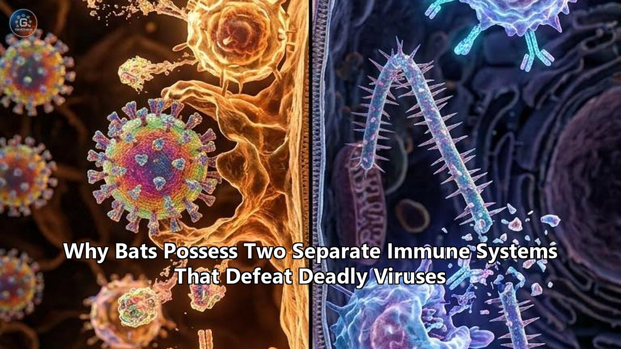
That framework is now being rewritten. A series of genomic discoveries, culminating in an investigation published in Science Advances led by evolutionary biologist Dr. Hannah Frank of Tulane University alongside collaborators at Stanford University and the Centers for Disease Control and Prevention, has exposed an unprecedented reality: bats possess a bifurcated, two-tier immune architecture.
Where other mammals deploy a single connected defensive network, bats operate two distinct, decoupled systems. The first is a perpetually active, ultra-rapid frontline antiviral shield that suppresses viral replication without pause. The second is an entirely separate, heavily dampened anti-inflammatory apparatus engineered to prevent the immune system from destroying its host. Furthermore, in more than 500 species of vesper bats, researchers discovered an unexpected duplication of the adaptive machinery itself: two distinct, functional sets of antibody-producing genes—a configuration never before documented in any other mammal.
┌─────────────────────────────────────────────────────────────────────────┐
│                    THE BIFURCATED BAT IMMUNE ARCHITECTURE               │
├───────────────────────────────────┬─────────────────────────────────────┤
│     OPERATING SYSTEM 1:           │     OPERATING SYSTEM 2:             │
│     PERPETUAL FRONTLINE DEFENSE   │     IRONCLAD INFLAMMATORY BRAKE     │
├───────────────────────────────────┼─────────────────────────────────────┤
│ • Constitutive basal IFN-α/ω      │ • Mutated STING (loss of S358)      │
│ • Unbroken ISG transcription      │ • Deletion of PYHIN/AIM2 gene family│
│ • Antiviral ISG15 protein conjug. │ • Hyper-potent ASC2 dampening NLRP3 │
│ • Dual IGH antibody gene loci     │ • Subdued Caspase-1, IL-1β, IL-18   │
│ • Continuous autophagy & repair   │ • Tolerance to cytosolic self-DNA   │
└───────────────────────────────────┴─────────────────────────────────────┘
These findings explain a long-standing biological paradox: how these winged mammals can harbor many of the world's deadliest viruses without developing clinical symptoms, while shedding light on the mechanics of viral reservoirs, pandemic origins, and potential treatments for human autoimmune and inflammatory disorders.
The Field Evidence: Viral Reservoirs Without Disease
The road to uncovering this dual architecture began not in high-throughput sequencing facilities, but in the dense limestone karst caves of Southeast Asia, Central Africa, and Northern Australia.
For more than twenty years, field researchers netting bats noticed an anomaly that clashed with classical pathology. When an animal carries a high viral titer of an aggressive pathogen, its physiology typically deteriorates. In humans, non-human primates, and rodents, viral replication triggers high fevers, massive tissue necrosis, vascular leakage, and organ failure—most of which is caused not by the virus itself, but by the body’s uncontrolled immune response.
In bats, field biopsies told a completely different story.
When researchers tested wild colonies of Egyptian fruit bats (Rousettus aegyptiacus) harboring Marburg virus, or Australian flying foxes carrying Hendra virus, the animals showed zero signs of lethargy, respiratory distress, or weight loss. Blood panels revealed minimal markers of systemic inflammation. The viruses were present, replicating, and metabolically active, but the host tissues remained unharmed.
Typical Mammalian Host vs. Chiropteran Host Response to Lethal RNA Viruses:

Mammals (Human/Rodent):
  Virus Ingress ──> Delayed Recognition ──> Hyper-Interferon Wave ──> Cytokine Storm (IL-6, TNF-α) ──> Organ Damage / Death

Bats (Chiroptera):
  Virus Ingress ──> Basal IFN Pre-Clearance ──> Controlled Viral Plateau ──> ASC2/STING Suppression ──> Asymptomatic Reservoir
"For a long time, the dominant hypothesis was simply that bat cells lacked the specific surface receptors required for these viruses to inflict severe cell damage," explains Dr. Linfa Wang, Professor of Emerging Infectious Diseases at Duke-NUS Medical School. "What our molecular work proved was the opposite. The viruses infect bat cells efficiently. The difference is in how bats fight viruses: they do not fight them by burning down the entire house to kill the intruder. They maintain a permanent, low-level antiviral firebreak while actively shutting down the biological explosive charge that we call inflammation".
When virologists examined bat blood plasma and tissue transcriptomes, they observed two phenomena operating simultaneously that seemed mutually exclusive by mammalian standards:
- High baseline levels of intracellular antiviral defenses, keeping viral replication tightly throttled.
- A total absence of downstream inflammatory markers such as interleukin-1 beta (IL-1β), interleukin-6 (IL-6), and tumor necrosis factor-alpha (TNF-α).
In human biology, turning up antiviral defenses automatically ignites inflammation. In bats, these two biological programs operate on entirely independent regulatory tracks.
The Frontline Engine: Constitutive Antiviral Readiness
In human cells, the innate immune response functions like a sleeping sentry. Under normal baseline conditions, our production of Type I interferons—the master signaling proteins that instruct neighboring cells to block viral replication—is kept near zero. Only after a pathogen's viral RNA or DNA is detected by pattern recognition receptors (such as RIG-I, MDA5, or Toll-like receptors) does the cell sound the alarm, triggering a sudden, massive surge of interferon-alpha (IFN-α) and interferon-beta (IFN-β).
This delayed response creates a dangerous window of vulnerability. By the time human cells release interferons, the virus has already replicated billions of times. The subsequent interferon surge is often violent, causing widespread bystander tissue damage.
Frontline Innate Interferon Dynamics:

Human Infection Timeline:
Hour 0 ──────────────> Hour 24 ───────────────────────> Hour 48+
[Baseline: 0 IFN]      [Viral Proliferation]            [Massive IFN Spike & Cytokine Storm]

Bat Infection Timeline:
Hour 0 ──────────────> Hour 24 ───────────────────────> Hour 48+
[Constitutive IFN-α]   [Immediate Viral Throttling]     [Tight Plateau, Zero Hyperinflammation]
When comparative immunologists sequenced the interferon loci of bats, they discovered that evolution had fundamentally rewired this circuit. In species like Pteropus alecto, the interferon-alpha gene locus has shrunk down to just three functional genes (compared to 13 in humans), yet these genes are never fully switched off. They are constitutively transcribed at steady baseline levels throughout the bat’s body, even in healthy, uninfected tissues.
"Bat cells live in a state of perpetual antiviral readiness," says Dr. Aaron Irving, an infectious disease researcher specializing in bat comparative immunology. "Because their baseline interferon signaling is always idling at an active RPM, the virus never gets the multi-day head start that it gets in a human patient. The frontline antiviral infrastructure is already deployed before the virus even enters the respiratory or digestive tract."
This perpetual baseline defense is not limited to interferons alone. A multi-institutional project led by the international Bat1K consortium and published in Nature conducted a systematic genomic analysis of 115 mammalian genomes, including high-coverage chromosome-level bat genomes. The investigation identified critical modifications in interferon-stimulated genes (ISGs), most notably ISG15.
ISG15 Pathway Divergence:
Human Mechanism:
  ISG15 Induction ──> Extracellular Secretion ──> Pro-inflammatory Cytokine Amplification ──> Lung Tissue Injury

Bat Mechanism:
  Constitutive ISG15 ──> Intracellular Protein Conjugation (ISGylation) ──> Viral Structural Disruption (90% Drop in Viral Yield)
In humans, ISG15 is secreted outside the cell during infections, where it acts as a cytokine that amplifies hyperinflammation, a primary driver of lung tissue destruction during severe respiratory infections like COVID-19.
In bats—particularly horseshoe bats (Rhinolophidae) and roundleaf bats (Hipposideridae)—specific amino acid changes alter how the protein functions. Bat ISG15 is directed internally to perform intracellular protein conjugation (ISGylation), directly binding to viral proteins and disrupting their structural assembly.
Laboratory assays conducted on infected cell lines showed that bat ISG15 reduced SARS-CoV-2 viral production by up to 90 percent purely through direct protein disruption, without triggering the pro-inflammatory extracellular signaling observed in humans.
Disarming the Host: The Silenced Inflammatory Circuit
If a human or mouse maintained constant interferon and antiviral gene activation, the biological cost would be catastrophic: chronic systemic inflammation, microvascular thrombosis, autoimmune destruction, and rapid mortality.
To make this persistent antiviral baseline viable, bats evolved an equally sophisticated, independent counter-system: an ironclad genetic brake on the molecular pathways that trigger systemic inflammation.
INTRACELLULAR SENSING AND INFLAMMATORY BRAKING MECHANISM

 [ Cytoplasmic DNA / Viral Particles / Damaged Self-mtDNA ]
                           │
           ┌───────────────┴───────────────┐
           ▼                               ▼
   [ Human Pathway ]               [ Bat Adaptation ]
           │                               │
   cGAS-STING Activation          cGAS-STING Activation
   (S358 Phosphorylation)         (Serine-to-X Mutation at S358)
           │                               │
           ▼                               ▼
    TBK1 / IRF3 Hyper-Spike         Subdued Signal Output
           │                               │
           ▼                               ▼
  NLRP3 Inflammasome Surge        Bat ASC2 Active Inhibition
  Caspase-1 Cleavage              (Dampened Caspase-1 Cleavage)
           │                               │
           ▼                               ▼
 Massive IL-1β / IL-18 Lysis      Zero Pyroptosis / Intact Tissues
   (Cytokine Storm / Death)          (Healthy Viral Tolerance)
The evidence for this second system emerged when researchers investigated how bats process cytosolic DNA. In all mammals, the presence of double-stranded DNA inside the cell's cytoplasm is an emergency indicator of trauma or viral infection.
The primary sensor for this is the cGAS-STING (cyclic GMP-AMP synthase – stimulator of interferon genes) pathway. When STING is activated in humans, it triggers downstream kinases that spark massive cytokine release and cell death.
In a key study published in Cell Host & Microbe, a team led by Duke-NUS researchers sequenced the STING protein across multiple bat genera and identified an evolutionary mutation found in every bat examined: a loss of the crucial serine residue at position 358 (S358).
In human and rodent STING, phosphorylation at S358 is the biochemical switch required to initiate the inflammatory chain reaction. Bats replaced this serine, dulling the downstream activation of inflammatory cascades without abolishing their basic pathogen-sensing ability.
STING Protein Amino Acid Sequence Alignment at Critical Activation Motif:

Human (H. sapiens):      - - - P L R T E E D S³⁵⁸ H N - - -  ==> Phosphorylation triggers massive cytokine storm
Mouse (M. musculus):     - - - P L R T E E D S³⁵⁸ H N - - -  ==> High inflammatory cascade
Black Flying Fox:        - - - P L R T E E D P³⁵⁸ H N - - -  ==> Mutation suppresses hyper-activation
Horseshoe Bat:           - - - P L R T E E D A³⁵⁸ H N - - -  ==> S358 lost; inflammation prevented
The anti-inflammatory modifications do not end with STING. Genomic screens have confirmed additional structural changes across bat inflammatory machinery:
- Complete Loss of the PYHIN Gene Family: While humans carry multiple PYHIN genes (such as AIM2 and IFI16) that detect cytosolic DNA and trigger rapid cell death (pyroptosis), bats have completely lost the entire PYHIN cluster from their genomes.
- Hyper-Potent ASC2 Suppression: In a study published in Cell, Dr. Matae Ahn and Prof. Linfa Wang identified that bats express a modified variant of the ASC2 protein. Four specific amino acid substitutions in bat ASC2 turn it into a high-affinity competitive antagonist that blocks the assembly of the NLRP3 inflammasome.
- Subdued Caspase-1 Cleavage: Because the bat NLRP3 inflammasome is suppressed, the activation of Caspase-1—the enzyme responsible for processing pro-IL-1β and pro-IL-18 into active, tissue-destroying cytokines—is reduced to a tiny fraction of human levels.
Molecular Comparison of Inflammatory Machinery:

Component                Human Architecture               Bat Architecture
────────────────────────────────────────────────────────────────────────────────
STING S358               Preserved (High Inflamm.)        Mutated/Lost (Suppressed)
PYHIN / AIM2             Active (Pyroptotic Lysis)        Completely Deleted
ASC2 Inflammasome Brake  Low Basal Affinity               Mutated for High Inhibition
NLRP3 Activation         Rapid, Hyper-Reactive            Severely Attenuated
Caspase-1 Processing     Explosive Cytokine Release       Controlled, Low-Level Output
"When you observe bat immune cells under the microscope during a viral challenge, they don't lyse and rupture like human macrophages do," notes Dr. Ahn. "The bat cell retains its membrane stability, suppresses the inflammasome, keeps the virus contained, and refuses to trigger the self-inflicted damage that kills human patients".
The Tulane Discovery: A Duplicated Adaptive Arsenal
For decades, immunology assumed that this bifurcated response was purely an innate adaptation. That assumption was broken when researchers at Tulane University, working in collaboration with Stanford University and the CDC, examined the genetic foundations of bat adaptive immunity.
Publishing in Science Advances, corresponding author Dr. Hannah Frank and her team uncovered an adaptive arrangement never before found in any other mammalian order: vesper bats (Vespertilionidae) possess two completely separate, functional sets of immunoglobulin heavy chain (IGH) gene loci—the master genetic blueprints used to assemble antibodies.
MAMMALIAN VS. VESPER BAT IMMUNOGLOBULIN ARCHITECTURE

Standard Mammalian Model (Humans, Rodents, Ungulates, Primates):
┌───────────────────────────────────────────────────────────────┐
│ SINGLE IGH LOCUS:                                             │
│ ──[ V-Segments ]──[ D-Segments ]──[ J-Segments ]──[ C-Region ] │
│ (Single genetic recombination engine for all antibodies)      │
└───────────────────────────────────────────────────────────────┘

Vesper Bat Model (Discovered in Science Advances):
┌───────────────────────────────────────────────────────────────┐
│ IGH LOCUS 1: Primary Repertoire                               │
│ ──[ V1-Cluster ]──[ D1-Cluster ]──[ J1-Cluster ]──[ C1-Region]│
├───────────────────────────────────────────────────────────────┤
│ IGH LOCUS 2: Specialized Secondary Repertoire                 │
│ ──[ V2-Cluster ]──[ D2-Cluster ]──[ J2-Cluster ]──[ C2-Region]│
└───────────────────────────────────────────────────────────────┘
In every other mammal studied to date—from mice and elephants to humans—the adaptive immune system builds the heavy chains of its antibodies from a single genetic locus through V(D)J recombination.
The Tulane investigation revealed that more than 500 species of vesper bats, spanning every continent except Antarctica, carry a complete duplication of this critical locus. They operate two distinct antibody-generating engines simultaneously.
"We've never seen anything like this in a mammal before," said Dr. Frank. "The only other vertebrates where anything analogous has been observed are certain lineages of cartilaginous and ray-finned fish. This completely changes our understanding of how mammalian immune systems can be organized and raises exciting new questions about why bats have been so evolutionarily successful and how they respond to viruses".
Structural Characteristics of the Dual Antibody System:

Locus System      Repertoire Profile          Proposed Functional Specialization
────────────────────────────────────────────────────────────────────────────────
IGH Locus 1       High Baseline Polyspecific  Broad, cross-reactive viral binding;
                  Diversity                   immediate low-affinity neutralization.
────────────────────────────────────────────────────────────────────────────────
IGH Locus 2       Rapid Somatic               Precision high-affinity pathogen
                  Hypermutation Cluster       clearance without chronic clonal fatigue.
The presence of two independent antibody systems provides vesper bats with an expanded adaptive repertoire.
While the first locus can generate broad, cross-reactive antibodies that bind to conserved viral structural motifs across diverse viral families, the second locus is free to diversify through somatic hypermutation to handle specific, persistent viral variants.
This gives the bat an adaptive dual-track system that mirrors its innate defense: broad, continuous surveillance coupled with targeted neutralization, entirely isolated from destructive systemic pathology.
The Evolutionary Catalyst: Why Flight Demanded a Two-Tier System
How did such a bifurcated immune system evolve in a single order of mammals? To trace the origin of this architecture, evolutionary biologists look back 65 million years to the moment bats became the only mammals capable of powered flight.
Flight is the most metabolically demanding form of locomotion in the animal kingdom. When a bat takes to the air, its metabolic rate surges between 15- and 30-fold above resting levels.
Its heart rate can exceed 1,000 beats per minute, and its internal core body temperature routinely reaches 40°C to 41°C (104°F to 106°F)—temperatures that mirror a life-threatening fever in a human.
Metabolic Stress Comparison: Flight vs. Rest

Resting Mammal (Rodent/Human):
  Metabolism: 1.0x Baseline
  Core Temp: ~37.0°C
  ROS Production: Baseline
  Cytosolic DNA Leakage: Minimal

Flying Bat (Chiroptera):
  Metabolism: 15.0x – 30.0x Baseline
  Core Temp: 40.0°C – 41.0°C
  ROS Production: Massive Free Radical Surges
  Cytosolic DNA Leakage: High (Continuous Mitochondrial / Nuclear Shear)
At these metabolic extremes, a bat's cells experience significant physiological wear and tear. High oxygen consumption in flight muscle tissue generates continuous waves of reactive oxygen species (ROS), causing oxidative stress that damages mitochondria and cell nuclei.
This cellular stress causes fragments of mitochondrial DNA (mtDNA) and nuclear DNA to constantly leak through broken membranes into the cytoplasm.
In any standard terrestrial mammal, this continuous leakage of self-DNA into the cytoplasm would be fatal:
- The cellular DNA sensors (cGAS, STING, AIM2) would detect the host's own leaking mtDNA.
- The immune system would mistake this damaged self-DNA for a massive systemic infection.
- The NLRP3 inflammasome would fire continuously, releasing a cytokine storm that causes systemic auto-inflammatory destruction and death.
THE FLIGHT-IMMUNITY EVOLUTIONARY FEEDBACK LOOP

   [ Origin of Powered Flight (65 Million Years Ago) ]
                          │
                          ▼
   [ Extreme Metabolic Rate (15–30x) & Core Temp (41°C) ]
                          │
                          ▼
   [ Severe Oxidative Shear & Continuous mtDNA Cytosolic Leakage ]
                          │
         ┌────────────────┴────────────────┐
         │                                 │
         ▼                                 ▼
[ Without Immune Rewiring ]      [ Evolutionary Solution ]
Catastrophic Autoimmunity       1. Mutate STING (S358 lost)
Constant NLRP3 Inflammasome     2. Delete PYHIN / AIM2 Genes
Lethal Auto-Inflammatory Death  3. Boost ASC2 Inflammasome Brake
                                4. Turn On Basal Frontline IFN
                                                   │
                                                   ▼
                                [ Unintended Evolutionary Byproduct ]
                                Immune system rendered impervious to
                                viral inflammation; bats become ideal
                                asymptomatic reservoirs for pathogens.
To achieve powered flight, ancestral chiropterans were forced to evolve an ironclad tolerance mechanism: they dismantled the molecular pathways linking cytosolic DNA detection to systemic inflammation. They removed the PYHIN gene cluster, modified STING, and upgraded the ASC2 brake.
However, shutting down these inflammatory alarms created a new hazard: it left them vulnerable to real intracellular viral pathogens.
To offset this gap, evolutionary pressure drove the expansion of an independent frontline defense: constitutive interferon-alpha production, internal ISG15 antiviral tagging, and duplicated antibody loci.
The decoupling was complete. Powered flight forced bats to construct an immune system split into two independent tracks: one to continuously block pathogens, and another to prevent the host's own metabolism from killing itself.
As an unintended evolutionary byproduct, bats became virtually impervious to viral disease.
Inside the BSL-4: Human vs. Bat Cells in Direct Combat
To demonstrate this dual-track system in action, scientists set up direct comparative challenges inside high-containment biosafety facilities.
Using stem-cell-derived organoids and primary endothelial cell lines, researchers exposed human tissue cultures and bat (Rousettus aegyptiacus and Pteropus alecto) tissue cultures to identical viral challenges, including Middle East Respiratory Syndrome coronavirus (MERS-CoV), Marburg virus, and Bat SARS-like coronaviruses.
The divergence in cell fate begins within hours of infection:
Human vs. Bat Cellular Trajectory During High-Titer Viral Challenge:

HUMAN CELLS:
Hour 0-12  ──> Silent viral entry; intracellular viral genome replication spikes.
Hour 12-24 ──> Sudden detection triggers delayed Type I IFN wave; massive cGAS-STING signaling.
Hour 24-48 ──> NLRP3 inflammasome assembly; Caspase-1 activation; pyroptosis ruptures cell membrane.
Hour 48+   ──> Massive release of IL-1β, IL-6, TNF-α; vascular breakdown and runaway tissue lysis.

BAT CELLS:
Hour 0-12  ──> Viral entry meets pre-existing baseline IFN-α and conjugated ISG15; viral replication stalls.
Hour 12-24 ──> Low-level viral presence detected; modified STING prevents downstream hyper-activation.
Hour 24-48 ──> Bat ASC2 prevents NLRP3 assembly; Caspase-1 remains inactive; membranes stay sealed.
Hour 48+   ──> Viral replication hits steady low-level plateau; zero pro-inflammatory cytokine release.
Quantitative transcriptomic and proteomic data from these experiments show a sharp divergence:
| Metric Measured | Human Endothelial / Lung Tissue | Bat Endothelial / Lung Tissue |
|---|---|---|
| Peak Viral Load Reduction | Baseline (Standard Growth) | 70% to 90% Throttled (via ISG15/IFN-α) |
| Interleukin-1 Beta (IL-1β) Output | 100x to 1,000x Increase | < 2x Basal Change (Near-Zero) |
| Caspase-1 Activity Level | Hyper-Elevated (Pyroptotic) | Strongly Suppressed (ASC2 Brake) |
| Cellular Viability at 72 Hours | < 15% Surviving Intact Cells | > 92% Surviving Intact Cells |
| Nitric Oxide / ROS Damage | Severe Lipid Peroxidation | Minimal (Up-regulated Autophagy) |
"When you observe human cells infected with these viruses, the destruction is chaotic," explains Dr. Cara Brook, an evolutionary ecologist and virologist at the University of Chicago. "The virus triggers a cellular panic response. The cell rips itself apart to sound the alarm, which recruits waves of immune cells that destroy the host tissue."
"In bat cells, the response is controlled," Brook continues. "The cell-intrinsic antiviral pathways manage the infection locally, while the anti-inflammatory systems prevent the cell from rupturing. It changes how we think about disease. We realize that the disease is not an inevitable outcome of viral infection; it is a failure of immune regulation. The bat shows us how to separate fighting the virus from suffering from the pathology".
┌─────────────────────────────────────────────────────────────────────────┐
│              VIRAL PATHOLOGY VS. HOST IMMUNE DAMAGE                     │
├─────────────────────────────────────────────────────────────────────────┤
│                                                                         │
│  [VIRUS INVASION]                                                       │
│          │                                                              │
│          ├──────────────────────────────┐                               │
│          ▼                              ▼                               │
│  [Human Immune System]          [Bat Two-Tier System]                   │
│          │                              │                               │
│     (Delayed IFN)              (Basal IFN-α & ISG15)                    │
│          ▼                              ▼                               │
│   Massive Viral Spike            Viral Growth Halted                    │
│          ▼                              ▼                               │
│  (Uncontrolled STING)          (Mutated STING & ASC2)                   │
│          ▼                              ▼                               │
│  NLRP3 Hyperactivation          Inflammasome Blocked                    │
│          ▼                              ▼                               │
│  Lethal Cytokine Storm          Zero Tissue Pathology                   │
│          ▼                              ▼                               │
│    ORGAN FAILURE                   HEALTHY HOST                         │
│                                                                         │
└─────────────────────────────────────────────────────────────────────────┘
This dynamic explains how bats fight viruses without succumbing to the illnesses that devastate other mammals. They do not possess a single super-charged immune system; they operate two distinct, counter-balanced systems.
One side acts as a constant, non-destructive antiviral suppressant, while the other maintains strict control over the inflammatory pathways that drive human organ failure.
Translational Medicine: Borrowing the Bat Blueprint
The discovery of this decoupled, two-tier immune architecture is shifting from evolutionary biology into clinical drug development.
For decades, modern pharmacology attempted to treat severe viral infections by focusing on antiviral compounds designed to inhibit specific viral enzymes (like remdesivir or paxlovid), or by using blunt anti-inflammatory drugs like corticosteroids (such as dexamethasone), which suppress the entire immune system and leave patients vulnerable to secondary infections.
Chiropteran immunology offers a third way: targeted inflammatory dampening paired with preserved, targeted frontline antiviral defense.
THERAPEUTIC PARADIGM SHIFT

Current Human Approach:
┌───────────────────────────┐     ┌───────────────────────────┐
│ Antiviral Inhibitors      │  +  │ Broad Corticosteroids     │
│ (Blocks one specific bug) │     │ (Suppresses entire immune │
│                           │     │  system; risk of sepsis)  │
└───────────────────────────┘     └───────────────────────────┘

Bat-Inspired Approach (Translational Pipeline):
┌─────────────────────────────────────────────────────────────┐
│ 1. Engineered Bat-ASC2 Peptides                             │
│    -> Selectively blocks NLRP3 inflammasomes without        │
│       impairing basic cellular defenses                     │
├─────────────────────────────────────────────────────────────┤
│ 2. Small-Molecule STING S358 Mimetic Antagonists            │
│    -> Clamps pro-inflammatory cytokine release (IL-1β/IL-6) │
│       while leaving baseline Type I IFN intact              │
├─────────────────────────────────────────────────────────────┤
│ 3. Dual-Locus Synthetic Monoclonal Antibodies               │
│    -> Derived from bat IGH locus architecture for broad     │
│       pan-sarbecovirus and pan-filovirus neutralization     │
└─────────────────────────────────────────────────────────────┘
Several clinical and preclinical programs are already working to translate these bat adaptations into human therapeutics:
1. Engineered Bat-ASC2 Peptides
At Duke-NUS and associated spin-off biotechnology ventures, researchers synthesized peptides based on the four critical amino acid changes found in bat ASC2.
When administered to mouse models suffering from lethal influenza-induced lung inflammation, SARS-CoV-2 acute respiratory distress syndrome (ARDS), or crystalline-induced gouty arthritis, the engineered bat ASC2 molecules suppressed NLRP3 inflammasome assembly.
The treatment prevented fatal lung hyperinflammation while allowing the animals' standard immune systems to clear the virus naturally.
2. STING S358 Pharmacological Clamps
Small-molecule inhibitors designed to selectively block the phosphorylation site at serine 358 on human STING are currently in preclinical testing.
By mimicking the natural mutation carried by bats, these compounds prevent cytosolic self-DNA (leaked from damaged mitochondria during physical trauma, sepsis, or infection) from triggering uncontrolled systemic cytokine release, without shutting down upstream interferon signaling.
3. Dual-Locus Synthetic Antibody Libraries
Utilizing the structural insights from the Tulane discovery of the duplicated IGH loci in vesper bats, immunologists are engineering bi-specific antibody platforms.
These platforms pair an ultra-broad polyreactive binding arm (derived from the first locus architecture) with an affinity-matured neutralizing arm (derived from the second), creating antibodies capable of neutralizing rapidly mutating RNA viruses across entire viral families.
"This marks a new approach to infectious disease and inflammatory medicine," says Dr. Ahn. "Rather than discovering a new drug for every emerging virus that spills over into humans, we can adjust our own immune response using the evolutionary blueprint already perfected by bats over 65 million years".
Longevity, Cancer, and the Inflammaging Shield
The implications of this decoupled immune architecture extend beyond emerging zoonotic viruses.
One of the longest-standing questions in mammalian zoology is why bats live so long. In the animal kingdom, lifespan is tightly linked to body size and metabolic rate: small mammals with rapid heart rates and high metabolisms, such as mice and shrews, typically live two to three years.
Bats break this biological rule. The Brandt's bat (Myotis brandtii), which weighs just four to eight grams, can live for more than 40 years in the wild—nearly ten times longer than a rodent of comparable size.
Body Mass vs. Maximum Lifespan Discrepancy:

Species                 Body Mass     Max Lifespan     Metabolic Rate (Flight/Active)
────────────────────────────────────────────────────────────────────────────────
House Mouse (M. musculus)   20-30 g       3-4 years        Standard Terrestrial
Shrew (Sorex araneus)       5-12 g        1-2 years        High Terrestrial
Brandt's Bat (M. brandtii)  4-8 g         40+ years        Extreme (Powered Flight)
Gerontologists recognize that a primary driver of aging and age-related chronic disease in humans is a process known as inflammaging: the slow, continuous, low-grade systemic inflammation driven by cellular senescence, accumulation of damaged mitochondrial fragments, and continuous NLRP3 inflammasome activation over decades.
In humans, this chronic low-level inflammation degrades tissues over time, contributing to atherosclerosis, neurodegenerative diseases (like Alzheimer's and Parkinson's), type 2 diabetes, and cancer.
The Mechanism of Human "Inflammaging" vs. The Bat Longevity Shield:

Human Aging Pathway:
Years of Metabolism ──> Damaged mtDNA Leakage ──> Chronic NLRP3/STING Activation ──> Low-Grade Systemic Inflammation ──> Tissue Degradation / Cancer

Bat Aging Pathway:
Decades of Flight ──> Damaged mtDNA Leakage ──> STING/ASC2/PYHIN Deletions ──> Zero Chronic Inflammation ──> Maintained Proteostasis / Extreme Lifespan
Because bats decoupled their innate immune sensors from inflammatory cascades to survive the metabolic demands of flight, they eliminated inflammaging at its source:
- Their modified STING does not trigger chronic inflammation in response to damaged self-DNA.
- Their enhanced ASC2 prevents the low-grade assembly of NLRP3 inflammasomes as they age.
- Their constitutive baseline interferon expression and high rates of autophagy continuously clear misfolded protein aggregates and damaged organelles before they can trigger senescence.
As a result, bats rarely develop age-related inflammatory illnesses, and their rates of malignant tumors are among the lowest recorded across Mammalia.
The same bifurcated immune architecture that allows them to tolerate lethal viral infections provides an internal shield against aging and cellular degeneration.
The Ecological Paradox and the Spillover Threat
While the bat's dual immune system is an evolutionary success, it creates a persistent ecological challenge for other species, including humans.
Because how bats fight viruses relies on high-speed frontline suppression rather than complete viral clearance, bat colonies often maintain persistent, low-level viral infections indefinitely.
Viruses co-evolving inside this environment are subject to unique evolutionary pressures. To survive in a host with active, baseline interferon defenses, these viruses must adapt to replicate rapidly and develop mechanisms to counter immune signaling.
THE SPILLOVER PARADOX

 [ Inside the Bat Host ]
  • Persistent, baseline interferon pressure (IFN-α)
  • Viruses adapt to replicate efficiently against active defenses
  • Host inflammatory brake prevents clinical disease
                     │
                     ▼
 [ Spillover Event into Humans / Livestock ]
  • Virus encounters an un-primed, resting immune system
  • Rapid initial viral replication outpaces delayed defenses
  • Delayed human immune system overreacts with massive cytokine surge
  • Result: Catastrophic immunopathology, vascular collapse, death
When an adapted bat virus—such as Marburg, Nipah, or a novel coronavirus—spills over into an intermediate host or directly into a human, the balance is lost.
The human immune system, built on a single interconnected track, cannot handle the virus's replication speed without firing its inflammatory alarms at full blast. The resulting cytokine release, lung damage, and vascular breakdown are the direct consequence of a virus evolved to withstand the bat's frontline defense meeting a host that lacks the bat's inflammatory brakes.
┌─────────────────────────────────────────────────────────────────────────┐
│                      ZOONOTIC DIVERGENCE SUMMARY                        │
├─────────────────────────────────────────────────────────────────────────┤
│                                                                         │
│   BAT RESERVOIR                              HUMAN SPILLOVER            │
│  ┌────────────────────────┐                 ┌────────────────────────┐  │
│  │ Frontline: Always On   │  Pathogen Jumps │ Frontline: Delayed     │  │
│  │ Inflammation: Dampened │ ──────────────> │ Inflammation: Violent  │  │
│  │ Result: Zero Disease   │                 │ Result: Lethal Sepsis  │  │
│  └────────────────────────┘                 └────────────────────────┘  │
│                                                                         │
└─────────────────────────────────────────────────────────────────────────┘
This dynamic is further complicated by environmental stressors. When bat colonies experience habitat fragmentation, climate stress, or nutritional deprivation, their tight metabolic regulation weakens.
Under metabolic stress, the delicate balance between their frontline antiviral shield and their inflammatory brakes fluctuates, leading to temporary spikes in viral shedding.
Understanding the molecular mechanics of these two systems is essential for predicting when, where, and why viral spillover occurs.
The Next Horizon in Bat Immunology
The confirmation of this bifurcated immune architecture—from the loss of STING phosphorylation and the enhancement of ASC2 to the discovery of duplicated antibody loci in vesper bats—has opened new directions for molecular immunology.
Several key questions remain at the center of ongoing research:
- How do non-vesper bat families configure their adaptive genomes?
While the Tulane study confirmed duplicated IGH loci across the 500+ species of Vespertilionidae, genome sequencing efforts are underway via the Bat1K project to determine whether similar adaptive duplications exist across other bat families, such as the Pteropodidae (fruit bats) and Phyllostomidae (New World leaf-nosed bats).
- What regulates the balance between frontline baseline interferons and metabolic exhaustion?
Maintaining active baseline interferon expression requires continuous energy expenditure. Researchers are investigating the epigenetic switches that allow bats to sustain this frontline state without suffering metabolic exhaustion.
- Can bat-derived anti-inflammatory molecules clear human safety trials?
With synthetic bat ASC2 peptides and STING-dampening small molecules advancing through preclinical animal testing, the first human safety and pharmacokinetic trials are preparing to evaluate whether these mechanisms can control severe human auto-inflammatory conditions.
Upcoming Milestones in Chiropteran Immunology:

Timeline       Target Investigation / Clinical Milestone
────────────────────────────────────────────────────────────────────────────────
2026–2027      Full chromosome-level assembly of 1,000+ bat species via Bat1K.
2027           Phase 1 human clinical trials of bat-ASC2-derived anti-inflammatory
               peptides for acute viral lung injury and sepsis.
2027–2028      In vivo testing of engineered bi-specific dual-locus humanized
               monoclonal antibodies targeting pan-coronavirus variants.
2028+          Application of bat inflammatory-brake mimetics to chronic human
               neurodegenerative and autoimmune disorders (Alzheimer's, Lupus).
For more than a century, science viewed the immune system through a single framework: a unified network that uses inflammation to drive its antiviral weapons.
By tracking the biology of the world's only flying mammals, researchers have discovered an entirely different way nature solves the problem of infection.
By separating the weapon from the blast wave—maintaining a persistent frontline defense while muting destructive inflammatory pathways—bats have shown that the key to surviving the world's deadliest pathogens is not destroying the virus at all costs, but mastering the art of immune tolerance.
Reference:
- https://pmc.ncbi.nlm.nih.gov/articles/PMC11876839/
- https://sse.tulane.edu/how-bats-tolerate-viruses-tulane-study-sheds-light-one-natures-biggest-immune-mysteries
- https://www.sciencedaily.com/releases/2026/09/260901010713.htm
- https://academic.oup.com/gbe/article/15/9/evad148/7259420
- https://www.freethink.com/health/bat-protein-inflammasomes
- https://www.livescience.com/health/viruses-infections-disease/a-unique-antibody-system-may-help-explain-bats-unusual-tolerance-of-viruses
- https://www.facebook.com/DukeNUSMedicalSchool/posts/-what-can-bats-teach-us-about-staying-healthy-for-longerresearchers-have-been-st/1436713218486888/
- https://www.proimmune.com/mi2025-linfa-wang
- https://pubmed.ncbi.nlm.nih.gov/39880942/
- https://bsky.app/profile/did:plc:2tjdireh5paxwxdjak2qs7xg
- https://researcherslinks.com/current-issues/Current-Understanding-and-Future/6/8/10095/html
- https://scitechdaily.com/bats-have-a-secret-immune-system-scientists-have-never-seen-in-any-other-mammal/
- https://www.youtube.com/watch?v=ifRp7VeL5GI
- https://bat1k.com/