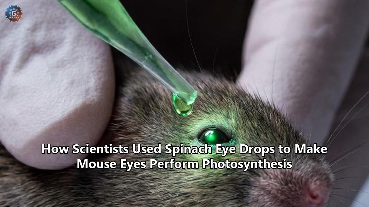
In a remarkable fusion of botany and ophthalmology, researchers have successfully transplanted the light-harvesting machinery of spinach leaves into the eyes of living mice.
The experimental treatment, developed by a collaborative team led by researchers at the National University of Singapore (NUS), does not turn the rodent eyes green, nor does it grant them the ability to survive entirely on sunlight. Instead, it introduces a highly specialized, nanoscale photosynthetic system directly into the ocular cells. Once there, this system absorbs ambient indoor light to generate a continuous stream of cellular energy and natural antioxidants, rapidly curing dry eye disease.
The study, published in the journal Cell, represents a profound paradigm shift in bioengineering. By crossing the evolutionary barrier that has separated plants and animals for over a billion years, the scientists demonstrated that animal cells can temporarily adopt plant-like functions to repair tissue damage and soothe inflammation. Called LEAF (Light-reaction Enriched thylAkoid NADPH-Foundry), this plant-derived nanotechnology has outperformed clinical-standard pharmaceuticals in animal models, offering a cheap, non-invasive, and highly elegant therapeutic alternative for a condition that plagues over a billion people worldwide.
With clinical trials already on the horizon, this discovery could redefine how we treat chronic inflammatory disorders, demonstrating that the very light that allows us to see our environment can also be harnessed to heal us from within.
The Clinical Nightmare of Dry Eye Disease
To understand why scientists turned to spinach to heal mammalian eyes, one must first appreciate the complex, painful, and surprisingly stubborn nature of dry eye disease, medically known as keratoconjunctivitis sicca.
Far from a minor inconvenience or temporary irritation, dry eye disease is a chronic, progressive condition that affects approximately 1.5 billion people across the globe. In our modern landscape—dominated by relentless screen time, air conditioning, contact lens use, and environmental pollutants—the prevalence of this ocular misery is skyrocketing.
The disease targets the delicate ocular surface, which relies on a highly sophisticated, three-layered tear film to remain healthy:
- The Outer Lipid (Oily) Layer: Produced by the meibomian glands, this prevents tear evaporation.
- The Middle Aqueous (Watery) Layer: Produced by the lacrimal glands, this hydrates the eye, supplies oxygen, and washes away debris.
- The Inner Mucin (Mucus) Layer: Produced by conjunctival goblet cells, this binds tears to the corneal surface.
When any of these components are compromised, the tear film breaks down, leaving the corneal epithelium exposed. This exposure triggers a cascade of cellular distress. Without the protective barrier of tears, the ocular surface suffers from localized hyperosmolarity, meaning the remaining fluid becomes highly salty and concentrated. This salt imbalance draws water out of the corneal cells, causing them to shrink, warp, and trigger a massive inflammatory response.
The Role of Oxidative Stress and the Metabolic Bottleneck
At the heart of dry eye disease lies oxidative stress. When corneal cells are dried out and chronically inflamed, their internal energy generators—the mitochondria—begin to malfunction. Instead of cleanly producing adenosine triphosphate (ATP), the primary energy currency of the cell, the damaged mitochondria start leaking highly reactive, unstable oxygen molecules known as reactive oxygen species (ROS).
These ROS, including hydrogen peroxide ($H_2O_2$) and superoxide radicals, behave like chemical wrecking balls inside the cell. They mutate DNA, destroy cellular membranes through lipid peroxidation, and degrade the proteins that hold the corneal epithelium together. The result is a cycle of destruction:
$$\text{Tear Film Instability} \longrightarrow \text{Mitochondrial Distress} \longrightarrow \text{ROS Accumulation} \longrightarrow \text{Inflammation} \longrightarrow \text{Further Tear Loss}$$
To combat this onslaught, healthy cells rely on a natural antioxidant defense system. The master key to this defense is a coenzyme called nicotinamide adenine dinucleotide phosphate (reduced form), or NADPH. NADPH acts as a critical donor of electrons (a reducing agent), powering the enzymes that neutralize ROS and restore redox balance.
However, in chronic dry eye disease, the cell’s natural pathways for generating NADPH and ATP are completely overwhelmed. The cells run out of metabolic "fuel" to fight off the oxidative stress, leading to cell death, corneal scarring, chronic pain, and blurred vision.
Why Traditional Ophthalmic Drugs Fall Short
The current pharmaceutical arsenal against dry eye disease is notoriously inadequate. Clinicians typically prescribe anti-inflammatory eye drops, such as cyclosporine A (commercially known as Restasis) or lifitegrast (Xiidra). While these drugs can help manage symptoms, they suffer from major limitations:
- Slow Onset of Action: Cyclosporine A often takes weeks, or even months, of twice-daily applications before patients experience any significant relief.
- Narrow Therapeutic Target: These drugs are primarily immunosuppressants. They block specific T-cell activation pathways to reduce inflammation, but they do nothing to directly repair the underlying metabolic damage, restore ATP levels, or actively neutralize ROS.
- Ocular Side Effects: Many patients complain of severe burning, stinging, and localized redness upon applying these drops.
- High Economic Cost: Biologic and advanced small-molecule eye drops are incredibly expensive, placing a massive financial burden on healthcare systems and patients alike.
Faced with this therapeutic bottleneck, the research team at NUS asked a radical question: What if, instead of trying to chemically patch up the damaged mammalian metabolic pathway, we bypassed it entirely? What if we introduced an independent, light-powered engine that could manufacture NADPH and ATP right inside the inflamed corneal cells?
To find such an engine, they had to look outside the animal kingdom.
Nature’s Sol Power: The Evolutionary Inspiration
To appreciate the conceptual leap of utilizing spinach eye drops photosynthesis to heal animals, it is helpful to step back and look at the fundamental divide between how plants and animals generate energy.
For more than a billion years, eukaryotic life has been broadly split into two camps:
- Autotrophs (Plants and Algae): Organisms that use solar energy, water, and carbon dioxide to synthesize their own organic molecules via photosynthesis.
- Heterotrophs (Animals and Fungi): Organisms that must ingest organic matter and break it down through cellular respiration to generate energy.
Plants carry out this solar-to-chemical conversion within specialized organelles called chloroplasts. These organelles are believed to have originated over 1.5 billion years ago through endosymbiosis—a process where an ancient, non-photosynthetic eukaryote engulfed a photosynthetic cyanobacterium, forming a permanent, mutually beneficial relationship. Over evolutionary time, this cyanobacterium evolved into the modern chloroplast.
Animals, however, completely lost or never acquired this capability. We rely entirely on mitochondria to burn the sugars we eat. Yet, there are a few striking exceptions in nature that suggest the boundary between plant-like and animal-like energy production is more porous than we think.
The Solar-Powered Sea Slug
The most famous natural rule-breaker is the sacoglossan sea slug, specifically Elysia chlorotica.
[Algal Food Source] ---> [Sea Slug Ingests Algae] ---> [Chloroplasts Extracted]
                                                                │
                                                                ▼
                                                    [Stored in Gut Cells]
                                                    (Kleptoplasty Process)
                                                                │
                                                                ▼
                                                    [Months of Solar Energy]
This remarkable marine animal feeds on the intertidal alga Vaucheria litorea. Instead of fully digesting the algal cells, the sea slug carefully extracts the intact chloroplasts and stores them inside its own digestive tract cells. This phenomenon, known as kleptoplasty (literally "organelle theft"), allows the sea slug to turn bright green and perform photosynthesis for up to nine months. When food is scarce, the slug can survive entirely on the sugars generated by its stolen, light-harvesting cellular organelles.
Prior Attempts at Cross-Kingdom Photosynthetic Engineering
Inspired by the solar-powered sea slug, synthetic biologists have spent years trying to engineer photosynthetic capabilities into mammalian tissues.
In 2022, a pioneering study by researchers at the Zhejiang University School of Medicine in China made headlines when they successfully transplanted thylakoid membranes from spinach into the knee joints of mice with osteoarthritic cartilage damage. Published in Nature, that research demonstrated that the transplanted plant machinery could generate ATP and NADPH inside mammalian cartilage cells, slowing joint degradation.
However, that cartilage-targeting experiment faced a steep physical hurdle: light penetration. Skeletal joints are buried deep beneath layers of skin, fat, and muscle. Getting visible light to penetrate these tissues to power the photosynthetic reactions required invasive light-delivery systems or prolonged, high-intensity light exposure, limiting its clinical practicality.
Similarly, in 2024, researchers at the University of Tokyo successfully inserted functional chloroplasts extracted from red and green algae into cultured hamster cells in vitro, maintaining photosynthetic activity for roughly 48 hours. But keeping these delicate organelles functional in a living, breathing mammal remained a profound challenge. The mammalian immune system is highly aggressive, and raw, foreign plant organelles are typically recognized as hostile invaders and destroyed before they can perform any useful work.
The Eye: A Natural Window to the Light
This is where the NUS team, led by Associate Professor David Tai Wei Leong and Dr. Xing Kuoran, made their masterstroke.
Instead of targeting deep, dark, internal tissues like joints or organs, they focused their attention on the eye. The mammalian eye is a physiological marvel specifically designed to let light in. The cornea—the clear, dome-shaped outer surface of the eye—is completely transparent, allowing visible light to pass through unobstructed so we can see.
"The eye is uniquely suited for this type of strategy, since light is already an intrinsic component of its normal physiological function," explained Dr. Xianfeng Lin, an orthopedic surgeon at Zhejiang University who co-authored the 2022 knee-joint study but was not directly involved in the eye-drop project. "The work extends the role of light in the eye from being purely sensory to potentially contributing to local metabolic support and tissue repair."
By designing a specialized formulation of spinach eye drops photosynthesis could occur naturally on the surface of the eye using nothing more than ordinary, ambient indoor light. The eye is the perfect natural laboratory for solar-powered medicine.
The Molecular Architecture of the LEAF Nanotechnology
So, how exactly do you turn a piece of grocery-store spinach into a high-tech, medical-grade eye drop? The process requires an elegant blend of cellular disassembly, selective filtration, and nano-encapsulation.
Step 1: Isolating the Thylakoids
To perform photosynthesis, plants rely on chloroplasts. But a whole, intact chloroplast is too large (typically 5 to 10 micrometers across) to be easily absorbed by mammalian cells. Furthermore, chloroplasts contain a watery internal fluid called the stroma, which houses enzymes like Rubisco.
In a plant cell, these stromal enzymes run the "dark reactions" of photosynthesis (the Calvin cycle), using up ATP and NADPH to build sugars. For treating dry eye, the researchers didn’t want the cells to build sugars; they wanted to maximize the availability of free ATP and NADPH to act as antioxidants and cellular fuel.
To achieve this, the NUS team developed a patented, ultra-gentle chemical and mechanical extraction method to isolate only the thylakoids—the membrane structures within chloroplasts that are the actual sites of the light-dependent reactions.
  [Whole Spinach Leaf]
           │
           ▼ (Mild mechanical grinding)
  [Intact Chloroplasts]
           │
           ▼ (Gentle osmotic shock)
  [Ruptured Chloroplast Envelope] ──> [Discard Stroma] (Removes NADPH-consuming enzymes)
           │
           ▼ (Purified Thylakoid Grana Stacks)
  [Encapsulation with Pluronic F127]
           │
           ▼
  [400 nm LEAF Nanoparticles] (Transparent & Bio-compatible)
By subjecting the chloroplasts to a gentle osmotic shock, they ruptured the outer envelopes. They discarded the stroma and collected the thylakoid grana—the coin-like stacks of membrane disks containing the active photosynthetic machinery.
These grana contain:
- Photosystem II (PSII): The light-absorbing protein complex that splits water molecules ($H_2O$) into oxygen ($O_2$), protons ($H^+$), and free electrons.
- The Cytochrome $b_6f$ Complex and Plastocyanin: The molecular "wire" that pumps protons across the membrane, creating a proton gradient.
- Photosystem I (PSI): The second light-absorbing complex that uses light energy to transfer those free electrons to $NADP^+$, reducing it into the highly prized antioxidant NADPH.
- ATP Synthase: The molecular turbine that uses the proton gradient to convert ADP into ATP.
Step 2: Streamlining and Optimizing the Machinery
By stripping away the stroma, the team created a highly streamlined, dedicated NADPH factory. Without the Calvin cycle enzymes constantly consuming the newly generated molecules, the isolated thylakoids were freed to dump their chemical energy directly into their surrounding environment.
According to the study, this selective purification boosted the production of NADPH by approximately 20 percent compared to native, unprocessed chloroplasts.
Step 3: Nano-Encapsulation and the Mystery of the Non-Green Eye Drops
A major engineering challenge was preserving the highly complex, three-dimensional structure of the thylakoid membranes. These membranes are incredibly delicate. If they are bent, torn, or misaligned, the photosynthetic electron transport chain breaks down, resulting in a catastrophic loss of function. Traditional physical grinding methods, such as high-frequency sonication or high-pressure homogenization, completely destroy this fragile architecture.
To solve this, the NUS team used a gentle surfactant processing technique. They encapsulated the purified thylakoid grana inside a protective, biocompatible coating made of Pluronic F127, an FDA-approved non-ionic surfactant.
This surfactant self-assembles around the thylakoid fragments, stabilizing their structure and packaging them into highly uniform, spherical nanoparticles measuring roughly 400 nanometers (nm) in diameter. This size is critical: it is small enough to be easily absorbed by mammalian cells through standard endocytic pathways, yet large enough to keep the complex photosynthetic proteins structurally intact.
One might expect that an eye drop derived from spinach would be a deep, murky green, potentially tinting the user's eyes. However, the LEAF formulation is transparent. Because the nanoparticles are incredibly small (400 nm is at the lower limit of visible light wavelengths) and the overall chlorophyll concentration is kept extremely low, the drops do not scatter or absorb light in a way that is visible to the naked eye. When applied, the drops look like standard clear saline.
How LEAF Operates Inside the Mammalian Eye: The Dual-Domain Defense
Once the LEAF nanoparticles are administered to the eye, they perform a highly coordinated, two-pronged rescue mission. This "dual-domain defense" works both inside and outside the mammalian cells to halt the destructive cycle of dry eye disease.
                     [LEAF Eye Drop Administered]
                                  │
         ┌────────────────────────┴────────────────────────┐
         ▼                                                 ▼
[Intracellular Domain]                            [Extracellular Domain]
  • Absorbed by corneal cells & macrophages        • Released into tear film
  • Generates ATP & NADPH via light                • Directly neutralizes ROS (H2O2)
  • Restores cellular redox balance                • Boosts natural eye enzymes
  • Reprograms macrophages to anti-inflammatory     • Restores tear film stability
1. The Intracellular Domain: Functioning as a "Neo-Organelle"
When the 400 nm LEAF nanoparticles make contact with the ocular surface, they are quickly recognized and absorbed by two primary cell types: corneal epithelial cells (which form the clear outer layer of the eye) and macrophages (large immune cells that patrol the cornea and drive inflammation).
Because they are wrapped in the biocompatible Pluronic F127 coating, these nanoparticles evade immediate destruction by cellular lysosomes. Instead, they enter the cell's cytoplasm and begin to function as temporary, light-driven neo-organelles.
When ambient visible light enters the eye, it strikes the embedded thylakoids. The pigments—chlorophylls and carotenoids—absorb this light energy and kick off the light-dependent reactions of photosynthesis. The electron transport chain springs to life, splitting water molecules, pumping protons, and churning out NADPH and ATP directly into the animal cell’s cytoplasm.
This is a profound metabolic shortcut. Normally, a starving or inflamed animal cell must rely on glycolysis and mitochondrial respiration to produce energy, a slow and multi-step process that requires oxygen and is often broken in diseased states. With LEAF, the cells can skip the entire food-to-energy pathway, directly harvesting environmental light to restore their energy reserves and rebuild their antioxidant defenses.
2. The Extracellular Domain: Healing the Tear Film
While many of the LEAF nanoparticles are absorbed intracellularly, a significant portion remains in the extracellular space, suspended in the eye's tear film. Here, they act as a highly effective biochemical shield.
In patients with dry eye, the thin layer of tear fluid coating the cornea is highly toxic, packed with hydrogen peroxide ($H_2O_2$) and other inflammatory oxidants that continuously irritate and erode the corneal surface. When the extracellular LEAF particles are exposed to light, they pump NADPH directly into this tear fluid.
This photosynthesized NADPH acts in two ways:
- Direct Chemical Neutralization: It directly reduces and neutralizes reactive free radicals in the local environment.
- Enzymatic Support: It acts as a necessary fuel source for the eye's endogenous antioxidant enzymes (such as glutathione reductase), boosting their natural capacity to clear out toxic hydrogen peroxide.
The 30-Minute Turnaround: Reprogramming the Immune System
The impact of this dual-domain defense is staggeringly fast. In laboratory experiments utilizing inflamed human corneal cells and immune cells, the researchers observed that within just 30 minutes of light exposure, the introduction of LEAF caused a dramatic drop in intracellular and extracellular ROS levels.
Even more impressively, the sudden surge in light-driven NADPH reprogrammed the local immune system. Macrophages, which are typically locked in a highly destructive, "angry" pro-inflammatory state (M1 phenotype) in dry eye patients, rapidly transitioned into a soothing, "healing" anti-inflammatory state (M2 phenotype).
Instead of pumping out inflammatory proteins (cytokines) that worsen tissue damage, the reprogrammed immune cells began secreting molecules that promote tissue repair and cellular survival.
The Breakthrough Mouse Experiments: Restoring Corneal Health in 5 Days
To test whether this plant-derived machinery could perform in a living organism, the NUS researchers and their clinical collaborators at the Eye Centre of the Second Affiliated Hospital of Zhejiang University conducted extensive animal trials on mice.
[Mice with Induced Dry Eye Disease]
  │
  ├─► [Control Group: Saline Eye Drops] ──► 30% Corneal Thickness Loss (Severe Damage)
  │
  ├─► [Standard Group: Restasis (Cyclosporine)] ──► Slow, Partial Recovery & Irritation
  │
  └─► [Experimental Group: LEAF Eye Drops] ──► 100% Recovery in 5 Days, No Side Effects
The Experimental Setup
The researchers used a well-established animal model for dry eye disease. Mice were treated to induce a severe, chronic form of keratoconjunctivitis sicca, resulting in dry, inflamed eyes, damaged corneal tissue, and a dramatic drop in natural tear production.
The mice were then divided into three treatment groups:
- The Negative Control Group: Treated with standard, sterile saline eye drops.
- The Positive Control Group: Treated with cyclosporine A (Restasis), the current gold standard prescription drug for human dry eye.
- The Experimental Group: Treated with LEAF eye drops twice daily and exposed to normal, ambient indoor lighting.
The Staggering Results
Over the course of a five-day trial, the researchers monitored the mice's eye health using a variety of diagnostic tools, including corneal thickness measurements, tear volume assessments, and cellular staining. The results were unequivocal:
- Corneal Damage and Thickness: In the saline-treated control group, the chronic inflammation caused the cornea to continuously erode, resulting in a 30 percent loss of corneal thickness. In stark contrast, the mice treated with LEAF eye drops showed a rapid, complete restoration of the corneal epithelium. Within just five days, their corneal thickness and cellular structure had returned to completely normal, healthy levels.
- Tear Secretion: One of the hallmark struggles of dry eye is the inability to produce enough tears. While saline drops provided only temporary, fleeting lubrication, the LEAF treatment successfully repaired the damaged tear-producing cells. By day five, the LEAF-treated mice showed a doubling of tear film stability and a massive increase in natural tear secretion, leaving their eyes continuously hydrated and healthy.
- Outperforming the Gold Standard: When compared directly, the LEAF eye drops significantly outperformed Restasis. While Restasis showed modest, slow anti-inflammatory effects, it did not match the rapid tissue regeneration and profound reduction in oxidative stress achieved by the light-powered plant nanoparticles.
Safety and Tolerability
A critical concern in any cross-kingdom biological experiment is safety. Introducing plant-derived proteins and membranes into an animal's eye could easily trigger a massive immune rejection, allergic reaction, or localized toxicity.
To assess this, the researchers conducted rigorous safety evaluations over a two-month observation period. They screened for:
- Local Eye Irritation: The drops caused no redness, swelling, abnormal discharge, or discomfort in the mice.
- Skin Sensitization: Dermal tests showed no allergic or hypersensitivity reactions.
- Systemic Organ Toxicity: Detailed pathological exams of the mice’s major internal organs (heart, liver, lungs, kidneys, and spleen) showed absolutely no accumulation of the nanoparticles or systemic toxic side effects.
- Behavioral Issues: The treated mice scurried about, ate, drank, and slept normally, completely unaffected by the temporary photosynthetic activity happening on the surface of their eyes.
This exceptional safety profile is likely due to the highly transient nature of the treatment. The LEAF nanoparticles do not integrate into the mouse’s DNA, nor do they replicate. Instead, they act as a temporary therapeutic template. Once they have performed their light-driven work, they are safely degraded and cleared out by the eye's natural cellular recycling machinery, leaving no trace behind.
Extending the Breakthrough to Humans: Patient Tear Samples
While the success in mice was an extraordinary milestone, the researchers needed to know if this plant-inspired biotechnology would hold up in the complex biochemical environment of a human patient.
To bridge this gap, the NUS team conducted ex vivo experiments utilizing real tear fluid harvested from human clinical patients suffering from diagnosed dry eye disease.
Human dry eye tears are vastly different from healthy tears; they are highly inflammatory, acidic, and loaded with toxic concentrations of hydrogen peroxide ($H_2O_2$). This creates a harsh, hostile environment that typically deactivates many delicate proteins and therapies.
The researchers introduced the LEAF nanoparticles into these human tear samples and exposed them to ambient light. The results were spectacular:
| Metric | Control (Untreated Tears) | LEAF-Treated Tears (Under Light) | Change (%) |
|---|---|---|---|
| NADPH Levels | Depleted / Baseline | ~20-Fold Increase | $+1,900\%$ |
| Hydrogen Peroxide ($H_2O_2$) | High / Destructive | Slashed to Near Zero | $-95\%$ |
| Extracellular ROS | High / Inflammatory | Slashed to Baseline | $-500\%$ (approx. 5-fold reduction) |
By boosting NADPH levels roughly 20-fold and slashing damaging hydrogen peroxide by over 95 percent, the experiment proved that the spinach-derived photosynthetic engine is fully capable of operating inside human ocular secretions.
This crucial ex vivo validation strongly suggests that the therapy will translate successfully from laboratory rodents to human clinical patients.
The Economics of Leafy Green Medicine
In the world of modern biotechnology, some of the most exciting laboratory breakthroughs never reach patients because they are simply too expensive to manufacture. Advanced gene therapies, synthetic biologics, and engineered antibodies often require complex, sterile bioreactors, highly specialized cell lines, and eye-watering budgets to produce, resulting in retail prices that can reach tens of thousands of dollars per dose.
This is where the spinach eye drops photosynthesis approach stands out as a true game-changer for affordable global healthcare.
The Cost Breakdown: Farm to Pharmacy
The raw material for this cutting-edge nanotechnology is not a rare, genetically modified organism or a synthesized chemical compound. It is ordinary, culinary spinach (Spinacia oleracea). Spinach was chosen by the NUS team because it is cheap, widely available, grows rapidly, and boasts an incredibly high concentration of easily extractable chloroplasts.
               [Raw Material] -> Handful of Spinach (~$0.21 USD)
                                     │
                                     ▼ (Simple Extraction Process)
               [Yield] ---------> Enough LEAF Nanoparticles for:
                                  • 50+ Patients
                                  • Treated 2x Daily
                                  • For 1 Full Month
Associate Professor David Leong Tai Wei highlighted the staggering economic efficiency of the extraction process:
"Because the spinach-derived material is administered like eye drops and works with regular lighting, the potential for clinical application in patient treatment is very high. A single handful of spinach, worth about 300 South Korean won (approximately $0.21 USD), can produce enough LEAF nanoparticles to treat more than 50 patients twice daily for an entire month!"
The Global Financial Impact of Dry Eye
To appreciate the scale of this economic breakthrough, it is helpful to look at the current financial burden of dry eye disease:
- The US Market Alone: Dry eye syndrome costs the United States economy an estimated $3.84 billion annually in direct healthcare costs, prescription medications, and lost workplace productivity.
- The Global Scale: With over a billion people affected, the global economic toll reaches into the hundreds of billions of dollars.
- Democratizing Care: For developing nations and low-income communities, expensive daily prescription drops like Restasis are completely out of reach. A highly effective, light-powered treatment derived from a common leafy green could democratize ocular care, providing premium, regenerative relief at a fraction of a cent per dose.
Furthermore, the LEAF formulation exhibits excellent stability. The researchers reported that the stabilized nanoparticles can be stored for up to one year without losing their structural integrity or light-driven photosynthetic performance. This long shelf-life makes the treatment highly practical for global shipping, storage in remote clinics, and over-the-counter retail distribution.
The Next Frontiers: Where Else Can We Put Chloroplasts?
While curing dry eye disease is a massive achievement, the successful integration of plant-derived photosynthetic machinery into mammalian tissue opens up mind-bending possibilities for the future of medicine.
Because chronic inflammation and oxidative stress are the root causes of a vast array of human illnesses, the NUS team envisions adapting their LEAF technology to target other areas of the body.
                   [The Future of Photosynthetic Medicine]
                                      │
         ┌────────────────────────────┼────────────────────────────┐
         ▼                            ▼                            ▼
   [The Retina]                  [The Skin]                [Internal Organs]
• Diabetic Retinopathy        • Wound Healing             • Ischemic Heart Disease
• Macular Degeneration        • Psoriasis & Eczema        • Powered by Bio-luminescence
• Combats Hypoxia with Light  • Anti-Aging Therapies      • Deep-Tissue Energy Recovery
1. Advanced Ocular Diseases: The Retina and Macula
The surface of the eye is not the only light-accessible tissue that suffers from metabolic failure. The retina—the light-sensitive layer of tissue at the back of the eye—is prone to devastating, blinding conditions such as diabetic retinopathy and age-related macular degeneration (AMD).
Both of these diseases are characterized by severe hypoxia (a lack of oxygen) and subsequent oxidative stress. Because the retina is naturally exposed to the light entering the eye, delivering a specialized LEAF formulation to the back of the eye could help supply oxygen, ATP, and antioxidant support directly to struggling photoreceptors, preserving vision in millions of aging patients.
2. Dermatology: Solar-Powered Skin Healing
Our skin is our largest organ and is constantly exposed to environmental visible light. This makes it an ideal target for photosynthetic therapies.
- Chronic Wounds: Diabetic ulcers and non-healing wounds are severely oxygen-depleted, halting the cellular division needed for tissue repair. Applying a light-activated, thylakoid-based gel could flood the wound bed with oxygen and ATP, rapidly accelerating the healing process.
- Inflammatory Dermatitis: Conditions like psoriasis and eczema, which are driven by runaway inflammation and immune dysregulation, could be calmed by using the same macrophage-reprogramming mechanisms demonstrated in the corneal cells.
- Anti-Aging and Sun Damage: While it sounds paradoxical, using light-activated plant particles to actively neutralize the ROS generated by UV exposure could lead to revolutionary, highly advanced skincare formulations.
3. Internal Organs: Photosynthesis in the Dark?
Perhaps the most ambitious frontier the NUS researchers are exploring is how to deploy photosynthetic therapies in internal organs that never see the light of day, such as the heart, liver, or kidneys.
How do you run a light-dependent reaction in the dark? The team is actively developing two ingenious strategies:
- Bioluminescence Integration: By co-delivering the LEAF nanoparticles with biocompatible, light-emitting proteins (similar to the luciferase enzymes found in fireflies or deep-sea jellyfish), they can create a self-contained, light-producing system. The bioluminescent reaction serves as the "sunlight," driving the thylakoids to produce ATP and NADPH deep inside internal organs without any external light source.
- Upconversion Nanoparticles (UCNPs): These specialized, synthetic nanoparticles can absorb harmless, deeply penetrating near-infrared (NIR) light (which can easily pass through human skin and muscle tissue) and convert it into the visible light required to trigger the photosynthetic thylakoids.
This could allow doctors to treat conditions like ischemic heart disease (where a heart attack deprives cardiac tissue of oxygen and energy) by injecting LEAF particles and shining an NIR light over the patient's chest to instantly reboot energy production in the struggling heart muscle.
Scientific Challenges and the Path to the Pharmacy
Despite the dazzling success of the preclinical trials, there are substantial scientific hurdles and rigorous regulatory steps that must be navigated before spinach eye drops photosynthesis can make the leap from mouse models to your local pharmacy shelf.
1. The Immunogenicity Hurdle
The human immune system is a highly complex defense network designed to destroy anything that is not "self." Plant proteins are highly foreign to mammals. While the two-month mouse trials showed no adverse immune responses or localized inflammation, repeating these safety profiles in humans is a much more difficult task.
Before human clinical trials can begin, researchers must thoroughly map out the long-term immunogenicity of the LEAF formulation. They need to answer:
- Will repeated daily dosing over months or years eventually sensitize the human immune system, triggering an allergic reaction or an autoimmune response against the ocular surface?
- Can we further modify the surfactant shell (Pluronic F127) or introduce immune-evasive coatings to completely hide the plant-derived proteins from human immune surveillance?
2. The Lifespan of the "Neo-Organelles"
Because LEAF functions as a temporary neo-organelle, it does not possess the cellular machinery to repair or replicate itself. In a plant cell, chloroplast proteins are constantly damaged by light exposure (photoinhibition) and are continuously repaired and replaced by the plant's nuclear and plastid DNA.
In a mammalian cell, this repair mechanism does not exist. This means that once the proteins inside the LEAF nanoparticles are worn out or damaged by their own light-driven reactions, they will permanently stop functioning. The NUS study indicates that the particles degrade safely, which is excellent for safety.
However, researchers must determine the precise functional lifespan of these particles inside human eye cells. If they only function for a few hours, patients may require frequent, inconvenient dosing. Optimizing the formulation to maximize cellular residence and functional longevity is a key focus of current research.
3. Light Optimization and Environmental Variables
The efficiency of photosynthesis is highly dependent on the quality and intensity of the light source. The NUS study demonstrated that LEAF works remarkably well under standard "ambient indoor lighting". However, real-world conditions vary wildly:
- What happens to a patient using the drops in a dark room, or at night? Will the therapy simply lie dormant until morning, or will the lack of light cause the cells to experience a sudden drop in energy?
- Conversely, what happens under intense, direct sunlight? Could an overload of light energy cause the thylakoids to produce an excess of singlet oxygen, paradoxical worsening oxidative stress instead of curing it?
Establishing clear, safe operational parameters for patients—such as recommended ambient lux levels and guidelines for sun exposure after application—will be a critical component of clinical safety trials.
4. Regulatory Navigation
The regulatory pathway for a therapy like LEAF is uncharted territory. It is not a traditional small-molecule drug, nor is it a standard genetic therapy or biologic. It is a "cross-kingdom endosymbiotic-like nanomedicine."
Agencies like the US Food and Drug Administration (FDA) and the European Medicines Agency (EMA) will need to establish entirely new frameworks to evaluate the safety, purity, and consistency of plant-derived nanoconstructs.
Issues such as ensuring the complete absence of plant-based endotoxins, bacterial contaminants, or residual genomic DNA from the spinach leaves during the manufacturing process will require ultra-strict Quality Assurance (QA) and Good Manufacturing Practice (GMP) protocols.
Conclusion: A Solar-Powered Future for Medicine
The image of a mouse scurrying about a laboratory, its eyes quietly performing photosynthesis using spinach particles, feels like something lifted directly from the pages of speculative science fiction.
Yet, this elegant, cross-kingdom biotechnology is a very real, incredibly promising development.
By looking to nature's oldest solar collectors, researchers at the National University of Singapore have bypassed the traditional limits of pharmacology. Instead of introducing synthetic chemicals to force mammalian cells to heal, they have given those cells the raw, light-driven tools to heal themselves. The result is a treatment that is highly effective, remarkably safe, and potentially so cheap to manufacture that it could change the lives of over a billion people suffering from dry eye disease.
Beyond the immediate clinical benefits, the LEAF technology challenges our very understanding of biological boundaries. It proves that the deep evolutionary chasm between plants and animals is not an impassable wall, but rather a frontier waiting to be bridged.
As the NUS team prepares to take this research into human clinical trials, we are standing on the threshold of a new medical era. A future where the cure for our most painful, chronic conditions might not be found in a synthetic laboratory, but in the grocery store produce aisle—powered entirely by the very light that lets us see the world.
Reference:
- https://finance.biggo.com/news/4RoeOJ4BNl__-4_GX6k3
- https://www.livescience.com/health/scientists-got-mouse-eyes-to-perform-photosynthesis-and-no-they-didnt-turn-green
- https://timesofindia.indiatimes.com/science/green-breakthrough-mouse-eyes-performed-photosynthesis-in-a-remarkable-experiment-that-could-transform-eye-care/articleshow/131435294.cms
- https://www.optometrytimes.com/view/spinach-derived-nanoparticles-outperform-cyclosporine-a-in-preclinical-dry-eye-m
- https://www.optica-opn.org/home/newsroom/2026/may/transplanting_photosynthetic_machinery_into_the_eye/
- https://www.youtube.com/watch?v=5gu8zlzdA80
- https://news.nus.edu.sg/eyes-that-photosynthesise/
- https://www.aop.org.uk/ot/news/2026/05/26/scientists-develop-photosynthesis-therapy-for-dry-eye-disease
- https://www.gktoday.in/photosynthesis-achieved-in-mouse-eye-cells/
- https://www.biotechniques.com/bioengineering-biophysics/photosynthes-eyes-spinach-based-therapy-offers-hope-for-dry-eyes/
- https://www.sciencealert.com/spinach-based-eye-drops-could-help-treat-a-common-eye-condition
- https://finance.biggo.com/news/4RoeOJ4BNl__-4_GX6k3
- https://decodingbio.substack.com/p/biobyte-160-transplant-allows-mouse?utm_source=substack&utm_medium=email&utm_content=share&action=share
- https://gigazine.net/gsc_news/en/20260519-spinach-mouse-eye-photosynthesis/
- https://biz.chosun.com/en/en-science/2026/05/18/MYYP33PQBFDC7CFPTD74LI7RAI/
- https://singularityhub.com/2026/05/26/photosynthetic-drops-soothe-dry-eyes-with-sunlight/
- https://www.livescience.com/health/scientists-got-mouse-eyes-to-perform-photosynthesis-and-no-they-didnt-turn-green
- https://www.sciencealert.com/spinach-based-eye-drops-could-help-treat-a-common-eye-condition
- https://elifesciences.org/reviewed-preprints/105496
- https://www.eurekalert.org/news-releases/1128550
- https://biz.chosun.com/en/en-science/2026/05/18/MYYP33PQBFDC7CFPTD74LI7RAI/
- https://pubmed.ncbi.nlm.nih.gov/42143020/
- https://en.wikipedia.org/wiki/Thylakoid
- https://www.newsbytesapp.com/news/science/singapore-researchers-create-spinach-eye-drops-enabling-mouse-eye-photosynthesis/tldr
- https://www.medicaldaily.com/spinach-inspired-eye-drops-may-offer-new-hope-treating-common-vision-condition-475444