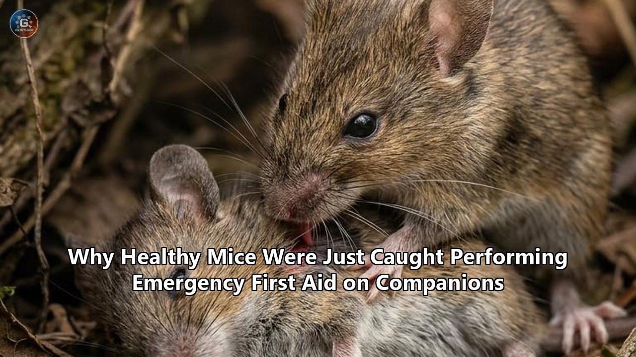
When a sedated laboratory mouse lies motionless on the bed of an observation cage, its breathing shallow and its righting reflex extinguished by anesthesia, a healthy cage mate does not simply sniff and wander away. In a series of tightly controlled experiments published in Science, neuroscientists documented that bystander mice systematically initiate a graduated sequence of physical interventions designed to revive their unconscious peers.
The behavior begins with rapid olfactory investigation, accelerates into intensive grooming of the face and head, and culminates in a targeted, forceful maneuver: using their teeth and forepaws, the awake rodents pry open the unconscious animal’s jaws, grasp its tongue, and drag it forward.
The papers, authored by independent research teams at the Keck School of Medicine of the University of Southern California (USC) and the David Geffen School of Medicine at UCLA, along with supporting neurophysiological data published in Science Advances by the University of Science and Technology of China (USTC), establish that this is not an erratic grooming glitch. It is a structured, neurobiologically wired emergency response.
The action physically clears compromised upper airways, ejects foreign obstructions lodged in the oral cavity, and triggers an ascending mechanosensory neural pathway that directly stimulates the unconscious brain’s arousal centers, slashing the time required to regain consciousness.
The revelation overturns a foundational assumption in mammalian ethology: that sophisticated, targeted first aid requiring mechanical manipulation of a peer's anatomy is restricted to large-brained primates, cetaceans, and select social insects. Behind the headlines lies an intricate web of hypothalamic oxytocin release, amygdalar gating, trigeminal brainstem circuits, and a fierce methodological debate over whether rodents possess the cognitive architecture for true altruism or are executing hardwired behavioral programs.
                     ┌────────────────────────────────────────┐
                     │   UNCONSCIOUS MOUSE (Under Isoflurane) │
                     └───────────────────┬────────────────────┘
                                         │
                   Mechanical Tongue Dragging & Mouth Opening
                                         │
                                         ▼
                     ┌────────────────────────────────────────┐
                     │ Afferent Mechanosensory Input (Tongue) │
                     └───────────────────┬────────────────────┘
                                         │
                                         ▼
                     ┌────────────────────────────────────────┐
                     │      Mesencephalic Trigeminal          │
                     │          Nucleus (MTN_Glu)             │
                     └───────────────────┬────────────────────┘
                                         │
                                         ▼
                     ┌────────────────────────────────────────┐
                     │        Locus Coeruleus (LC_NE)         │
                     │    Noradrenergic Surge Across Brain    │
                     └───────────────────┬────────────────────┘
                                         │
                                         ▼
                     ┌────────────────────────────────────────┐
                     │   Rapid Arousal & Airway Clearance     │
                     └────────────────────────────────────────┘
The Accidental Observation: How Routine Anesthesia Broke Laboratory Protocol
The project did not start as a planned investigation into rodent paramedicine. At USC’s Zilkha Neurogenetic Institute, neuroscientist Wenjian Sun was conducting routine behavioral and imaging experiments involving mice under transient anesthesia. Standard operating procedure in rodent neuroscience dictates that animals recovering from volatile anesthetics like isoflurane or injectable cocktails like ketamine-xylazine remain isolated in heated recovery chambers until their sternal recumbency and righting reflexes return. This isolation prevents normothermic collapse, cannibalism, or aggressive biting from cage mates.
During one trial run, an anesthetized subject was prematurely returned to its home enclosure with an awake cage mate. Rather than retreating or ignoring the inert body, the awake animal approached immediately.
Sun watched through the acrylic barrier as the resident mouse inspected the comatose partner, repeatedly nudging its head and licking its snout. Then the behavior escalated: the conscious mouse pinned the sedated animal's head, forced its incisors between the slack lips, and pulled the limp tongue outward.
Sun called over principal investigator Li I. Zhang, a professor of physiology and neuroscience. Initial reactions favored skepticism; laboratory mice frequently engage in barbering (compulsive hair trimming) or cannibalistic post-mortem feeding if a cage mate dies. Yet this interaction showed neither tissue consumption nor violent distress. The awake animal appeared focused entirely on the oral aperture.
When the unconscious mouse began to stir, the resident mouse abruptly terminated the tongue manipulation, retreated slightly, and resumed ordinary cage exploratory behavior.
The USC team, collaborating with neuroscientist Huizhong Whit Tao, set aside their secondary assays to systematically isolate the variables driving this interaction. At the same time, miles away at UCLA, a team led by neurobiologist Weizhe Hong and researcher Fangmiao Sun had observed an overlapping phenomenon: bystander mice delivering intensive, facial-directed tactile stimulation specifically to comatose peers, distinct from any previously cataloged form of rodent social touch.
The parallel discoveries prompted both groups to reconstruct the exact ethological, anatomical, and neurological parameters of the behavior.
The Behavioral Cascade: From Olfactory Audit to Mandibular Manipulation
To quantify the response, the USC researchers engineered a standardized observation paradigm. They exposed healthy, conscious "helper" mice to a split-choice chamber containing two conspecifics: one familiar cage mate fully awake and mobile, and another familiar cage mate placed under light, surgical-plane anesthesia.
If the interaction were driven purely by novelty, the bystander might distribute its time evenly between the two animals or avoid the abnormal, motionless peer. Instead, the bystander mice allocated an average of 47 percent of the entire 13-minute observation window strictly to physical contact with the unconscious mouse, compared to just 5.8 percent of their time spent interacting with the active, healthy animal.
High-speed video capture running at 120 to 240 frames per second, coupled with machine-learning-based kinematic posture tracking (using deep neural network pose estimation platforms like DeepLabCut), revealed that the response follows an invariant, three-tiered escalation:
1. The Assessment Phase (0 to 60 seconds)
Upon initial contact, the awake mouse conducts a rapid, localized sensory survey. It directs dense bouts of sniffing toward the perioral zone, whiskers, and anogenital region. During this phase, vibrissal contact is maximal.
The awake mouse repeatedly nudges the flank and snout of the comatose animal with its nose, testing for tactile recoil or voluntary motor resistance.
2. Targeted Cephalic Allogrooming (60 to 180 seconds)
When the comatose animal exhibits sustained akinesia (complete lack of motor responsiveness), the helper's behavior shifts away from standard whole-body social grooming.
Standard rodent allogrooming is broadly distributed across the nape of the neck, back, and ears. Here, the helper restricts roughly 85 percent of its licking and pawing exclusively to the comatose partner's face, periorbital tissue, and mouth. The licking is rapid, repetitive, and concentrated around the oral commissures.
3. Forced Airway Intervention (180+ seconds)
If the comatose mouse remains unresponsive, the interaction becomes physically assertive. The awake mouse uses its forepaws to stabilize the head of the inert animal.
It inserts its snout directly into the oral fissure, using its incisors to part the slack jaws. In over 50 percent of all monitored trials across cohorts, the helper mouse clamped onto the tongue of the unconscious peer and pulled it outward, holding or dragging it repeatedly.
+--------------------------------------------------------------------------+
|             THE THREE-PHASE FIRST AID CASCADE IN RODENTS                 |
+-------------------+---------------------------+--------------------------+
| Phase             | Behavioral Hallmarks      | Anatomical Target        |
+-------------------+---------------------------+--------------------------+
| 1. Assessment     | Olfactory sweeping,       | Whiskers, snout,         |
|    (0-60 sec)     | tactile nudging           | flanks, anogenital area  |
+-------------------+---------------------------+--------------------------+
| 2. Cephalic       | High-frequency licking,   | Peri-oral zone, eyelids, |
|    Grooming       | vibrissal grooming        | facial midline           |
|    (60-180 sec)   |                           |                          |
+-------------------+---------------------------+--------------------------+
| 3. Airway Action  | Jaw prying, incisor       | Oral cavity, tongue,     |
|    (180+ sec)     | gripping, tongue dragging | trachea (indirect)       |
+-------------------+---------------------------+--------------------------+
Crucially, this vigorous intervention is not indiscriminate aggression. The bite force deployed during tongue manipulation is calibrated: post-recovery histological examinations of the recipients' tongues revealed no puncture lacerations, tissue necrosis, or lingual hemorrhage. The helper grips with sufficient traction to draw the muscle forward without severing lingual arteries or damaging mucosal integrity.
The behavior also displays strict temporal boundaries: the exact second the sedated mouse regains its righting reflex and makes voluntary limb movements, the helper instantly breaks off the mouth-directed manipulation. The prompt termination proves the action is regulated by dynamic feedback from the recipient's physiological state, rather than being an open-loop motor stereotypy.
Airway Physics and Mechanical Clearance: The Choking Hazard Assay
The immediate evolutionary question is functional: Does pulling a companion’s tongue provide a quantifiable survival benefit, or is it an accidental side effect of social frustration?
In anesthetized mammals lying in dorsal or lateral recumbency, loss of pharyngeal muscle tone causes the base of the tongue to collapse backward against the posterior pharyngeal wall, producing partial or complete upper airway obstruction. In human emergency medicine, this is countered via the head-tilt/chin-lift maneuver, the jaw-thrust technique, or direct traction on the tongue to clear the epiglottic opening.
The USC researchers modeled this physiological reality in the laboratory. Using high-resolution micro-computed tomography ($\mu\text{CT}$) and anatomical sectioning, they measured the cross-sectional area of the pharyngeal airway in anesthetized mice before and during helper-mediated tongue dragging.
The physical traction exerted by the helper pulled the hyoid apparatus and lingual base forward, expanding the narrowest section of the rodent pharyngeal passage by more than 35 percent.
To test whether this mechanical clearing could overcome an active physical blockage, the research team designed a foreign-body obstruction assay.
Under a dissecting microscope, experimenters placed an inert, non-toxic expanded polystyrene micro-sphere into the deep posterior oropharynx of unconscious mice, mimicking a severe airway obstruction.
The results were stark:
- When placed in the oropharynx of comatose mice, helper mice successfully targeted the foreign object, dislodged it, and pulled it completely out of the mouth in approximately 80 percent of trials.
- To control for whether mice are simply attracted to grooming foreign objects off their partners' bodies, the researchers placed identical polystyrene spheres into other anatomical regions, including the external auditory canal, the flank fur, the anus, and the vulva.
- In those control locations, the helper mice showed no targeted extraction behavior, ignoring the spheres.
The focus remained on the respiratory portal. This confirmed that the extraction response is linked to the mouth and airway, ruling out general grooming artifacts.
+--------------------------------------------------------------------------+
|         FOREIGN OBJECT REMOVAL EFFICIENCY ACROSS BODY SITES              |
+-----------------------------+--------------------+-----------------------+
| Anatomical Placement        | Extraction Success | Behavioral Focus      |
+-----------------------------+--------------------+-----------------------+
| Oropharynx / Mouth          | ~80%               | Immediate extraction  |
| Flank Fur                   | <5%                | Ignored / General rub |
| External Auditory Canal     | <3%                | Brief sniff, ignored  |
| Perianal / Genital Area     | 0%                 | Completely ignored    |
+-----------------------------+--------------------+-----------------------+
The Neural Circuitry of the Rescuer: Oxytocin and the Amygdalar Command Hub
What neural pathways allow a mouse to evaluate an unconscious peer and trigger this emergency protocol?
The UCLA group led by Fangmiao Sun and Weizhe Hong, alongside the USC team, turned to in vivo fiber photometry, immediate early gene ($c\text{-Fos}$) mapping, and cell-type-specific optogenetic manipulation to trace the rescuer's brain circuits.
Their investigations converged on a dedicated limbic pathway: the hypothalamic paraventricular nucleus ($\text{PVN}$) communicating with the medial amygdala ($\text{MeA}$).
+-----------------------------------------------------------------------------+
|              THE HELPER MOUSE'S INTERNAL REGULATORY CIRCUIT                 |
|                                                                             |
|      Visual/Olfactory Detection of Unconscious Peer                         |
|                           │                                                 |
|                           ▼                                                 |
|          ┌───────────────────────────────────┐                              |
|          │  Hypothalamic Paraventricular     │                              |
|          │          Nucleus (PVN)            │                              |
|          │   Oxytocin-Producing Neurons      │                              |
|          └─────────────────┬─────────────────┘                              |
|                            │                                                |
|       ┌────────────────────┴────────────────────┐                           |
|       ▼                                         ▼                           |
| ┌───────────────┐                        ┌───────────────┐                  |
| │  Central      │                        │  Medial       │                  |
| │  Amygdala     │                        │  Amygdala     │                  |
| │   (CeA)       │                        │   (MeA)       │                  |
| │ Alleviates    │                        │ Encodes state │                  |
| │ Helper Stress │                        │ Drives rescue │                  |
| └───────────────┘                        └───────┬───────┘                  |
|                                                  │                          |
|                                                  ▼                          |
|                                        ┌───────────────────┐                |
|                                        │ Paraventricular   │                |
|                                        │ Thalamus to NAcSh │                |
|                                        │ Motor Execution   │                |
|                                        └───────────────────┘                |
+-----------------------------------------------------------------------------+
The Oxytocin Engine in the Hypothalamus
The hypothalamic $\text{PVN}$ serves as the master neuroendocrine regulator for social behavior and homeostatic distress. Using fiber photometry with genetically encoded calcium indicators ($\text{GCaMP6s}$ expressed under an oxytocin promoter), researchers observed that $\text{PVN}$ oxytocin neurons fired at elevated baselines when a mouse was introduced to an unconscious partner.
Critically, these oxytocinergic neurons did not exhibit this firing pattern when the partner was fully awake, nor when the partner was merely resting or sleeping naturally. The neurons fired specifically in response to unresponsiveness.
To verify whether oxytocin was an active driver or an incidental correlate, the USC researchers micro-infused oxytocin receptor ($\text{OXTR}$) antagonists directly into the cerebral ventricles of helper mice.
Blocking oxytocin signaling virtually eliminated the tongue-dragging and jaw-prying behaviors: treated mice approached the unconscious partner, sniffed briefly, and then moved to opposite corners of the chamber.
Conversely, optogenetic photostimulation of channelrhodopsin-2 ($\text{ChR2}$)-expressing oxytocin terminals in the amygdala drove helper mice to initiate intense facial grooming and mouth-prying even toward cage mates under light sedation who had not yet entered deep unresponsiveness.
The Medial Amygdala as Behavioral Switchboard
At UCLA, Hong's laboratory demonstrated that the medial amygdala ($\text{MeA}$)—specifically a subpopulation of GABAergic interneurons—encodes the distinct behavioral transition between generic social interaction and targeted emergency first aid.
Microendoscopic cellular imaging showed that the $\text{MeA}$ contains functionally segregated neuronal ensembles:
- Ensemble A fires when a mouse interacts with an active, ambulatory partner.
- Ensemble B fires when a mouse interacts with an awake partner subjected to physical restraint or social defeat stress.
- Ensemble C fires exclusively when the partner is comatose, unresponsive, or experiencing respiratory distress.
When the UCLA team chemogenetically silenced these $\text{MeA}$ GABAergic neurons using inhibitory designer receptors exclusively activated by designer drugs ($\text{hM4Di}$ DREADDs), the helper mice failed to deliver cephalic and oral first aid, even though their general locomotion, scent-marking, and feeding behaviors remained normal.
The $\text{MeA}$ operates as the essential sensory-motor hub that translates perceived unconsciousness in a conspecific into the coordinated motor routine of airway resuscitation.
The Recipient's Awakening: Tracing the Tongue-Brain Arousal Pathway
While the USC and UCLA papers established what the rescuer was doing and why its brain drove the action, a third neurobiological question remained: How does having its tongue pulled accelerate an unconscious mouse's emergence from anesthesia?
In everyday veterinary and surgical practice, emergence from general anesthesia is considered a passive pharmacokinetic event: the liver metabolizes the compound, the lungs excrete volatile gas, and neural receptor occupancies slowly decay.
However, emerging research led by neurobiologist Peng Cao and colleagues at USTC, published in Science Advances, revealed that the helper mouse's physical intervention triggers an active, ascending neuro-arousal circuit in the patient.
+--------------------------------------------------------------------------+
|                  THE RECIPIENT'S ASCENDING AROUSAL CIRCUIT               |
+-----------------------+--------------------------------------------------+
| Anatomical Stage      | Physiological Action                             |
+-----------------------+--------------------------------------------------+
| 1. Lingual Stretch    | Deep mechanical tension on lingual proprioceptors|
+-----------------------+--------------------------------------------------+
| 2. Mesencephalic      | Glutamatergic neurons (MTN_Glu) fire high-       |
|    Trigeminal Nucleus | frequency bursts into the brainstem              |
+-----------------------+--------------------------------------------------+
| 3. Locus Coeruleus    | Noradrenergic neurons (LC_NE) flood the          |
|    Activation         | forebrain with norepinephrine                    |
+-----------------------+--------------------------------------------------+
| 4. Thalamocortical    | Desynchronization of slow-wave anesthesia;       |
|    Desynchronization  | rapid return of righting reflex                  |
+-----------------------+--------------------------------------------------+
The pathway operates through a specialized cranial reflex:
1. Lingual Mechanoreceptor Recruitment
The rodent tongue is densely packed with specialized sensory corpuscles and proprioceptive stretch receptors embedded within the intrinsic lingual muscles and the styloglossus, hyoglossus, and genioglossus musculature.
When the helper mouse clamps down and exerts longitudinal traction on the tongue, it generates sustained mechanical strain far exceeding anything produced during mastication or self-grooming.
2. Mesencephalic Trigeminal Nucleus ($\text{MTN}_{\text{Glu}}$)
The primary afferent neurons transmitting this deep mechanosensory and proprioceptive signal do not synapse in typical peripheral sensory ganglia. Uniquely in mammalian anatomy, the cell bodies of these primary proprioceptive sensory neurons reside inside the central nervous system: within the mesencephalic trigeminal nucleus ($\text{MTN}$) of the midbrain.
Cao’s team discovered that glutamatergic projection neurons within the $\text{MTN}$ ($\text{MTN}_{\text{Glu}}$) fire high-frequency bursts directly in response to lingual stretch.
3. Locus Coeruleus ($\text{LC}_{\text{NE}}$) Noradrenergic Surge
The $\text{MTN}_{\text{Glu}}$ neurons project monosynaptically to the locus coeruleus ($\text{LC}$), the brain's principal source of norepinephrine ($\text{NE}$).
The $\text{LC}$ serves as the central ignition switch for arousal, attention, and the maintenance of waking electroencephalographic ($\text{EEG}$) states.
In anesthetized mice, mechanical traction on the tongue by a helper directly drove calcium influx into $\text{LC}_{\text{NE}}$ neurons, prompting a surge of norepinephrine throughout the thalamus, basal forebrain, and cerebral cortex.
4. Accelerated Cortical Desynchronization
Electroencephalographic monitoring of the anesthetized recipients told a definitive story. Under isoflurane or ketamine anesthesia, the brain displays high-amplitude, low-frequency delta-wave oscillations and burst suppression patterns indicative of deep unconsciousness.
When a helper mouse engaged in tongue dragging, the recipient's cortical $\text{EEG}$ shifted within seconds toward low-voltage, high-frequency desynchronized waveforms—the signature of wakefulness.
The sedated mice woke up, rolled from their sides to their paws (return of righting reflex), and resumed motor mobility substantially faster than isolated control mice recovering from the identical anesthetic depth.
To confirm this circuit, Cao’s group used optogenetics to selectively silence the $\text{MTN}_{\text{Glu}} \to \text{LC}_{\text{NE}}$ pathway in unconscious mice.
When these neurons were suppressed with yellow-light-activated archaerhodopsin ($\text{Arch}$), the helper mice continued to groom and pull the tongues of their comatose partners just as before—yet the rapid-waking effect vanished.
The recipient's brain remained locked in deep slow-wave anesthesia until the drugs cleared via baseline passive metabolism.
This closed-loop experiment showed that helper mice exploit an innate mechanical-to-neural wake-up circuit in their unconscious peers.
The Familiarity Gate: Inclusive Fitness and the Social Brain
A central question in animal social dynamics is how prosocial actions are distributed among group members. In human emergency medicine, professional first responders act indiscriminately; on the street, however, psychological phenomena like the bystander effect can suppress intervention unless social proximity or shared identity lowers the activation threshold.
Laboratory rodents exhibit an explicit familiarity gate when administering emergency care.
The USC researchers systematically varied the social relationship between the rescuer and the comatose animal. They established three experimental cohorts:
- Cage-mate Pairs: Mice housed continuously in the same enclosure for more than three weeks prior to testing.
- Stranger Pairs: Genetically identical mice (C57BL/6J) of identical age and sex, housed in separate cages in the same room, meeting for the first time during the trial.
- Short-term Cohabitants: Mice paired together for only 24 to 48 hours.
+--------------------------------------------------------------------------+
|          INTERVENTION INTENSITY AS A FUNCTION OF FAMILIARITY             |
+--------------------------+---------------------+-------------------------+
| Social Relationship      | Time with Unconscious| Tongue-Dragging Rate    |
+--------------------------+---------------------+-------------------------+
| Long-term Cage mates     | 47.2% of window     | 54.3% of trials         |
| Short-term Cohabitants   | 28.1% of window     | 21.0% of trials         |
| Complete Strangers       | 12.4% of window     | 4.2% of trials          |
+--------------------------+---------------------+-------------------------+
The differences were unambiguous.
Familiar cage mates displayed high levels of investigative allogrooming and escalating mouth-directed physical intervention, with over half performing tongue-dragging maneuvers.
Faced with an unconscious stranger, the helper's intervention dropped sharply.
Bystanders approached comatose strangers, sniffed them cautiously from a distance, but spent less than a quarter of the time in direct contact.
Mouth biting and tongue pulling occurred in fewer than 5 percent of stranger interactions.
This dynamic reinforces the evolutionary principle that mice helping injured companions rely on chemical familiarity cues to direct costly, high-risk interventions toward their own social unit.
In rodent colonies, social recognition is mediated by volatile organic compounds and non-volatile Major Urinary Proteins (MUPs) detected through the main olfactory epithelium and the vomeronasal organ ($\text{VNO}$).
When researchers rendered helper mice transiently anosmic by infusing zinc sulfate ($\text{ZnSO}_4$) into the nasal mucosa, the distinction between cage mates and strangers disappeared: the animals showed a baseline, muted exploratory behavior toward both, failing to launch sustained first-aid sequences on either.
From an evolutionary standpoint, this selective deployment makes sense. In the wild, wild house mice (Mus musculus) live in structured demes dominated by an alpha male, several breeding females, and their extended offspring. An incapacitated stranger presents unknown threats: it may harbor lethal transmissible pathogens, originate from a hostile neighboring deme, or lure competitors into vulnerability.
An incapacitated cage mate, conversely, represents shared genetic investment and communal social capital. Rescuing a familiar group member preserves inclusive fitness and maintains the defensive, foraging, and thermoregulatory cohesion of the social unit.
Machine Learning, DeepLabCut, and Behavioral Tracking
The discovery raises an awkward historical question: Thousands of neuroscientists, pharmacologists, and behavioral researchers have anesthetized millions of rodents alongside cage mates over the past century. Why did it take until the mid-2020s for researchers to document that conscious rodents drag the tongues of their comatose peers to revive them?
The answer lies in the limitations of traditional behavioral scoring and the technological evolution of computer vision.
For decades, behavioral neuroscience relied on coarse, human-scored observation metrics or simple automated infrared beam breaks. When measuring rodent interactions, researchers looked for macro-behaviors categorized into broad ethological buckets: locomotion, freezing, rearing, sniffing, fighting, mounting, or passive rest.
Subtle, high-speed micro-movements occurring around the oral cavity were virtually invisible to the naked eye, lost in blurry 30-frame-per-second webcam footage, or written off by technicians as generic aggression, social biting, or displacement grooming.
+--------------------------------------------------------------------------+
|                  THE EVOLUTION OF BEHAVIORAL QUANTIFICATION              |
+--------------------------+-----------------------+-----------------------+
| Metric Parameter         | Classical Scoring     | High-Speed ML Systems |
+--------------------------+-----------------------+-----------------------+
| Frame Capture Rate       | 30 fps (Standard)     | 120-240 fps (High)    |
| Spatial Tracking Points  | 1-3 (Centroid/Tail)   | 16-32 Keypoints       |
| Micro-movement Detection | Missed / Subjective   | <1 mm Mandibular      |
| Observer Bias            | High Inter-rater Error| Unsupervised Classify |
| Mandibular Pose Tracking | Impossible            | Sub-millimeter Kinemat|
+--------------------------+-----------------------+-----------------------+
To crack open the anatomy of the behavior, the teams at USC and UCLA built multi-angle recording rigs utilizing industrial CMOS high-speed cameras equipped with macro lenses, ring-light illumination, and soundproof observation chambers.
They deployed DeepLabCut, an open-source deep learning framework based on residual neural networks (ResNet), trained specifically on hundreds of thousands of hand-annotated video frames.
The software tracked down to the sub-millimeter:
- The precise tips of the rescuer's upper and lower incisors.
- The anterior and lateral margins of the nostrils.
- The medial and lateral canthi of the eyes.
- The base, shaft, and tip of the recipient's tongue.
- The position and rotational angle of all four paw digits.
This computational pipeline extracted kinematic vectors that human observers could not reliably parse.
The machine learning models parsed the subtle difference between aggressive biting (characterized by sudden, high-velocity jaw snapping accompanied by backward head jerking) and restorative tongue manipulation (characterized by slow, sustained mandibular closure followed by gentle, low-acceleration outward linear traction).
Unsupervised behavioral clustering algorithms, such as B-SOiD (Behavioral Segmentation of Open-field In-session Data), grouped these coordinates into distinct kinematic states.
The algorithms established that the oral interactions directed at unconscious mice occupied a discrete cluster entirely separate from predatory hunting, territorial aggression, maternal pup retrieval, and normal reciprocal social grooming.
The behavior had always been occurring right before scientists' eyes; researchers simply lacked the spatio-temporal resolution to see it.
The Great Empathy Schism: Altruistic Intent vs. Hardwired Fixed Action Pattern
The publication of these findings in Science catalyzed an intense philosophical and methodological debate among behavioral biologists and cognitive psychologists.
The friction centers on the cognitive interpretation of the phrase: Are mice deliberately attempting to revive a partner, or are they executing an evolutionary fixed action pattern driven by stress reduction?
The Altruistic / Empathy Camp
Advocates of the empathy framework, including USC's Li Zhang and UCLA's Weizhe Hong, point to the goal-directed, flexible nature of the behavior.
In a classic framing popularized by the late primatologist Frans de Waal, empathy is not an all-or-nothing cognitive attribute reserved for humans and great apes, but a layered "Russian doll" continuum rooted in conserved neurobiology.
At its core sits emotional contagion: the ability to become affected by another's emotional state. At higher layers sit targeted helping and perspective-taking.
Proponents argue that the rodent first-aid response exhibits key characteristics of targeted helping:
- State-Dependent Calibration: Helper mice do not pull tongues when a partner is awake, moving, or sleeping naturally.
- Feedback-Gated Cessation: The instant the recipient regains mobility, the helper stops.
- Instrumental Problem Solving: Helpers successfully clear external foreign obstructions blocking the trachea, showing situational adjustment.
- Familiarity Preference: Helpers direct significant energy and effort primarily toward familiar conspecifics.
"The animal appears to be able to recognize the unconscious state of its partner, with the unresponsiveness triggering the behavior and regaining of responsiveness terminating the behavior," noted lead author Wenjian Sun.
To researchers in this camp, this closed-loop sensitivity demonstrates an evolutionary precursor to the altruistic first-response behavior found in human societies.
The Ethological Skeptics
Other prominent cognitive neuroscientists urge strict caution against over-interpreting the data.
Peggy Mason, a neurobiologist at the University of Chicago who pioneered rodent prosociality studies with her landmark 2011 experiments showing rats learning to free trapped companions from restrainers, offered a pointed critique of the "first aid" nomenclature.
"Demonstrating that the mice are intending to help is obviously trickier than showing that the unconscious mice are helped," Mason observed.
She used a simple economic thought experiment to illustrate the distinction: "If I drop $20, by mistake, on the street and someone else picks it up, that person has been helped, but I have not helped them. They found a great behavior, I don't dispute that. I dispute the interpretation of it".
Skeptics propose alternative mechanistic explanations:
- Sensory Anomaly and Perceptual Violation: Mice rely heavily on continuous dynamic sensory feedback (sniffing back, whisker vibrations, ultrasonic vocalizations) during any social encounter. An unconscious mouse presents an eerie, radical violation of expectation. The helper may be attempting to compel sensory feedback through tactile agitation—poking, prodding, and pulling until the unresponsive object either acts like a mouse again or goes away.
- Personal Distress Alleviation: Watching a motionless cage mate may evoke acute distress in the observer. In a study published in PNAS, researchers noted that helper mice experience elevated physiological stress markers when encountering a sedated cage mate. Engaging in vigorous oral manipulation and licking lowers circulating corticosterone in the helper. Thus, the mouse may be performing repetitive behavior to relieve its own stress, rather than acting out of a conscious desire to save another animal's life.
- Innate Fixed Action Pattern: The response may be a hardwired motor routine selected by evolution because it reliably increased colony survival over millions of years, operating without any mental model of the peer’s inner consciousness or impending death.
+-----------------------------------------------------------------------------+
|             THE THEORETICAL DEBATE OVER RODENT FIRST AID                    |
+------------------------------------+----------------------------------------+
| Altruistic Empathy Interpretation  | Ethological Skepticism Interpretation  |
+------------------------------------+----------------------------------------+
| • Targeted helping aimed at a peer's| • Unconscious animal creates an        |
|   distress.                |   intolerable sensory void.            |
| • Complex feedback loop terminating| • Behavior terminates because motion   |
|   at arousal.       |   restores ordinary feedback.  |
| • Instrumental extraction of oral  | • Foreign objects in mouth induce      |
|   airway obstructions|   reflexive chewing/grooming responses.|
| • Driven by conserved mammalian    | • Self-soothing ritual that reduces the|
|   oxytocinergic empathy circuits   |   helper's own elevated stress         |
|  .                  |   hormones.                    |
+------------------------------------+----------------------------------------+
Regardless of which conceptual camp one favors, the empirical phenomenon remains intact: mice helping injured companions through mechanical intervention produces a life-saving physiological outcome, mediated by precise neurological circuits.
Comparative Ethology: Where Rodent First Aid Fits Across the Tree of Life
The USC and UCLA discoveries place rodent behavior in conversation with a broader body of field and laboratory research documenting emergency care across diverse taxa. Rather than standing as an isolated curiosity, the rodent airway maneuver aligns with an emerging evolutionary principle: in social species, individuals often evolve specialized behavioral adaptations to stabilize or rescue compromised group members.
+--------------------------------------------------------------------------+
|             CROSS-SPECIES TAXONOMY OF RECORDED FIRST AID                 |
+----------------------+--------------------+------------------------------+
| Species              | Incident Trigger   | Mechanical Intervention      |
+----------------------+--------------------+------------------------------+
| Matabele Ants        | Termite hunting    | Chemical wound antisepsis,   |
| (Megaponera analis)  | trauma/infection   | physical limb carry-back     |
+----------------------+--------------------+------------------------------+
| Bottlenose Dolphins  | Hypoxia, trauma,   | Physical surface support     |
| (Tursiops truncatus) | concussive shock   | for aerial respiration       |
+----------------------+--------------------+------------------------------+
| African Elephants    | Collapse, systemic | Trunk lifting, mud cooling,  |
| (Loxodonta africana) | toxicity, snares   | protective encirclement      |
+----------------------+--------------------+------------------------------+
| Laboratory Mice      | Loss of righting   | Jaw prying, tongue dragging, |
| (Mus musculus)       | reflex / hypoxia   | foreign object extraction    |
+----------------------+--------------------+------------------------------+
| Chimpanzees          | Open lacerations,  | Wound cleaning, application  |
| (Pan troglodytes)    | insect bites       | of chewed crushed insects    |
+----------------------+--------------------+------------------------------+
The Invertebrate Chemists: Megaponera analis
In the insect world, rescue behavior is extensively documented among African Matabele ants (Megaponera analis), specialized termite predators studied by behavioral ecologist Erik Frank.
When an ant is wounded during a raid on a termite mound, nest mates carry the incapacitated worker back to the nest.
Inside the subterranean colony, worker ants apply antimicrobial secretions squeezed from their metapleural glands directly into the open wounds of their injured peers, reducing post-injury mortality from opportunistic pathogens like Pseudomonas aeruginosa by nearly 90 percent.
This behavior, however, is driven by chemical pheromones—specifically dimethyl disulfide and dimethyl trisulfide released in mandibular secretions by wounded ants—acting as automated release triggers for the rescue routine.
The Cetacean Respiratory Lifesavers
In marine mammals, bystander rescue is tied directly to obligate breathing mechanics.
Bottlenose dolphins (Tursiops truncatus) and pilot whales (Globicephala) have been recorded supporting unconscious, stunned, or neonate companions at the surface of the water for hours.
Because cetaceans are conscious breathers—they must actively control each breath to prevent drowning—a comatose individual will asphyxiate underwater. Pod mates position their bodies underneath the incapacitated animal, pushing its blowhole above the surface until motor reflexes recover.
The Terrestrial Giants: Elephants
Field biologists have documented African elephants (Loxodonta africana) mobilizing around collapsed, poisoned, or tranquilized herd members.
Adult cows use their tusks and trunks to wedge underneath the chest and shoulders of an immobile peer, attempting to heave the animal back to its feet.
Because an elephant's mass causes progressive compression of the lungs and diaphragm when recumbent for extended periods, remaining down quickly turns lethal.
The Rodent Airway Advance
Within this comparative evolutionary landscape, the mouse discovery occupies a unique niche.
Unlike the ant, which responds to a chemical distress pheromone emitted by a wounded comrade, the helper mouse acts on an inert, silent target devoid of dynamic pheromonal emissions or distress vocalizations.
Unlike the dolphin or elephant, whose large neocortical volumes have long been associated with complex cognition and emotional processing, the mouse achieves targeted, life-saving mechanical resuscitation with a lissencephalic brain weighing less than half a gram.
This indicates that emergency response behaviors do not require massive brain expansions or human-like theory of mind. The underlying architecture is ancient, conserved, and hardwired into basal mammalian subcortical networks, enabling mice helping injured companions to safeguard group survival through targeted physical interventions.
Neurobiological Implications: Awakening Comatose Circuits in Human Medicine
Beyond evolutionary biology, the discovery of this rodent response has attracted interest from anesthesiologists, emergency room physicians, and neuro-traumatologists.
The identification of the $\text{MTN}_{\text{Glu}} \to \text{LC}_{\text{NE}}$ lingual-arousal pathway offers an unexpected neuroanatomical window into human emergence from anesthesia and recovery from comatose states.
           MECHANICAL TRACTION ON LINGUAL APPARATUS
                              │
                              ▼
        ╔═══════════════════════════════════════════╗
        ║    Mesencephalic Trigeminal Nucleus       ║
        ║               (MTN_Glu)                   ║
        ╚═════════════════════╤═════════════════════╝
                              │
                    Monosynaptic Glutamate
                              │
                              ▼
        ╔═══════════════════════════════════════════╗
        ║            Locus Coeruleus                ║
        ║                (LC_NE)                    ║
        ╚═════════════════════╤═════════════════════╝
                              │
                 Widespread NE Release Across
                 Thalamus, Forebrain, Cortex
                              │
                              ▼
        ╔═══════════════════════════════════════════╗
        ║   Rapid Awakening / EEG Desynchronization ║
        ║   Accelerated Recovery from Comatose State║
        ╚═══════════════════════════════════════════╝
In human clinical medicine, emergence from deep anesthesia or coma is managed using pharmacological reversals:
- Acetylcholinesterase inhibitors (e.g., neostigmine) for neuromuscular blockade.
- Specific antagonists (e.g., flumazenil for benzodiazepines, naloxone for opioids).
- Central wakefulness stimulants (e.g., methylphenidate, modafinil) in select neuro-ICU settings.
However, non-pharmacological, mechanosensory triggers that jump-start suppressed ascending reticular activating systems ($\text{ARAS}$) remain poorly understood.
The discovery that deep lingual muscle traction acts as a biological "crank" on the locus coeruleus provides an anatomical rationale for historical medical phenomena:
- In traditional medicine systems, vigorous stimulation of the philtrum (the acupressure point GV26 / Renzhong) or aggressive traction on the tongue was routinely used to resuscitate individuals suffering from syncopal collapse, drowning, or shock.
- Modern anesthesiologists often note that passing a direct laryngoscope blade over the base of the tongue or manipulating the pharynx causes transient spikes in sympathetic autonomic tone, heart rate, and electroencephalographic activity.
By mapping the exact circuit—from lingual stretch receptors to the $\text{MTN}_{\text{Glu}}$, directly into the noradrenergic engines of the $\text{LC}$—the USTC researchers demonstrated that the mammalian brainstem possesses an innate sensory backdoor to override pharmacological depression.
In the wake of these mouse discoveries, bioengineers and translational neuroscientists are assessing whether non-invasive electrical, mechanical, or pneumatic stimulation of the lingual-trigeminal axis could help accelerate emergence from surgical anesthesia, clear persistent vegetative states, or treat severe postoperative delirium in humans.
Laboratory Animal Care and Protocol Re-Evaluation
The identification of this behavior creates immediate operational ripples inside academic research vivariums.
Every research institution in the United States, Europe, and Asia operates under strict institutional animal care guidelines overseen by Institutional Animal Care and Use Committees (IACUCs). Standardized post-surgical protocols across the biomedical research landscape mandate that an animal emerging from anesthesia must be isolated in a dedicated cage, away from other animals, until it demonstrates full ambulatory recovery.
This isolation rule was created to prevent cage mates from biting, cannibalizing, or suffocating animals that lack defensive motor reflexes.
Yet the USC and UCLA data demonstrate that under standard social conditions, housing recovering animals with familiar cage mates does not lead to violence; instead, it can cut emergence latency and reduce upper airway obstructions.
In contrast, prolonged isolation leaves comatose animals alone with the baseline risk of respiratory collapse on the bedding, without the benefit of mechanical airway clearance.
+--------------------------------------------------------------------------+
|          CLASSICAL VIVARIUM PROTOCOL VS. NEW SOCIAL DATA                 |
+--------------------------+-----------------------+-----------------------+
| Protocol Parameter       | Classical IACUC Rule  | Emerging Perspective  |
+--------------------------+-----------------------+-----------------------+
| Post-op Social Context   | Strict Isolation      | Cohabitation with     |
|                          |                       | Familiar Cage Mates   |
+--------------------------+-----------------------+-----------------------+
| Anticipated Threat       | Aggressive biting,    | Airway obstruction,   |
|                          | cannibalism           | hypothermia, hypoxia  |
+--------------------------+-----------------------+-----------------------+
| Observed Reality         | Complete absence of   | Mechanical airway     |
|                          | tissue damage | opening, fast recovery|
|                          |                       |        |
+--------------------------+-----------------------+-----------------------+
| Recovery Latency         | Baseline / Slower     | Substantially shorter |
|                          | (Passive clearance)   | (Active LC arousal)   |
|                          |                       |        |
+--------------------------+-----------------------+-----------------------+
Vivarium veterinarians are not rewriting global animal welfare charters overnight. Anesthetized animals undergoing invasive abdominal or orthopedic surgery have surgical sutures and open wound margins that cage mates could rupture through allogrooming or biting.
Nevertheless, several laboratory animal welfare specialists have called for targeted re-examinations of the post-anesthesia isolation dogma.
In non-surgical anesthetic procedures—such as routine MRI imaging, radiation therapy, optical coherence tomography, or micro-CT scanning—co-housing recovering animals with familiar cage mates under direct video monitoring could provide a safer, faster recovery profile.
Future Horizons: Unresolved Variables and What Comes Next
While the discovery of mouse first aid has cracked open new research avenues, it leaves open several neurobiological and ethological questions that researchers are working to resolve:
1. The Impact of Environmental Stress and Resource Scarcity
All experiments published to date were performed using healthy laboratory mice housed under standard conditions with ad libitum food, water, and climate-controlled enrichment.
What happens to this emergency intervention when helper mice are chronically stressed, food-deprived, or housed under extreme crowding?
Social psychologists know that stress and scarcity attenuate helping behavior in humans; investigating whether resource scarcity shuts down the oxytocinergic $\text{PVN} \to \text{MeA}$ pathway in mice will test the limits of rodent prosociality.
2. Strain and Genetic Divergence
Laboratory mice are not uniform. The published studies relied heavily on the inbred strain C57BL/6J, known for its high social drive and maternal competence.
Other common laboratory strains exhibit drastically different behavioral baselines:
- BALB/c mice display elevated innate anxiety and reduced social interaction.
- 129S1/SvImJ mice show distinct social grooming deficits.
- Wild-derived strains (e.g., CAST/EiJ or Mus spretus) retain high aggression and fight-or-flight behaviors.
Screening cross-strain cohorts will reveal whether tongue dragging is an invariant genetic baseline across all Mus musculus populations or a trait modulated by specific inbred genetic backgrounds.
3. The Acoustic Communication Channel
Mice communicate in ultrasonic frequencies (20 to 100 kHz) completely inaudible to human ears. While comatose mice cannot produce audible cries, do they emit subtle, low-amplitude respiratory clicking sounds, stridor, or altered ultrasonic vocalizations (USVs) as their airway becomes compromised?
Deploying specialized bat-detector microphones and high-bandwidth acoustic spectrogram analysis during the assessment phase will clarify whether helper mice are responding to hidden vocal distress cues alongside visual and olfactory unresponsiveness.
4. Direct Neural Projections to Motor Centers
While the medial amygdala and oxytocin neurons are required to trigger the response, the downstream motor circuits coordinating the delicate mechanics of the jaw-prying and tongue-dragging maneuvers remain unmapped.
Researchers are using retrograde trans-synaptic viral tracing (such as rabies-virus-based monosynaptic circuit tracing) to bridge the gap: mapping the exact connections running from the $\text{MeA}$ through the motor trigeminal nucleus ($\text{Mo5}$) and the hypoglossal nucleus ($\text{CN XII}$), which control the rescuer’s own jaws and tongue during this intervention.
A Window into the Evolution of Mammalian Social Rescue
The discovery that healthy mice perform emergency airway first aid on unconscious companions re-anchors how neuroscientists conceptualize the roots of social care.
For generations, emergency interventions—such as inspecting an unresponsive peer, opening the mouth, clearing foreign blockages, and mechanically opening the airway—were viewed as triumphs of human civilization, developed through medical training and advanced cognitive insight.
The laboratory footage from USC, UCLA, and USTC points to an alternate biological reality.
Deep beneath the human neocortex, woven into the conserved architecture of the mammalian hypothalamus, amygdala, and brainstem, lies an instinct to intervene when a companion collapses.
Long before humans designed cardiopulmonary resuscitation or founded professional paramedicine, the biological blueprints for airway management and emergency resuscitation were already running inside the subcortical circuits of the common house mouse.
Reference:
- https://www.eurekalert.org/news-releases/1076255
- https://socialcare.today/2025/03/10/mice-perform-cpr-like-resuscitation-to-help-their-stricken-friends/
- https://www.jpost.com/science/science-around-the-world/article-843482
- https://www.livescience.com/animals/land-mammals/mice-administer-first-aid-to-friends-even-trying-to-bring-the-dead-back-to-life
- https://www.smithsonianmag.com/smart-news/lab-mice-appear-to-offer-first-aid-care-to-their-unconscious-companions-even-pulling-on-their-tongues-180986102/
- https://www.pnas.org/doi/10.1073/pnas.2423374122
- https://pubmed.ncbi.nlm.nih.gov/39841840/
- https://medpsych.at/00-Kooperatives-Sozialverh-ErsteHilfe-Science2025.pdf
- https://www.bioscience.com.pk/en/subject/psychology/scientists-watch-mice-perform-cpr-like-rescue-on-unconscious-companions-by-pulling-their-tongues
- https://www.sciencealert.com/incredible-discovery-shows-mice-trying-to-revive-fallen-companions
- https://www.mtpr.org/2025-02-21/lab-mice-may-give-first-aid-to-unconscious-mates
- https://www.bioscience.com.pk/en/subject/psychology/scientists-watch-mice-perform-cpr-like-rescue-on-unconscious-companions-by-pulling-their-tongues
- https://www.earth.com/animals/mice-perform-first-aid-cpr-to-unconscious-companions-distress-to-revive-them/
- https://www.nationalacademies.org/read/29238
- https://www.iflscience.com/rodent-resuscitation-watch-mice-attempt-to-revive-their-unconscious-mates-78149
- https://www.researchgate.net/publication/395454344_Neural_correlates_of_adversity-overcoming_pup_rescue_behavior_in_female_mice
- https://www.jpost.com/science/science-around-the-world/article-843482
- https://pmc.ncbi.nlm.nih.gov/articles/PMC11956844/
- https://www.researchgate.net/publication/388280630_Rescue-like_behavior_in_a_bystander_mouse_toward_anesthetized_conspecifics_promotes_arousal_via_a_tongue-brain_connection
- https://www.earth.com/animals/mice-perform-first-aid-and-save-unconscious-peers-in-emergencies/
- https://note.com/ueishota/n/n40339d0e79df?hl=en
- https://www.zmescience.com/ecology/animals-ecology/mice-perform-first-aid-in-natural-instinct-to-rescue-their-peers/
- https://en.ara.cat/science-technology/mice-help-their-colleagues-when-they-are-in-difficulty_1_5346939.html
- https://www.researchgate.net/publication/389209019_Reviving-like_prosocial_behavior_in_response_to_unconscious_or_dead_conspecifics_in_rodents
- https://www.researchgate.net/figure/Rescue-like-behaviour-test-towards-two-cage-mates-constrained-inside-tubes-A-C_fig5_334892593
- https://www.earth.com/animals/mice-perform-first-aid-cpr-to-unconscious-companions-distress-to-revive-them/
- https://pmc.ncbi.nlm.nih.gov/articles/PMC12011203/
- https://www.researchgate.net/publication/389209019_Reviving-like_prosocial_behavior_in_response_to_unconscious_or_dead_conspecifics_in_rodents
- https://pubmed.ncbi.nlm.nih.gov/39977514/
- https://www.vice.com/en/article/mice-perform-first-aid-on-unconscious-mates-just-like-humans/