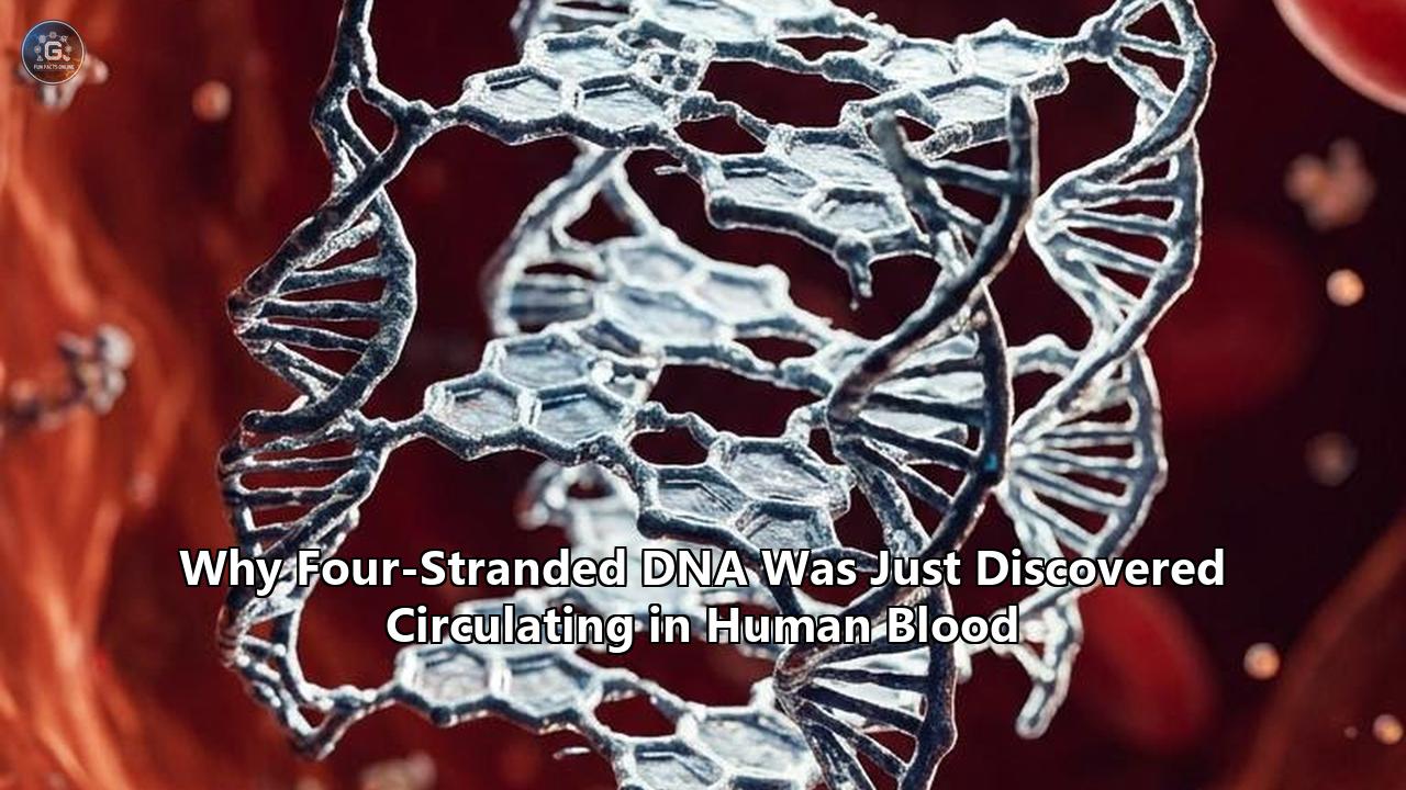
A research team led by molecular biologists at the University of Cologne has captured the first direct biochemical evidence of folded four-stranded DNA circulating freely within human blood plasma. The discovery, reported by Dr. Robert Hänsel-Hertsch and his colleagues, establishes that cell-free genetic material in circulation does not exist solely as the classic double helix described by James Watson and Francis Crick in 1953. Instead, segments of circulating nucleic acids fold into dense, four-stranded structures known as G-quadruplexes (G4s).
"Our study provides, to our knowledge, the first direct biochemical evidence that folded G-quadruplex structures are present in nucleic acids captured directly from human plasma," Hänsel-Hertsch stated regarding the findings. "Structure is a layer of information, and potentially of biology, that sequencing alone cannot see."
For decades, liquid biopsy technologies and genomic diagnostics have operated on a sequence-first assumption: cell-free DNA (cfDNA) was treated as a collection of linear, double-stranded fragments circulating as passive biological debris. Routine blood-based genomic tests denature and sequence this material, translating physical molecules into digital strings of A, T, C, and G.
The identification of stable, non-canonical folds in human plasma upends that single-molecule perspective. It creates a direct divide between two diagnostic philosophies: one that relies strictly on reading primary genetic sequences, and another that analyzes the physical geometry and topological folding of circulating molecules. Understanding how and why four-stranded structures survive in blood reveals the technical bottlenecks of conventional sequencing and exposes competing approaches to diagnostic medicine.
Molecular Architecture: Watson-Crick Duplexes vs. Hoogsteen G-Quadruplexes
The structural architecture of four-stranded DNA differs fundamentally from the standard Watson-Crick double helix. The double helix depends on anti-parallel strands joined by complementary base pairs—adenine binding with thymine through two hydrogen bonds, and cytosine binding with guanine through three. This creates a flexible, uniform, linear ladder optimized for packaging around histone octamers inside the cell nucleus.
By contrast, G-quadruplexes arise within guanine-rich regions of nucleic acids. When single strands are exposed during transcription, replication, or cellular breakdown, four guanine bases coordinate in a single plane through Hoogsteen hydrogen bonding. This planar arrangement is called a G-quartet (or G-tetrad). Multiple G-quartets stack vertically on top of one another to generate a compact, four-stranded architecture.
Canonical Double Helix (Watson-Crick)
   5' —- A === T —- 3'
   3' —- T === A —- 5'
   5' —- G === C —- 3'
   (Flexible, linear duplex held by standard base pairing)

Four-Stranded G-Quadruplex (Hoogsteen Tetrad)
        G ======= G
        ||       ||
        ||  (K+) ||  <-- Central channel stabilized by monovalent cations
        ||       ||
        G ======= G
   (Four guanine bases planar-bonded, stacking into a rigid 3D cylinder)
The thermodynamic stability of these two geometries is governed by distinct chemical factors:
- Ionic Dependence: Watson-Crick duplexes are destabilized by elevated temperatures and do not require specific central metal ions to maintain base pairing. G-quadruplexes require monovalent cations—most notably potassium ($K^+$), and to a lesser extent sodium ($Na^+$)—which sit inside the central channel between stacked tetrads to coordinate the eight oxygen atoms of the guanine rings and neutralize negative electrostatic charge.
- Enzymatic Susceptibility: In circulating blood, naked duplex DNA is rapidly degraded by serum nucleases, primarily deoxyribonuclease I (DNase I), which cleaves phosphodiester backbones along the minor and major grooves. In contrast, the tightly packed, cylindrical geometry of four-stranded G-quadruplexes sterically hinders active sites of many general endonucleases, providing intrinsic resistance to rapid enzymatic breakdown.
- Topology Diversity: While genomic double-stranded DNA remains predominantly in a uniform B-DNA right-handed conformation, four-stranded G4 structures adopt diverse topologies. Depending on the direction of the connecting phosphodiester loops, G-quadruplexes fold into parallel, anti-parallel, or hybrid conformations, creating distinct geometric targets for cellular proteins.
In cellular nuclei, G-quadruplexes act as transient structural switches, enriched in gene promoter regions (such as MYC, BCL2, and KRAS) and telomeric ends. They dynamically regulate how the genetic code is transcribed and replicated. However, discovering them intact in blood plasma indicates that these physical shapes are not merely fleeting intracellular regulatory states, but stable biological artifacts capable of surviving extracellular circulation.
Methodological Divide: Denaturing Spin Columns vs. Native Surface Capture
Why did it take decades to discover four-stranded structures in circulating blood? The answer lies in the conflicting goals of routine molecular biology workflows and non-denaturing structural chemistry.
Conventional liquid biopsy processing was built for high-throughput sequencing yield rather than structural preservation. The standard analytical pipeline systematically erases secondary and tertiary DNA conformations:
- Chemical Lysis and Denaturation: Plasma is treated with high concentrations of chaotropic salts (such as guanidinium thiocyanate), proteinase K, and elevated temperatures ($56^\circ\text{C}$ to $65^\circ\text{C}$) to strip off protective proteins and denature biological complexes.
- Solid-Phase Extraction: Nucleic acids are bound to silica membranes under conditions that favor linear molecules, washed with alcohol-heavy buffers, and eluted in low-salt, alkaline solutions that destabilize non-canonical structures.
- Double-Stranded Library Construction: Standard next-generation sequencing (NGS) preparation uses ligation enzymes that specifically target double-stranded DNA fragments with blunt or cohesive ends, routinely discarding single-stranded and non-duplex variants.
- Thermal Amplification (PCR): Samples undergo cycles of denaturation at $95^\circ\text{C}$, unwinding and linearizing folded structures before enzymatic sequencing.
Conventional Workflow: Destroys Secondary Structure
  Plasma Sample
    ──> Chaotropic Lysis & Heat (56-65°C)
    ──> Silica Column Extraction
    ──> Thermal Denaturation (95°C)
    ──> Standard NGS
  Result: Complete loss of 3D folds; only primary base sequence is recorded.

Native Capture Workflow: Preserves Secondary Structure
  Plasma Sample
    ──> Direct Poly(A)-Tailing (No heat, no chaotropic agents)
    ──> Solid-Phase Magnetic Immobilization
    ──> Native Affinity Probing (BG4 Antibodies / CD Spectroscopy)
  Result: Intact folded G-quadruplexes confirmed directly from circulation.
To capture folded structures without altering them, Hänsel-Hertsch's group designed a gentle native extraction protocol. They avoided harsh organic extractions, boiling steps, and standard silica elution. Instead, the team tagged endogenous cell-free nucleic acids directly within plasma using gentle enzymatic poly(A)-tailing, immobilizing the fragments onto solid magnetic beads without a single denaturation step.
This technical divergence illustrates a fundamental trade-off:
| Technical Parameter | Standard Liquid Biopsy Protocols | Native Nucleic Acid Capture |
|---|---|---|
| Primary Objective | Maximize total DNA yield and digital sequence accuracy | Preserve delicate 3D folding and native secondary structure |
| Extraction Chemistry | Chaotropic salts, alcohol washes, thermal lysis ($>56^\circ\text{C}$) | Non-denaturing physiological buffers, no thermal lysis |
| Library Preference | Mononucleosomal double-stranded fragments (~166 bp) | Size-agnostic; captures single-stranded and ultrashort DNA |
| Structural Integrity | Completely denatured into linearized single strands | Preserves endogenous Hoogsteen bonding and G-quartet stacks |
| Key Limitation | Blind to tertiary folding, epigenetic loops, and G4 states | Lower yield, prone to protein-binding contamination, lower throughput |
By avoiding thermal denaturation, the researchers prevented folded structures from melting, while simultaneously avoiding conditions that might cause linear, guanine-rich single strands to artificially fold in vitro during laboratory handling.
The Size Paradox: Mononucleosomal cfDNA vs. Ultrashort cfDNA
The discovery of folded structures in circulation is linked to an ongoing debate in fragmentomics: what size of DNA actually circulates in human blood?
For nearly twenty years, the standard model held that plasma DNA was structurally homogeneous. When healthy cells die through apoptosis, an endonuclease called caspase-activated DNase (CAD) cleaves chromatin into neat pieces between nucleosomes. Because a single histone core wraps roughly 147 base pairs of DNA, and linker segments account for an additional 20 base pairs, apoptotic cfDNA in human blood displays a consistent modal peak at approximately 166 to 167 base pairs. Clinical oncology tests, including Guardant360 and FoundationOne Liquid CDx, were designed around this mononucleosomal size profile.
Mononucleosomal cfDNA (~166 bp)
  [==== 147 bp wrapped around histone octamer ====]-- 20 bp linker --
  - High abundance in standard extractions
  - Double-stranded, apoptotic origin
  - Readily captured by commercial silica columns

Ultrashort cfDNA (~30–70 nt, modal peak ~50 nt)
  [~50 nt single-strand / folded G4 core]
  - Slips through standard size-selection cutoffs
  - Derived from open, non-nucleosomal regulatory chromatin
  - Contains high guanine content capable of non-canonical folding
However, recent studies by teams at Cancer Research UK and the University of Cologne identified an overlooked population: ultrashort cell-free DNA (us-cfDNA). These single-stranded fragments measure between 30 and 70 nucleotides in length, with a sharp mode centered near 50 nucleotides.
Standard clinical assays missed these short molecules because common purification columns discard fragments below 100 base pairs to avoid primer-dimer artifacts. When researchers applied single-stranded library preparation (ssDNA-seq), they found that us-cfDNA was not just random background degradation. In healthy individuals, ultrashort fragments accounted for a median of 19.1% of all sequenced cell-free DNA fragments. In cancer patients, this fraction fell significantly to 14.2%.
Crucially, genomic mapping showed that these ~50-nucleotide fragments did not originate from nucleosome-wrapped regions. Instead, they mapped to accessible, open chromatin in blood and tissue cells—specifically near transcription start sites, promoters, and active regulatory switches. These same loci contain dense concentrations of putative quadruplex sequences (PQS). Because these short strands are rich in guanine runs, they fold into compact, four-stranded structures that resist further degradation, allowing them to survive in the bloodstream as structural remnants of active cellular machinery.
Orthogonal Verification: Antibody Probes vs. Biophysical Spectroscopy
To confirm that these four-stranded complexes were genuinely folded in human plasma rather than random molecular aggregations, the Cologne researchers relied on two complementary analytical strategies: conformational antibody affinity and circular dichroism spectroscopy.
Antibody Affinity Probing (The BG4 Strategy)
Originally developed at the University of Cambridge by Shankar Balasubramanian and Giulia Biffi, the BG4 antibody is a single-chain variable fragment (scFv) engineered to recognize the three-dimensional geometry of a folded G-quadruplex. BG4 does not recognize single-stranded or double-stranded linear DNA; its binding pockets lock exclusively onto the planar surfaces and connecting loops of stacked G-quartets.
In the plasma study, BG4 successfully identified endogenous complexes pulled directly from blood samples. To ensure the antibody was not binding nonspecifically to random plasma components, the researchers ran competitive binding assays. When synthetic G4-forming oligonucleotides were added to compete for the antibody, the detection signal dropped proportionally. A control oligonucleotide that contained mutated sequences incapable of folding produced no reduction in binding, confirming the assay’s structural specificity.
Circular Dichroism (CD) Spectroscopy
While antibodies demonstrate binding affinity, circular dichroism spectroscopy measures physical structure. CD spectroscopy evaluates how chiral molecules absorb left- and right-circularly polarized light across ultraviolet wavelengths.
When synthetic oligonucleotides matching the most enriched plasma sequences were placed in potassium-containing physiological solutions, they produced distinct CD spectral signatures:
- A positive molar ellipticity peak near 264 nm.
- A negative molar ellipticity trough near 245 nm.
CD Ellipticity (mdeg)
      ^
 +20  |         /\  <-- Parallel G4 Signature (~264 nm)
      |        /  \
   0  |-------/----\------------------> Wavelength (nm)
      |             \      /
 -20  |              \____/  <-- Negative Trough (~245 nm)
      +--------------------------------
     220   240   260   280   300
This specific spectral profile matches a parallel G-quadruplex topology, in which all four strands of the tetrad run in the same 5'-to-3' chemical direction. This contrasts with anti-parallel quadruplexes, which show a positive peak at 295 nm and a negative trough at 260 nm. The CD spectra confirmed that the sequence tracts enriched in human blood spontaneously form stable, uniform, parallel four-stranded structures under physiological salt conditions.
Competing Diagnostic Approaches: Sequence-Centric vs. Structural Fragmentomics
The confirmation of folded four-stranded DNA circulating in human blood highlights a division between two diagnostic strategies: sequence-based mutation hunting versus conformational fragmentomics.
The Sequence-Centric Approach
Modern precision oncology relies heavily on identifying actionable somatic mutations in circulating tumor DNA (ctDNA). Techniques such as hybrid-capture next-generation sequencing, amplicon sequencing, and digital droplet PCR (ddPCR) examine blood samples for specific genetic alterations:
- Point mutations (e.g., EGFR L858R in non-small cell lung cancer, BRAF V600E in melanoma, KRAS G12D in colorectal cancer).
- Copy number variations (e.g., ERBB2/HER2 amplifications).
- Chromosomal rearrangements and fusions (e.g., ALK, ROS1).
While sequence-centric assays have become central to clinical oncology, they have clear trade-offs. They require high tumor shedding to reliably detect mutant alleles, which often makes them less sensitive for early-stage or low-burden cancers. They also demand high sequencing depth to distinguish genuine low-frequency variants from sequencing errors, and they provide little insight into the cell-of-origin if a mutation is shared across multiple tissue types.
Structural and Conformational Fragmentomics
Conformational fragmentomics, by contrast, shifts focus from individual base substitutions to the structural properties and folding patterns of cell-free molecules.
Instead of hunting for rare single-nucleotide variants, this approach assesses global features of chromatin regulation:
- Fragment length distributions (analyzing ratios of ultrashort vs. mononucleosomal fragments).
- End-motif fragmentation patterns (identifying preferences in how nucleases cut DNA).
- Preservation of non-canonical secondary folds (such as circulating G4s).
Because cancer cells undergo widespread epigenetic reprogramming—including chromatin remodeling, altered promoter accessibility, and genome-wide hypomethylation—their degradation patterns differ fundamentally from healthy blood cells.
Recent studies by Dr. Florent Mouliere and colleagues have shown that quantifying ultrashort fragments and their G4-forming potential can differentiate cancer patients from healthy controls, even when circulating tumor fractions are too low for conventional mutation-based sequencing to detect. Similarly, maternal plasma studies published in 2025 demonstrated that variations in ultrashort cell-free DNA containing potential G4 motifs could identify preeclampsia with an area under the curve (AUC) of 0.86 to 0.90, pointing toward diagnostic applications well beyond oncology.
| Diagnostic Feature | Sequence-Centric Liquid Biopsy | Structural / G4 Fragmentomics |
|---|---|---|
| Primary Target | Somatic single nucleotide variants, indels, gene fusions | Molecular length, 3D shape, and chromatin accessibility profiles |
| Detection Method | Deep targeted NGS, digital droplet PCR (ddPCR) | Native affinity capture, ssDNA-seq, conformational biosensors |
| Dependence on Tumor Burden | High; struggles when tumor fraction falls below 0.1% | Lower; evaluates systemic changes in chromatin and cell-death dynamics |
| Actionability | High; directly identifies targets for specific drugs (e.g., Osimertinib) | Developing; primarily serves as an early screening and classification biomarker |
| Processing Requirements | Standardized commercial extraction kits | Non-denaturing preservation and specialized isolation chemistries |
The Molecular Dilemma: DNA Quadruplexes vs. RNA Quadruplexes
Despite confirming folded four-stranded structures in human blood, the study revealed a key molecular ambiguity: current detection tools cannot easily tell whether these circulating structures are made of DNA or RNA.
Hänsel-Hertsch noted that the affinity probes used in the study bind directly to the three-dimensional G-quartet structure rather than the sugar-phosphate backbone. Both deoxyribonucleic acid (DNA) and ribonucleic acid (RNA) sequences rich in guanine can fold into G-quadruplexes. Consequently, an antibody like BG4 or a structure-specific chemical ligand will bind to both without distinguishing the ribose from the deoxyribose core.
This limitation highlights an important biochemical comparison:
DNA G-Quadruplexes (dG4)
- Backbone: 2'-deoxyribose sugar ring lacking a 2'-hydroxyl group.
- Structural Flexibility: Can adopt parallel, anti-parallel, or mixed hybrid configurations depending on cation concentrations and loop sequences.
- Origin: Derived from chromatin degradation, apoptotic cleavage of accessible promoter regions, and telomeric DNA.
- Biological Context: Indicates cell-death dynamics, chromatin unwinding, and genomic fragmentation.
RNA G-Quadruplexes (rG4)
- Backbone: Ribose sugar containing a bulky 2'-hydroxyl group (-OH).
- Structural Rigidity: The 2'-hydroxyl group enforces a C3'-endo sugar pucker and creates steric constraints that strongly favor parallel topologies, making RNA G4s thermodynamically more stable than their DNA counterparts.
- Origin: Derived from non-coding RNAs, ribosomal RNA degradation, transfer RNA halves, and messenger RNA untranslated regions (UTRs) released by dying cells or active extracellular vesicles.
- Biological Context: May reflect active cellular secretion, immune signaling, or platelet activation rather than passive genomic degradation.
Resolving this distinction requires rigorous enzymatic digestion protocols. The research team noted that follow-up validation will require treating plasma samples with selective nucleases—such as RNase A and RNase H to degrade RNA, alongside DNase I and Benzonase to digest DNA—prior to native capture.
Determining whether circulating quadruplexes are primarily DNA, RNA, or a combination of both will establish their physiological source. It will also influence how clinical blood samples must be collected, as RNA and DNA degrade at different rates and require distinct chemical stabilizers in collection tubes.
Circulating G4s: Passive Biomarkers vs. Druggable Targets
The discovery of stable quadruplex structures in circulation also bridges diagnostics and therapeutics. For over a decade, pharmaceutical developers have investigated four-stranded DNA as an intracellular target for cancer treatments.
Because G-quadruplexes appear frequently in the promoter regions of oncogenes such as MYC, KIT, and KRAS, researchers developed small-molecule ligands—including Quarfloxin (CX-3543), CX-5461, and Pyridostatin (PDS)—engineered to bind and stabilize these structures within cancer cells. When a small molecule locks a G4 into a rigid fold inside the nucleus, it acts as a physical barrier to RNA polymerase and DNA replication forks, inducing targeted DNA damage and driving rapidly dividing malignant cells into apoptosis.
Intracellular Therapeutic Targeting
  Small-Molecule Ligand (e.g., CX-5461)
    ──> Traps G4 in Oncogene Promoter (*MYC*)
    ──> Blocks RNA Polymerase & Stalls Replication Fork
    ──> Triggers DNA Double-Strand Breaks in Tumor Cells

Extracellular Diagnostic Sensing
  Endogenous Circulating G4 Fragment
    ──> Resists Serum Nucleases Due to Compact Fold
    ──> Captured via Conformation-Preserving Assays
    ──> Serves as a Biomarker for Disease Screening & Monitoring
Discovering these structures circulating in blood plasma creates a new point of comparison between intracellular drug targeting and extracellular monitoring:
- Pharmacodynamic Monitoring: If a patient receives a G4-stabilizing anticancer drug, does the treatment change the number of folded four-stranded complexes shed into the bloodstream? Circulating G4 levels could serve as a direct, non-invasive readout of whether a drug is successfully engaging its targets in tumor tissue.
- Immunogenicity and Inflammation: Naked extracellular DNA is recognized by the innate immune system via Toll-like receptor 9 (TLR9) and the cGAS-STING pathway, driving systemic inflammation. The degree to which folded four-stranded configurations trigger or evade these pattern recognition receptors compared to standard duplex DNA remains an open biological question.
- Targeted Clearance: While intracellular G4 targeting aims to cause localized genomic instability in tumor cells, circulating quadruplexes are free from chromatin packaging. If circulating G4s prove to be biologically active signaling molecules rather than inert debris, strategies designed to clear or neutralize them could open new therapeutic paths for managing autoimmune conditions such as systemic lupus erythematosus (SLE), where extracellular nucleic acids drive persistent inflammation.
Technical Hurdles and the Road to Clinical Use
Transitioning the detection of folded four-stranded DNA from an academic discovery into a validated clinical tool will require solving several engineering and biological challenges.
Direct Nanopore Structural Sensing
One promising path for reading structural DNA is solid-state and biological nanopore sequencing. In conventional sequencing pipelines, enzymes strip and unwind secondary structures so single-stranded DNA can thread through a protein pore one base at a time.
Next-generation nanopore platforms are being adapted to measure the physical shape of passing molecules directly. As a folded G-quadruplex passes through or docks at a pore opening, it generates distinct ionic current blockades that differ clearly from linear strands. Developing nanopore sensors capable of reading four-stranded DNA without thermal denaturation could enable fast, direct physical profiling of circulating nucleic acids in clinical laboratories.
Pre-Analytical Standardization
Liquid biopsy assays depend heavily on pre-analytical consistency. Factors such as the type of blood collection tube, the time elapsed before plasma centrifugation, and freeze-thaw cycles can alter sample quality:
- Standard K2-EDTA tubes allow white blood cells to lyse if processing is delayed beyond a few hours, flooding plasma with genomic duplex DNA that dilutes native cell-free signals.
- Specialized preservative tubes (e.g., Streck Cell-Free DNA BCT) use chemical fixatives that crosslink proteins to nucleic acids, which could distort delicate secondary structures.
- Establishing specialized handling guidelines that preserve native folded states without triggering artificial aggregation will be essential for reproducible clinical testing.
Prospective Cohort Validation
The University of Cologne study established proof of concept: folded four-stranded structures are present in human blood plasma. However, as the authors noted, the work was designed to demonstrate their physical presence, not to serve as an evaluated diagnostic test.
The next clinical phase will require prospective trials across large, diverse patient groups. Researchers must evaluate cohorts of healthy individuals alongside patients with early-stage cancers, autoimmune disorders, and metabolic diseases to establish baseline variations, clearance rates, and clinical sensitivity.
Confirming that genetic material in human blood circulates in three-dimensional, four-stranded conformations demonstrates that the architecture of cell-free nucleic acids is far richer than standard sequencing assays have captured. As analytical technologies evolve to measure molecular structure alongside primary sequence, liquid biopsy is poised to transition from reading DNA solely as an alphabetical code to analyzing it as a dynamic, physical biomarker.
Reference:
- https://m.economictimes.com/news/international/global-trends/scientists-find-dna-in-human-blood-can-fold-into-strange-four-stranded-shapes-a-hidden-layer-of-genetic-information-that-ordinary-sequencing-may-miss/amp_articleshow/134291394.cms
- https://www.sciencealert.com/scientists-discover-strange-4-stranded-genetic-structures-in-human-blood
- https://www.discovermagazine.com/four-stranded-dna-makes-human-debut-2311
- https://www.cmmc-uni-koeln.de/cmmc-news/show-news-detail/ultrashort-cell-free-dna-in-blood-identified-a-potential-new-diagnostic-target-for-precision-oncology
- https://www.zubairkhalid.com/knowledge/molecular-biology/g-quadruplex-dna
- https://en.wikipedia.org/wiki/G-quadruplex
- https://pubs.rsc.org/sc/article/doi/10.1039/d6sc04173f/1314650/Identification-of-a-G-quadruplex-forming-cell-free
- https://www.researchgate.net/publication/364726830_G-quadruplex_DNA_A_Longer_Story
- https://www.progress.org.uk/human-quadruple-stranded-dna-discovered/
- https://www.researchgate.net/publication/414222345_Nucleic_Acid_Capture_from_Human_Blood_Plasma_Uncovers_G-Quadruplex_Structures?_tp=eyJjb250ZXh0Ijp7InBhZ2UiOiJpbnN0aXR1dGlvbiIsInByZXZpb3VzUGFnZSI6bnVsbCwic3ViUGFnZSI6bnVsbH19
- https://genome.cshlp.org/content/genome%3A%3A%3Aearly%3A%3A%3A2021%3A%3A%3A12%3A%3A%3A20%3A%3A%3Agr.275691.121.full.pdf?implicit-login=true%26947
- https://scitechdaily.com/study-shows-four-stranded-quadruple-helix-dna-structure-in-human-cells/
- https://m.economictimes.com/news/international/global-trends/scientists-find-dna-in-human-blood-can-fold-into-strange-four-stranded-shapes-a-hidden-layer-of-genetic-information-that-ordinary-sequencing-may-miss/amp_articleshow/134291394.cms
- https://www.sciencealert.com/scientists-discover-strange-4-stranded-genetic-structures-in-human-blood
- https://pmc.ncbi.nlm.nih.gov/articles/PMC12255149/
- https://www.cam.ac.uk/research/news/four-stranded-quadruple-helix-dna-structure-proven-to-exist-in-human-cells