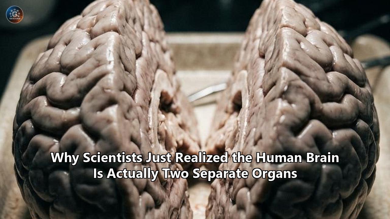
A Stanford Medicine research team published findings in Nature Neuroscience demonstrating that what science has long classified as a single vertebrate brain is developmentally and evolutionarily two distinct organs housed within a single skull. The study reveals that the brain does not originate from a solitary ancestral progenitor cell that branches outward. Instead, it forms during gastrulation from two parallel, non-overlapping cell lineages that never share a common neural ancestor: an anterior lineage that builds the forebrain and midbrain, and a posterior lineage that constructs the hindbrain and brainstem.
Led by developmental biologist Kyle Loh, with co-first authors Rayyan Jokhai and Carolyn Dundes, the team tracked these lineages in developing mouse embryos and confirmed the exact same parallel tracks in human pluripotent stem cells. For centuries, the foundational assumption of human brain anatomy maintained that the central nervous system emerges as a uniform sheet of tissue that rolls into a single tube, subsequently subdividing into regional compartments [PerQueryResult 1.2.1].
The Stanford team proved that this structural unity is an illusion created by embryonic spatial packing [PerQueryResult 1.1.2].
               ANCIENT BILATERIAN PROTO-NERVOUS SYSTEMS
                                  │
         ┌────────────────────────┴────────────────────────┐
         ▼                                                 ▼
Anterior Sensory Network                        Posterior Autonomic Core
(Otx2+ Lineage)                                 (Gbx2+ Lineage)
         │                                                 │
         ├────────────────────────┬────────────────────────┤
         │ ~550 Million Years Ago: Spatial Juxtaposition   │
         ▼                                                 ▼
[ORGAN 1: FOREBRAIN / MIDBRAIN]                 [ORGAN 2: HINDBRAIN / BRAINSTEM]
Executive Cognition & Language                  Autonomic Drive, Swallowing & Respiration
The anterior and posterior neural progenitors harbor mutually exclusive epigenetic landscapes that lock them into divergent fates from the moment they emerge. The front of the brain, responsible for language, consciousness, and abstract cognition, is generated by an Otx2-expressing lineage. The back of the brain, which orchestrates life-sustaining involuntary autonomic drives such as respiration, cardiac rhythm, swallowing, and sleep-wake cycles, is built by a Gbx2-expressing lineage.
"We've shown for the first time that the front of the brain arises from a totally different progenitor cell than the back of the brain," Loh said when presenting the data [PerQueryResult 1.1.2]. "Our research suggests that evolution took two existing neural systems and pushed them together spatially. Having the brain as one organ would probably be more efficient, but we rely on this primordial way to make the brain as two separate pieces [PerQueryResult 1.1.2]."
This realization solves a decades-old crisis in regenerative neurology. Because medical science assumed all brain cells descend from a single starter lineage, researchers spent thirty years using forebrain differentiation protocols to cultivate brainstem motor neurons, with consistent failure. By treating the hindbrain as an autonomous biological organ governed by its own developmental grammar, the Stanford team generated functional, electrophysiologically active human hindbrain motor neurons in vitro for the first time.
The achievement opens unhindered laboratory models for terminal conditions like amyotrophic lateral sclerosis (ALS) and spinal muscular atrophy (SMA). The path to this discovery was not instantaneous; it was a decades-long escalation through clinical roadblocks, evolutionary anomalies, and chromatin-mapping milestones.
The Monolithic Consensus (1890–1990)
To understand why the two-organ reality went undetected for so long, one must trace the structural assumptions that anchored twentieth-century embryology. Since the dawn of modern histology, the brain was interpreted as an undivided anatomical territory.
When Wilhelm His mapped the mechanical folding of the embryonic human nervous system in the late nineteenth century, he established the neural tube model [PerQueryResult 1.1.1]. In this framework, the primary neuroectoderm forms an undivided neural plate on the dorsal surface of the embryo. This plate folds inward along its longitudinal axis to produce a hollow tube. According to His and subsequent generations of morphologists, the rostral end of this tube balloons into three primary vesicles:
- The prosencephalon (forebrain)
- The mesencephalon (midbrain)
- The rhombencephalon (hindbrain)
Classical Monolithic Model:
Neural Ectoderm Progenitor ──► Unitary Neural Tube ──► Prosencephalon (Forebrain)
                                                   ──► Mesencephalon (Midbrain)
                                                   ──► Rhombencephalon (Hindbrain)
The classical model held that regional identity was stamped onto cells after the neural tube was already unified. Morphogen gradients—concentrations of signaling molecules like Sonic Hedgehog (SHH), Bone Morphogenetic Proteins (BMPs), Wnts, and Fibroblast Growth Factors (FGFs)—were thought to wash across a tabula rasa of uniform neural stem cells, instructing them where to place regional boundaries based on relative spatial coordinates.
Santiago Ramón y Cajal, the founder of cellular neuroscience, mapped the intricate connectivity of these brain divisions with silver-staining techniques. While he meticulously documented the radical cellular differences between cerebellar Purkinje cells, brainstem motor nuclei, and cerebral pyramidal neurons, his architectural diagrams positioned them within a single continuous highway network.
The physical continuity of the adult brain tissue reinforced this interpretation. When an anatomist dissects a preserved specimen, the cerebral peduncles flow directly into the pons, which tapers smoothly into the medulla oblongata and spinal cord. Meninges wrap all components within a common protective envelope. Blood supplies like the Circle of Willis weave forebrain and hindbrain vascularization into a unified thermodynamic network.
Because the adult organ looked unbroken, researchers assumed its developmental trajectory had to be unbroken as well. For an entire century, every textbook dealing with human brain anatomy opened with the same fundamental assertion: one neural plate, one progenitor pool, one organ.
The Culture Dish Bottleneck (1998–2012)
The first systemic crack in the unitary brain model appeared not in evolutionary biology or paleontology, but in petri dishes holding human embryonic stem cells (hESCs).
When James Thomson at the University of Wisconsin-Madison isolated the first human embryonic stem cell lines in 1998, and Shinya Yamanaka engineered induced pluripotent stem cells (iPSCs) in 2006, regenerative medicine envisioned a direct roadmap to engineering any human brain cell on demand. If the prevailing developmental theory was correct, coaxing a stem cell into a specific neuron required only recapitulating the progressive branching steps seen in the embryo.
Expected Linear Differentiation:
Pluripotent Stem Cell ──► Pan-Neural Progenitor ──► Signaling Gradient ──► Hindbrain Motor Neuron
Investigators developed protocols based on "default" neuralization. In 2009, Lorenz Studer’s laboratory at the Memorial Sloan Kettering Cancer Center refined dual-SMAD inhibition—using small molecules like Noggin and SB431542 to block BMP and TGF-beta pathways. This maneuver efficiently drove human stem cells into neuroectoderm. From that state, investigators added morphogen cues to steer cells toward particular fates.
For the forebrain, the protocol was a triumph. Within weeks, human stem cells routinely transformed into cerebral cortical projection neurons, striatal interneurons, and basal forebrain cholinergic cells. Midbrain dopaminergic neurons—critical for Parkinson's disease modeling—likewise materialized when researchers applied exact concentrations of SHH and FGF8.
1998-2012 Differentiation Outlier Results:
Pluripotent Stem Cells + Dual-SMAD Inhibition ──► Forebrain Cortical Neurons [High Success]
Pluripotent Stem Cells + Dual-SMAD + SHH/FGF8 ──► Midbrain Dopaminergic     [High Success]
Pluripotent Stem Cells + Rostrocaudal Factors ──► Authentic Hindbrain Nuclei [Failed / Atypical]
Yet, attempts to grow authentic human hindbrain neurons ran into an unyielding biological barrier.
Whenever teams attempted to produce the specific motor neurons of the caudal hindbrain—the somatic motor cells anchored in rhombomeres 5 and 6 that form the abducens, facial, and glossopharyngeal nerves—the cells resisted. They failed to express the correct combinations of Hox transcription factors, produced disordered firing patterns, or defaulted back to forebrain identities.
At the time, the scientific community blamed the culture media. The consensus held that researchers simply had not isolated the correct cocktail of caudalizing signals. Perhaps the timing was off by twenty-four hours; perhaps the concentration of retinoic acid was too weak or too toxic; perhaps cells required a three-dimensional scaffold or specialized shear stress.
For nearly fifteen years, laboratories tinkered with chemical concentrations, altered extracellular matrix coatings, and tweaked oxygen levels. The underlying assumption—that a human pluripotent stem cell can be pushed through a single, shared "neural stem cell" gate to reach any part of the brain—was never cross-examined. The field remained locked in an empirical dead end because it was relying on an inaccurate developmental map.
Evolutionary Anomalies in Ancient Lineages (2012–2019)
While stem cell biologists struggled in culture rooms, evolutionary developmental biologists ("evo-devo") were uncovering genetic contradictions across ancient phyla that did not fit the single-brain narrative.
If the vertebrate brain is a single organ that gradually evolved specialized lobes at its rostral tip, one would expect the earliest bilaterians to possess a primitive, singular neural mass that expanded uniformly. But comparative genetic mapping across deep time began telling the opposite story.
Evolutionary Lineage Split Across 600 Million Years:
Ancestral Metazoa / Cnidaria:
  ├── Anterior Nerve Ring / Aboral Sensory System   ──► Otx2 Lineage (Forebrain/Midbrain)
  └── Posterior Circular Plexus / Oral Motor System ──► Gbx2 Lineage (Hindbrain/Brainstem)
In cnidarians, such as Hydra and sea anemones—which diverged from the lineage leading to vertebrates more than 600 million years ago—the nervous system is arranged not as a singular central organ, but as two distinct, non-overlapping nervous systems situated at opposite physiological poles:
- An aboral nerve net, specialized for environmental sensation, light reception, and spatial orientation.
- An oral nervous ring, focused on autonomic motor functions, muscular contraction, and food intake.
When evo-devo researchers examined the transcriptomic profiles governing these separate cnidarian networks, they found orthologs of Otx expressed in the sensory aboral apparatus, while homeobox genes affiliated with Gbx and the Hox cluster were anchored to the oral, motor-regulatory network.
The anomaly deepened as researchers dissected hemichordates, such as the marine acorn worm (Saccoglossus kowalevskii). Acorn worms possess no internalized brain, living instead with a decentralized epidermal nervous system. Yet in 2015, detailed genetic lineage tracing revealed that the acorn worm's neurogenic ectoderm is split clean in two from the onset of development: an anterior territory driven by an Otx gene circuit and a posterior territory governed by a Gbx gene circuit. Between these zones laid an inviolable molecular boundary.
Similar observations accumulated in the study of early chordates, such as the lancelet (Branchiostoma lanceolatum) and tunicates. The genes that established the rostral sensory plate never intermingled with those creating the visceral, caudal motor network.
Despite these findings, the split was viewed as an evolutionary relic that vertebrates had long since outgrown and streamlined. The vertebrate brain, with its elegant skull, meninges, and myelinated tracts, was assumed to have synthesized these ancestral programs into a unified developmental process. The evolutionary anomalies were cataloged in zoology papers but were largely ignored by human neurologists and medical embryologists.
Single-Cell Resolution Breaks the Paradigm (2020–2024)
By 2020, advances in single-cell genomics transformed molecular biology from an era of bulk tissue analysis to single-cell resolution. Biologists were no longer forced to grind up entire mouse or human embryos to measure average gene expression. Instead, they could isolate individual embryonic cells at hourly intervals, tracking their transcriptomes and chromatin accessibility profiles in real time.
This technical shift set the stage for the definitive discovery led by the Stanford Medicine team.
Rayyan Jokhai and Carolyn Dundes, working in Kyle Loh’s laboratory, focused their investigation on the critical moment of gastrulation—the stage roughly 6.5 to 7.5 days after fertilization in mice, equivalent to the third week of human gestation. Gastrulation is the decisive morphological window when a flat, two-dimensional blastula reorganizes into three foundational germ layers: ectoderm, mesoderm, and endoderm.
According to classical dogma, a subset of the ectoderm is instructed by organizer tissue (the node) to become general neural ectoderm. In that paradigm, this general neural ectoderm acts as a universal parent stem cell pool for every neuron from the prefrontal cortex down to the caudal tip of the spinal cord.
Theoretical Dogma (Refuted):
Epiblast / Ectoderm ──► Universal Neural Progenitor ──► Diversification via Regional Signals

Stanford Discovery (Demonstrated):
                    ┌──► Anterior Neural Ectoderm (aNE)  [Otx2+] ──► Forebrain + Midbrain
Epiblast / Ectoderm ┤
                    └──► Posterior Neural Ectoderm (pNE) [Gbx2+] ──► Hindbrain / Brainstem
Jokhai and Dundes decided to test this universal parent pool concept at single-cell resolution. Using single-cell RNA sequencing (scRNA-seq) paired with single-cell Assay for Transposase-Accessible Chromatin using sequencing (scATAC-seq), they sampled thousands of individual ectodermal cells at the exact moment neural identity was first switched on.
The data did not reveal a single, uniform neural stem cell population that gradually branched.
Instead, the data revealed two completely separate populations of brain progenitor cells emerging at the exact same time point on opposite sides of a sharp molecular border:
- One cohort of progenitor cells expressed high levels of the transcription factor Otx2 and was entirely devoid of Gbx2.
- The other cohort expressed Gbx2 and was entirely devoid of Otx2.
There were no intermediate cells. There was no transitional hybrid state expressing both genes, nor was there an undifferentiated precursor that turned on both before choosing one.
Gastrulation Single-Cell Progenitor Segregation:
Gene Expression:  [Otx2+ / Gbx2-]   <--- Inviolable Boundary --->   [Otx2- / Gbx2+]
Chromatin State:  aNE Sites Open                                    pNE Sites Open
Target Organ:     Forebrain & Midbrain                              Hindbrain / Brainstem
Fate Locking:     Epigenetically Sealed                             Epigenetically Sealed
To determine whether these cells were merely regionally tuned variants of the same basic organ tissue or fundamentally different biological lineages, the researchers examined their chromatin structure. Chromatin is the complex of DNA and protein that acts as a cell’s molecular filing cabinet; regions wound tightly around histones are inaccessible and permanently silenced, while unwound, open loops can be actively transcribed.
The chromatin profiles of the Otx2 and Gbx2 cells diverged fundamentally. The anterior neural ectoderm (destined to become the forebrain and midbrain) had unwound regulatory sequences tied to telencephalic and cortical development, while keeping hindbrain gene promoters locked down.
Conversely, the posterior neural ectoderm (destined to form the hindbrain) possessed an open chromatin landscape around homeobox cluster loci, while the regulatory switches required to build cerebral structures were packaged away in dense, impenetrable heterochromatin.
The two cell types were not variations on a theme. They were two separate, lineage-restricted biological programs running parallel to one another from day one.
The Breakthrough: Replicating the Divergence in Human Stem Cells (2024–2026)
To confirm whether this biological segregation was preserved in human development, the Stanford team moved from mouse models to human pluripotent stem cells.
They designed an experiment to test lineage plasticity. If the brain is a single organ, exposing an early human neural progenitor to the right caudalizing signals should comfortably steer it into hindbrain territory. But if the brain consists of two separate organs with non-overlapping developmental lineages, an anterior progenitor should be epigenetically blocked from adopting a true hindbrain identity.
The team established conditions to differentiate human pluripotent stem cells into either pure anterior neural ectoderm (aNE) or posterior neural ectoderm (pNE) within a rapid 48-hour window. They then labeled the pNE population with fluorescent dyes and cultured both cell populations together in the presence of strong signaling molecules known to specify hindbrain tissue, including high-dose retinoic acid and Wnt agonists [PerQueryResult 1.2.2].
Lineage Challenge Experiment:
Condition: Mix aNE (Uncolored) + pNE (Fluorescently Labeled)
Add: Potent Caudal / Hindbrain Signaling Factors (Wnt Agonists + Retinoic Acid)
Result:
  ├── pNE (Labeled)   ──► Transformed readily into authentic Rhombomere 5/6 Motor Neurons
  └── aNE (Uncolored) ──► Epigenetically refractory; refused hindbrain fate; aborted or formed aberrant forebrain
The outcome verified the hypothesis:
- The posterior neural ectoderm cells responded immediately to the caudal cues, activating downstream hindbrain gene programs and differentiating into mature, functional motor neurons characteristic of hindbrain rhombomeres 5 and 6.
- The anterior neural ectoderm cells, exposed to the identical molecular soup in the exact same dish, were refractory. Their chromatin architecture was locked. They could not access the hindbrain transcriptional pathways and either aborted differentiation or produced misspecified, disorganized cell types.
The barrier was impenetrable. The two cell pools were fundamentally incompatible in their developmental competence.
Having identified the exact developmental parameters of the posterior lineage, the Stanford team bypassed the traditional forebrain default pathways entirely [PerQueryResult 1.2.1]. By cultivating human stem cells exclusively along the posterior track, they accomplished what had eluded stem cell researchers for three decades: they generated pure, authentic human hindbrain motor neurons.
In patch-clamp electrophysiology recordings, these lab-grown hindbrain neurons exhibited the precise electrical properties of native brainstem cells: firing sharp, rhythmic action potentials and generating neuromuscular synapses in co-culture [PerQueryResult 1.3.4]. They synthesized the structural proteins and neurotransmitters required to direct the muscular apparatus of the human face, pharynx, tongue, and diaphragm.
The study, published in September 2026, delivered concrete proof: the human brain is not a singular anatomical organ derived from one flexible parent lineage. It is a composite entity assembled from two distinct organs running on separate molecular operating systems.
Anatomy of the Composite Brain
With the discovery confirmed across murine lineages and human stem cell systems, the anatomical map of the vertebrate nervous system requires structural revision. The adult structure is not a monolithic processing unit, but an intricate physical union of two biological organs functioning in close coordination.
========================================================================================
                      THE DUAL-ORGAN MODEL OF THE HUMAN BRAIN
========================================================================================
FEATURE                   ORGAN 1: THE COGNITIVE-ASSOCIATIVE     ORGAN 2: THE AUTONOMIC-SOMATIC
                          COMPLEX                                SUBSTRATE
----------------------------------------------------------------------------------------
Embryonic Lineage         Anterior Neural Ectoderm (aNE)         Posterior Neural Ectoderm (pNE)
Master Genetic Drivers    Otx2, Pax6, Foxg1, Emx2                Gbx2, En1/2, Krox20, Hox Cluster
Adult Anatomical Zones    Telencephalon (Cortex, Basal Ganglia)  Rhombencephalon (Medulla, Pons,
                          Diencephalon (Thalamus, Hypothalamus)  Cerebellum)
                          Mesencephalon (Tectum, Tegmentum)
Primary Systemic Tasks    Abstract reasoning, conscious memory,  Cardiac regulation, respiratory rhythm,
                          language, symbolic thought, sensory    swallowing, sleep/wake, facial motor
                          integration, voluntary action          control, basal homeostasis
Epigenetic Configuration  Chromatin open for cortical/sensory    Chromatin open for segmented rhombomeric
                          promoters; caudal genes silenced       patterns; rostral promoters silenced
Phylogenetic Precedent    Aboral sensory apparatus in basal      Oral motor/feeding nerve plexus in
                          metazoans and early chordates          ancient bilateral organisms
========================================================================================
Understanding how these systems interact requires evaluating each organ's evolutionary lineage, genetic command network, and physiological focus within human brain anatomy.
Organ 1: The Cognitive-Associative Complex (Forebrain and Midbrain)
The first organ occupies the anterior and superior regions of the cranial vault [PerQueryResult 1.1.1]. It is derived exclusively from the Otx2-positive anterior neural ectoderm.
ANTERIOR PROGENITOR LINEAGE (aNE)
               │
               ▼
   [Otx2 / Pax6 / Foxg1]
               │
      ┌────────┴────────┐
      ▼                 ▼
Telencephalon     Diencephalon + Mesencephalon
(Neocortex,       (Thalamus, Hypothalamus,
 Basal Ganglia,    Substantia Nigra,
 Hippocampus)      Superior Colliculus)
Its physical domain encompasses:
- The neocortex, which contains the six-layered computational machinery that enables language, prospective planning, mathematics, artistic creation, and conscious perception.
- The hippocampal formation and amygdalar nuclei, coordinating episodic memory storage and high-order emotional valencing.
- The basal ganglia (striatum, globus pallidus), responsible for action selection and motor planning.
- The thalamus, functioning as the primary sensory relay and cortical gatekeeper.
- The midbrain tectum and tegmentum, coordinating auditory and visual orienting reflexes and providing ascending dopaminergic inputs.
The regulatory architecture of Organ 1 is governed by transcription factors including Pax6, Foxg1, and Emx2. Its chromatin state keeps caudal Hox genes locked down, insulating these cells from responding to visceral body-axis signals.
Functionally, Organ 1 is outward-facing. It samples sensory data from the external environment, models future scenarios, generates linguistic communication, and regulates voluntary motor strategies.
A mammal can lose substantial portions of Organ 1 and remain alive. A lesion in the association cortices or hippocampus alters personality, language, or memory, but vital life functions continue uninterrupted.
Organ 2: The Autonomic-Somatic Substrate (Hindbrain and Brainstem)
The second organ resides at the base of the skull, seated directly above the foramen magnum [PerQueryResult 1.3.2]. It is formed exclusively from the Gbx2-positive posterior neural ectoderm.
POSTERIOR PROGENITOR LINEAGE (pNE)
               │
               ▼
   [Gbx2 / Krox20 / Hox Cluster]
               │
      ┌────────┴────────┐
      ▼                 ▼
Metencephalon     Myelencephalon
(Pons,             (Medulla Oblongata,
 Cerebellum)       Pre-Bötzinger Complex,
                   Cranial Motor Nuclei)
Its physical domain encompasses:
- The myelencephalon (medulla oblongata), which houses the pacemaker networks of life: the Pre-Bötzinger complex that generates breathing rhythms, the rostral ventrolateral medulla controlling vasomotor tone and blood pressure, and the postrema chemoreceptors regulating metabolic safety.
- The metencephalon (pons and cerebellum), which coordinates fine motor adjustments, cerebellar predictive timing loops, and central relays for cranial nerves [PerQueryResult 1.1.1].
- The somatic and branchial motor nuclei (rhombomeres 1 through 8), which directly innervate the striated musculature of the vocal cords, tongue, pharynx, jaw, and eyes.
The regulatory architecture of Organ 2 is defined by the segmented activation of Hox genes (Hoxa1, Hoxb1, Hoxb2, through Hoxb4), arranged in collinear physical clusters that dictate the identity of each anatomical rhombomere.
Functionally, Organ 2 is inward-facing and preservation-driven. It maintains biological homeostasis, blood chemistry, gas exchange, and neuromuscular mechanics.
Unlike Organ 1, Organ 2 is immediately indispensable for organismal survival [PerQueryResult 1.4.1]. An infarction or structural damage measuring mere millimeters within the medullary reticular formation terminates respiration and cardiac tone instantly, causing brain death.
Organ 2 is the physiological anchor of life; Organ 1 is the computational apparatus superimposed upon it [PerQueryResult 1.3.2].
Organ Function Comparison:
Organ 1 (Cognitive-Associative)  ──► Inward: Evaluates internal states
                                 ──► Outward: Language, Abstract Thought, Future Planning
                                 ──► Clinical Damage: Cognitive Deficits, Intact Survival

Organ 2 (Autonomic-Somatic)      ──► Inward: Blood Pressure, Cardiac Rhythms, Gas Exchange
                                 ──► Outward: Visceral Motor, Swallowing, Vocal Cord Control
                                 ──► Clinical Damage: Apnea, Hemodynamic Collapse, Immediate Death
Overturning Clinical Neurology
The validation that the brain consists of two biologically distinct organs transforms experimental medicine. By establishing that the hindbrain is not a caudal offshoot of forebrain tissue, this work directly addresses multiple historical treatment dead ends.
Pathology Impacts:
  ├── Motor Neuron Disorders (ALS & SMA): Lab models shifted to true pNE Rhombomeric Lineages
  ├── Pediatric Neuro-Oncology (DIPG vs Supratentorial GBM): Explains distinct histone mutations
  └── Bioengineering / Organoids: Replaces monolithic culture with dual-assembloid modeling
1. Cracking the Hindbrain Motor Neuron Barrier in ALS and SMA
For decades, the search for targeted therapies for amyotrophic lateral sclerosis (ALS) and spinal muscular atrophy (SMA) was bottlenecked by cell culture limitations.
In patients suffering from bulbar-onset ALS, the earliest and most aggressive damage targets the branchial motor neurons situated in the brainstem—the cells that govern tongue movement, mastication, vocal fold closure, and swallowing. When these neurons die, patients develop progressive dysphagia, aspirate oral secretions, and succumb to respiratory failure.
For twenty years, researchers modeled ALS by using dual-SMAD inhibition to grow human motor neurons from stem cells. Because they followed forebrain protocols while attempting to caudalize the cells late in culture, they produced motor neurons with atypical transcriptional profiles that lacked the genuine identity of rhombomeric brainstem nuclei.
As a result, drug compounds that protected these synthetic cells in high-throughput laboratory screens regularly failed when tested in human clinical trials. The cells in the culture dishes were not using the molecular survival pathways native to true hindbrain neurons.
With the Stanford team’s lineage-specific protocol, researchers can now cultivate millions of authentic human rhombomere 5 and 6 motor neurons on demand. These lab-grown cells display the native chromatin accessibility landscapes, channel densities, and metabolic vulnerabilities found in human patients.
This breakthrough establishes an accurate platform for testing whether experimental antisense oligonucleotides, small molecules, or neuroprotective drugs can rescue vulnerable brainstem motor neurons from degeneration.
2. Resolving Pediatric Brain Cancer Epigenetics
Pediatric neuro-oncology has long faced a biological mystery: why do specific aggressive brain tumors occur almost exclusively in the brainstem, while others appear only in the cerebral cortex?
Consider Diffuse Intrinsic Pontine Glioma (DIPG), also known as diffuse midline glioma, an incurable pediatric cancer that infiltrates the pons of the hindbrain. Nearly 85% of DIPG tumors harbor a specific somatic mutation in histone H3: the H3K27M mutation, where lysine 27 is substituted with methionine, causing global hypomethylation of chromatin.
In sharp contrast, supratentorial pediatric glioblastomas arising in the cerebral cortex rarely exhibit this specific mutation, displaying instead alterations in IDH1, IDH2, or histone H3.3 G34R/V.
Anatomists struggled to explain why two areas of the same organ would select for mutually exclusive epigenetic driver mutations. The dual-organ reality clarifies the dynamic:
- The pons is assembled from the Gbx2 posterior lineage, where chromatin maintenance is tied to H3K27 trimethylation cascades that silence anterior gene expression programs.
- The cerebral cortex is built from the Otx2 anterior lineage, governed by different histone modification balances [PerQueryResult 1.1.1].
DIPG's H3K27M mutation is lethal specifically because it disrupts the precise epigenetic machinery that the posterior lineage relies upon to preserve its cellular identity. The oncogenic vulnerability is directly linked to the developmental lineage of the organ in which it arises.
3. Redesigning Brain Organoid Engineering
Over the past decade, 3D cerebral organoids—often popularized as "mini-brains"—have become widely adopted tools for studying neurodevelopment and neurodevelopmental disorders like autism and schizophrenia.
Yet organoid engineering has been plagued by high variability and aberrant spatial organization. When scientists attempt to grow a "whole-brain organoid" from a single cluster of stem cells in a single bioreactor, the tissue develops into disorganized mosaics. Cortical layers fold directly into chaotic clusters of midbrain, retinal, or cerebellar markers with no functional logic.
The Stanford study clarifies why these whole-brain protocols fail: it is developmentally impossible to generate both organs correctly using a single signaling bath [PerQueryResult 1.2.2].
Exposing a single cluster of pluripotent stem cells to neural induction factors forces individual cells to make chaotic local decisions between the Otx2 and Gbx2 pathways based on random diffusion noise. One part of the organoid locks its chromatin into an anterior state; a neighboring patch locks into a posterior state. The two populations possess divergent developmental trajectories and cannot coordinate tissue architecture.
Next-Generation "Assembloid" Production:
Bioreactor A: Pure aNE Pathway ──► Organoid 1 (Cortical / Forebrain Tissue)
                                                    │
                                                    ├──► Micro-Engineered In Vitro Junction
                                                    │
Bioreactor B: Pure pNE Pathway ──► Organoid 2 (Hindbrain / Brainstem Tissue)
The future of neural bioengineering lies in assembling distinct tissues:
- Growing pure Organ 1 (forebrain) organoids using explicit anterior lineage differentiation [PerQueryResult 1.2.2].
- Growing pure Organ 2 (hindbrain/brainstem) organoids using dedicated posterior lineage differentiation [PerQueryResult 1.2.2].
- Physically fusing the two mature organoids in vitro to form an "assembloid" that mirrors how embryonic development juxtaposes the two tissues during gastrulation.
The Evolutionary Escalation: 550 Million Years of Mechanical Convergence
How did human biology arrive at this dual-organ construction?
The explanation reaches back to the ocean floors of the Ediacaran and Cambrian periods, between 550 and 600 million years ago, when the ancestral lineages of bilaterian animals were first testing directional locomotion.
EVOLUTIONARY TIMELINE: 600 MILLION YEARS OF BRAIN CONVERGENCE
══════════════════════════════════════════════════════════════════════════════════════
600+ Ma   Early Eumetazoan Ancestors (Cnidarian split)
          Two uncoupled neural networks: Aboral sensory plexus (Otx) and
          Oral visceral ring (Gbx/Hox).
          ──────────────────────────────────────────────────────────────────
550 Ma    Early Bilaterian Emergence (Deuterostome-Protostome divergence)
          Cephalization drives directional movement. Sensory apparatus clusters
          rostrally; feeding and autonomic mechanics align underneath.
          ──────────────────────────────────────────────────────────────────
500 Ma    Primitive Chordate Archetypes (Early Agnatha / Stem Craniata)
          Anterior and posterior lineages are brought into physical contact.
          The isthmic organizer forms an anatomical junction between them.
          ──────────────────────────────────────────────────────────────────
350 Ma    Early Tetrapods
          Encasement in an unbroken cartilaginous / bony neurocranium.
          Common meningeal envelopes evolve, obscuring the developmental split.
          ──────────────────────────────────────────────────────────────────
Present   Mammalian Human Brain Anatomy
          Neocortical expansion buries the brainstem, leaving an outwardly
          unified structure derived from two independent embryonic origins.
══════════════════════════════════════════════════════════════════════════════════════
In an organism that moves through its environment in a consistent direction, evolution prioritizes cephalization: the concentration of sensory receptors and navigational computation at the leading end of the body.
However, early bilateral ancestors were already equipped with two operational neural subsystems [PerQueryResult 1.1.2]:
- A rostral sensory plexus, derived from the anterior neural plate, dedicated to analyzing chemical cues, light fields, and mechanical disturbances ahead.
- A caudal-visceral network, anchored around the pharynx and gut, managing peristalsis, rhythmic feeding, metabolic gas transfer, and directional propulsion.
As directional locomotion evolved, survival favored bringing these systems into close physical proximity. Fast motor responses required immediate transmission of sensory signals from the front-end sensors to the motor-driving hindbrain networks.
Evolution solved this engineering problem not by dismantling the two programs to engineer a single unified brain from scratch, but through spatial juxtaposition. It took the two existing neural systems and pushed them together inside the rostral pole of the animal [PerQueryResult 1.1.2].
"Having the brain as one organ would probably be more efficient, but we rely on this primordial way to make the brain as two separate pieces," Loh pointed out [PerQueryResult 1.1.2].
Evolution is a tinkerer, not a clean-slate engineer. Once gene regulatory circuits are established, rewriting the fundamental developmental code of a vital organ system carries prohibitive risk.
Attempting to fuse the anterior sensory network and the posterior visceral network into a single, unified embryonic progenitor would require rewiring hundreds of master transcriptional switches at once, with near-certain embryonic lethality.
Instead, evolution preserved both distinct progenitor lineages. It maintained their separate identities during gastrulation, let them emerge side-by-side, and then joined them mechanically within the cranial enclosure.
To bridge the gap between them, evolution developed specialized boundary signaling zones, most notably the isthmic organizer at the midbrain-hindbrain junction.
The isthmic organizer acts as a biological molecular suture, using balanced secretions of FGF8 and Wnt1 to stitch together the Otx2 world in front and the Gbx2 world behind, ensuring that axon tracts can traverse the border and establish functional communication [PerQueryResult 1.1.1].
The Isthmic Molecular Suture:
ANTERIOR (Organ 1)                                POSTERIOR (Organ 2)
Otx2 Territory      ──► [FGF8 / Wnt1 Boundary] ◄──  Gbx2 Territory
Forebrain / Midbrain    (The Isthmic Organizer)     Hindbrain / Brainstem
This explains why the brain appears continuous in adult human brain anatomy. Millions of ascending and descending axonal cables cross the isthmic boundary:
- Corticospinal projections extend downward from the neocortex through the brainstem to reach spinal motor neurons.
- Ascending reticular activating system fibers radiate upward from the brainstem to project diffuse arousal signals across the cortex.
The two organs are interconnected by dense neural wiring, but their cellular origins, structural blueprints, and genetic regulatory profiles remain separate.
This distinction clarifies an important point: this scientific discovery has nothing to do with popular concepts of "left brain versus right brain" [PerQueryResult 1.3.1]. The left and right cerebral hemispheres are symmetrical, bilateral halves of Organ 1 alone, sharing the identical Otx2 progenitor lineage and connected via the corpus callosum.
Nor does this validate Paul MacLean's mid-twentieth-century "triune brain" hypothesis—a model that claimed mammalian brains were assembled like Russian nesting dolls (reptilian, paleomammalian, and neomammalian layers).
MacLean's model was an anatomical approximation unsupported by developmental genetics. The true division runs not between reptiles and mammals, but between the deep embryonic programs that split the nervous system at gastrulation: anterior versus posterior, Otx2 versus Gbx2.
Structural Implications for Modern Medicine
Now that the biological division between these two cranial organs is established, scientific, clinical, and anatomical institutions are facing concrete operational revisions:
PRACTICAL RESEARCH AND CLINICAL PIVOTS
─────────────────────────────────────────────────────────────────────────────
1. Regenerative Medicine
   * Abandoning default dual-SMAD differentiation for brainstem modeling.
   * Applying lineage-restricted protocols for true pNE-derived motor neurons.
   * Synthesizing patient-specific rhombomere 5/6 cells for ALS and SMA screens.

2. Pharmacological Design
   * Developing dual-target assays to screen neuroprotective compounds.
   * Screening candidates independently against aNE and pNE chromatin states.
   * Preventing false negatives caused by testing brainstem drugs on cortical models.

3. Neuro-Oncological Therapeutics
   * Designing epigenetic drugs tailored to histone architectures (e.g., DIPG).
   * Modeling pediatric brainstem gliomas exclusively in pNE lineage backgrounds.
   * Avoiding non-specific chromatin-modifying therapies that destabilize aNE cells.

4. Formal Anatomical Classification
   * Submitting revisions to the Federative International Programme on
     Anatomical Terminology (FIPAT).
   * Updating Terminologia Anatomica to reflect the dual-lineage composite model.
─────────────────────────────────────────────────────────────────────────────
The ability to accurately model diseases like ALS is only the first step. A wide range of human neuropathologies selectively target hindbrain structures while leaving the forebrain unaffected:
- Central Sleep Apnea and Ondine's Curse (Congenital Central Hypoventilation Syndrome), driven by mutations in PHOX2B, can now be modeled in native, lab-grown medullary respiratory clusters.
- Pseudobulbar Affect, where patients experience uncontrolled bouts of laughing or crying due to disrupted connectivity between the cognitive forebrain and the autonomic hindbrain, can be investigated using dual-organ assembloids.
- Sudden Infant Death Syndrome (SIDS), long suspected to involve silent failures in the serotonin-producing raphe nuclei of the brainstem, can be evaluated using patient-derived cells grown through dedicated posterior lineage tracks.
Next Steps and Scientific Milestones
The discovery that the human brain is a composite of two distinct biological organs establishes several concrete frontiers for molecular neuroscience:
CRITICAL RESEARCH MILESTONES (2026–2030)
─────────────────────────────────────────────────────────────────────────────
Target Date     Objective
─────────────────────────────────────────────────────────────────────────────
Late 2026       Complete high-resolution mapping of the human isthmic organizer
                at single-cell resolution to identify the signaling factors that
                mechanically bind the two organs during week 4 of gestation.

2027            Determine the lineage origin of the spinal cord: verify whether
                spinal cord motor columns originate from a continuation of the
                pNE Gbx2 lineage or an independent neuromesodermal progenitor (NMP).

2028            Deploy high-throughput drug screening libraries using authentic
                human rhombomere 5/6 motor neurons to identify compounds capable
                of halting bulbar ALS progression.

2029            Engineer vascularized, integrated dual-organ assembloids capable
                of modeling the interface between the cerebral cortex and the
                brainstem in continuous, long-term microfluidic culture.

2030            Formulate proposed revisions to the international standards of
                human brain anatomy within the Terminologia Anatomica framework.
─────────────────────────────────────────────────────────────────────────────
A major unresolved question centers on the lineage origin of the spinal cord [PerQueryResult 1.3.4]. While the Stanford study confirmed that the hindbrain forms from the Gbx2-positive posterior neural ectoderm, the origin of the caudal spinal cord remains an area of active investigation.
Evidence suggests that the caudal-most nervous system may stem from a specialized population called neuromesodermal progenitors (NMPs), which generate both posterior neural tissue and paraxial mesoderm. If validated, the central nervous system may be revealed as an even more intricate composite: a tri-lineage structure composed of an anterior cognitive organ, a posterior autonomic organ, and a caudal somatic transmission column.
Simultaneously, international anatomical committees are reviewing the finding. The Federative International Programme on Anatomical Terminology (FIPAT), which regulates the global standard reference Terminologia Anatomica, has classified the encephalon as a singular organ for over a century. Revising this taxonomy to reflect a composite, dual-organ architecture will involve extensive debate among clinical anatomists, neurosurgeons, and developmental biologists.
The realization that human beings harbor two separate brain organs within their skulls resolves decades of contradictory data. It explains why the parts that make humans capable of language and abstract thought run on a fundamentally different molecular operating system than the parts that keep the heart beating and the lungs drawing air.
The brain is not an unbroken monolith. It is an evolutionary partnership—two distinct organs, forged across deep evolutionary time, cohabiting within a single cranial vault.
Reference:
- https://med.stanford.edu/news/all-news/2026/09/two-separate-brains.html
- https://neurosciencenews.com/brain-separate-organs-evolution-31219/
- https://www.labcompare.com/617-News/628244-Study-The-Human-Brain-is-Actually-Two-Separate-Organs/
- https://www.courthousenews.com/your-brain-may-actually-be-two-brains-in-one/
- https://www.sciencealert.com/the-human-brain-has-two-distinct-origins-scientists-discover
- https://www.techexplorist.com/human-brain-actually-two-separate-organs/104309/
- https://www.reddit.com/r/neurobiology/comments/1wjpk8m/human_brain_is_two_separate_organs_research_finds/
- https://timesofindia.indiatimes.com/etimes/wellness/not-one-but-two-stanford-university-scientists-find-the-brain-is-two-separate-organs/articleshow/134348588.cms
- https://nautil.us/you-have-two-brains-not-one-1285111
- https://www.wionews.com/science/think-twice-human-brain-is-not-one-but-two-separate-organs-1789737537017