In March 2026, researchers at Empa, the Swiss materials science institute, quietly solved one of the most stubborn problems in modern medicine. A team led by chemist Giacomo Reina and Peter Wick developed an ultra-thin, biocompatible coating that neutralizes dangerous hospital pathogens on command, requiring nothing more than a brief exposure to infrared light. In early testing, this light-activated material eliminated nearly all traces of specific drug-resistant bacterial strains. The active ingredient was not a newly synthesized chemical compound or a complex biological peptide. It was graphene.
The Empa breakthrough represents the crest of a massive, sudden wave of commercial and clinical applications for a material that spent the last two decades being characterized as a scientific curiosity in search of a practical problem. That narrative has decisively ended. Graphene is now being deployed as a physical weapon against the worsening crisis of antimicrobial resistance (AMR).
The scale of this deployment is moving faster than regulatory frameworks can easily track. High-profile sportswear infused with carbon nanomaterials debuted at the 2024 Paris Olympics, and similar textiles are already slated for the uniforms of the upcoming 2026 Asian Games. Commercial manufacturers report that over 10 million units of early-generation antibacterial toothbrushes utilizing carbon nanomaterials have already been sold globally. Meanwhile, industrial-scale producers like Black Swan Graphene have recently expanded their manufacturing facilities from 40 tonnes to over 140 tonnes annually, signaling a definitive shift from pilot-scale experimentation to mass commercialization.
To understand why the medical and materials science communities are suddenly pivoting toward carbon lattices to fight infections, you have to look past the chemistry of traditional drugs and into the brutal, microscopic physics of cell death.
The Economic Collapse of Chemical Warfare
The context for this shift is a global health infrastructure that is rapidly losing its primary chemical weapons. Since the discovery of penicillin, humanity has relied on a strategy of chemical warfare against bacteria. We introduce molecules that bind to specific bacterial proteins, inhibiting cell wall synthesis, protein translation, or DNA replication.
The bacteria, in response, evolve. They develop efflux pumps to actively spit the antibiotics back out of their cells. They mutate their binding sites. They produce enzymes like beta-lactamase that physically chew up the antibiotic molecules before they can do any damage.
This evolutionary arms race has reached a tipping point. The World Health Organization estimates that bacterial AMR was directly responsible for 1.27 million global deaths in 2019, a number that has only climbed. A July 2025 report from the Center for Global Development laid out the stark economic reality: if resistance rates continue to follow historical trends, superbugs will cost the global economy just under $2 trillion a year by 2050. By that same year, up to 39 million people could die annually from infections that were routinely curable just a decade ago.
The pharmaceutical pipeline is practically dry. Developing a new class of antibiotics takes an average of 10 to 15 years and costs roughly $1 billion. Because any new, highly effective antibiotic is immediately shelved by hospitals as a "drug of last resort" to prevent bacteria from developing resistance to it, pharmaceutical companies cannot sell enough volume to recoup their research and development costs. The traditional economic model for antibiotic development is fundamentally broken.
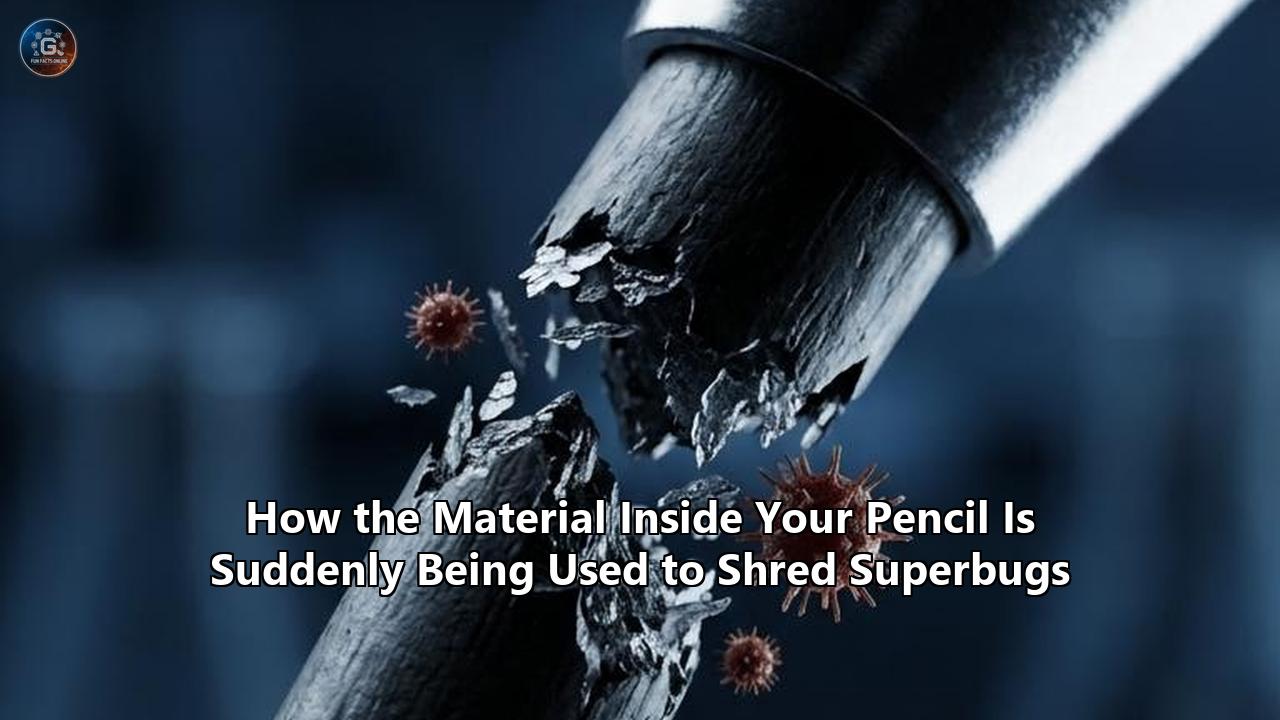
This is the exact void that carbon nanomaterials are rushing to fill. Instead of trying to outsmart bacterial evolution with complex chemistry, materials scientists are resorting to physical annihilation. A bacterium can mutate its DNA to change the shape of a protein receptor, rendering a chemical antibiotic useless. It cannot, however, mutate its way out of being physically shredded by a microscopic blade.
Anatomy of a Nano-Knife
The secret to graphene antimicrobial properties lies in its unique two-dimensional structure. Pure graphene is a single layer of carbon atoms arranged in a hexagonal, honeycomb lattice. It is highly conductive, incredibly strong, and entirely hydrophobic. However, in biological and medical applications, researchers rarely use pure, pristine graphene. Instead, they use Graphene Oxide (GO) or Reduced Graphene Oxide (rGO).
Graphene oxide is manufactured by treating graphite with strong oxidizers, a process that decorates the carbon lattice with oxygen-containing functional groups. Epoxide and hydroxyl groups attach to the flat basal planes of the sheet, while carboxyl groups attach to the edges. This subtle shift in chemistry changes everything. It makes the material dispersible in water—a strict requirement for anything interacting with the human body—and it turns the two-dimensional sheet into a highly effective microbial assassin.
When a bacterium, such as Staphylococcus aureus or Escherichia coli, comes into contact with a graphene oxide nanosheet, three distinct mechanisms of destruction occur simultaneously.
First is the "sharp edge effect," often referred to by physicists as the nano-knife. Because the material is only one atom thick, its edges are incomprehensibly sharp. When a bacterium interacts with the edge of a GO sheet, the intense localized physical stress physically punctures the outer phospholipid bilayer of the bacterial cell wall.
Once the membrane is breached, the second mechanism takes over: lipid extraction. The hydrophobic portions of the graphene sheet exert strong van der Waals forces on the hydrophobic lipid tails that make up the bacterial membrane. The graphene essentially acts as a molecular vacuum, pulling the lipids out of the cell wall and spreading them flat across its own surface. The bacterium literally dissolves as its structural integrity is stripped away molecule by molecule.
The third mechanism is oxidative stress. Even if the physical cutting does not immediately kill the pathogen, the graphene oxide acts as a highly conductive electron sink. It physically rips electrons away from the bacterium's antioxidant defense systems. This massive electron transfer disrupts the bacterium's internal respiratory chain, halting the production of ATP (the cell's energy currency) and flooding the interior of the cell with lethal Reactive Oxygen Species (ROS). The bacteria choke on their own metabolic exhaust.
This multi-pronged attack is what makes the development of resistance nearly impossible. To survive, a bacterium would need to simultaneously invent a cell wall impervious to physical cutting, immune to hydrophobic lipid extraction, and capable of functioning without electron transport.
Dialing the Oxygen: The Secret Geometry of Contact Killing
One of the historical challenges in utilizing carbon nanomaterials in medicine has been the lack of consistency. Early studies on the efficacy of GO against bacteria often yielded wildly contradictory results. One laboratory would report total eradication of Methicillin-resistant Staphylococcus aureus (MRSA), while another would report that the bacteria survived and even multiplied.
The key to resolving this contradiction—and unlocking the commercial viability we are seeing in 2026—was uncovered by a consortium of researchers from the University of Birmingham and other international institutions. They discovered that the exact mechanism of graphene antimicrobial properties depends entirely on a metric called Surface Oxygen Content (SOC).
The Birmingham researchers revealed that the amount of oxygen on the surface of the material acts as a physical toggle switch for its flexibility. When graphene oxide has a very high SOC, the sheet is highly flexible. In this state, it interacts with bacteria in a "parallel mode." The flexible sheets act like microscopic blankets, completely wrapping around the bacterial cells. This isolates the pathogen from its environment, preventing it from absorbing nutrients or proliferating, essentially starving it to death.
However, when the oxygen levels are reduced, the material becomes highly rigid. In this low-SOC state, the sheets interact with bacteria in a "perpendicular mode." Instead of wrapping the cell, the rigid sheets stand on edge, acting directly as nano-knives to slice the bacteria open.
Neither mode is inherently superior; their effectiveness depends entirely on the surrounding biomolecules and the specific target pathogen. By understanding how to precisely dial the oxygen content during the manufacturing process, materials scientists can now engineer specific forms of GO for specific tasks. For example, a highly flexible, wrapping GO might be optimal for a water filtration membrane, while a highly rigid, cutting GO is better suited for a surgical implant coating.
This tunability also solves the critical problem of selective toxicity. A weapon that shreds bacterial cells is only useful if it does not also shred human tissue. Recent work out of the Korea Advanced Institute of Science and Technology (KAIST) demonstrated that carefully engineered graphene oxide nanofibers can be tuned to selectively adhere to and destroy the membranes of prokaryotic "superbugs" while essentially ignoring the structurally distinct membranes of human eukaryotic cells. This allows the material to speed up wound healing in animal models without triggering the severe inflammatory cascades that plague earlier nanoscale interventions.
Zap and Trap: The Engineering of Laser-Induced Graphene
While chemists have been focused on tuning oxygen groups for liquid and surface applications, mechanical and electrical engineers have taken a radically different approach to harnessing the material against airborne and waterborne pathogens.
In hospital environments, patients face a 1-in-31 chance of acquiring a potentially life-threatening infection during their stay, often from pathogens circulating through the building's HVAC systems. Traditional HEPA filters trap these bacteria, but they do not kill them. The filter simply becomes a breeding ground for a highly concentrated colony of superbugs.
Enter Laser-Induced Graphene (LIG).
Discovered by chemist James Tour's laboratory at Rice University, LIG is manufactured by taking a common sheet of polyimide (a high-temperature plastic) and firing an industrial CO2 laser at its surface. The intense, localized photothermal conversion locally obliterates the chemical bonds of the plastic, releasing the non-carbon atoms as gas and leaving behind a highly porous, three-dimensional foam of pure, intertwined graphene sheets.
Under an electron microscope, LIG looks like a dense, microscopic forest. When airborne bacteria, fungi, or spores are pulled into this forest by a ventilation system, they become physically trapped in the jagged carbon matrix. But the true innovation is not the trapping; it is the zapping.
Because LIG is pure carbon, it is highly conductive. By applying a small electrical current to the filter, researchers can utilize a phenomenon called Joule heating. Within seconds, the localized temperature of the LIG filter spikes to over 350 degrees Celsius (662 degrees Fahrenheit).
This temperature is not arbitrary. It is precisely hot enough to completely obliterate the trapped pathogens. Crucially, it is also hot enough to vaporize their toxic byproducts. When normal filters trap bacteria, the bacteria eventually die and burst, releasing endotoxins into the air stream. If inhaled, these endotoxins can trigger severe immune responses and sepsis in vulnerable patients. The intense thermal pulse of the LIG filter decomposes these molecules into harmless trace gases. Once the pulse is over, the filter cools back to room temperature in seconds, completely sterilized and ready for the next cycle.
The research has rapidly expanded beyond basic hospital bacteria. Collaborative studies between institutions like the Indian Institute of Technology Bombay and Ben-Gurion University have demonstrated that these electrified LIG filters are highly effective at inactivating complex viruses, including prototype poxviruses and the SARS-CoV-2 virions responsible for COVID-19, particularly when applied to drinking water filtration. While viruses require higher voltages to inactivate than bacteria, the combination of physical trapping, irreversible electroporation (punching electrical holes in the viral envelope), and localized reactive oxygen species generation provides a comprehensive shield against viral contamination.
Further research by Christopher Arnusch and colleagues revealed that LIG does not always require intense heat to kill. They demonstrated a mechanism of "capacitive killing." Because the porous LIG acts like a supercapacitor, applying a minor voltage causes the material to store electrical charge. Even after the power source is disconnected, the surface retains an electrical potential that induces severe stress on any bacteria that land on it, rapidly degrading their viability without ever raising the temperature. This allows for the creation of continuously active antibacterial surfaces in environments where heating would be dangerous or energy-intensive.
The Empa Breakthrough: Light-Activated Assassins
Which brings us back to the March 2026 announcement in Switzerland. While the LIG filters rely on electrical currents, the team at Empa developed a way to trigger the antimicrobial properties of graphene using only light.
The challenge with implantable medical devices—like titanium dental implants or artificial joints—is that infections often develop weeks or months after the surgery. Biofilms, complex communities of bacteria encased in a protective slime, form on the surface of the implant. Once a biofilm is established, traditional systemic antibiotics cannot penetrate it. The only solution is often a highly traumatic surgery to remove the infected hardware.
Giacomo Reina and Peter Wick synthesized a highly specific coating combining graphene oxide with polyvinyl alcohol, a biocompatible polymer. This ultra-thin nanomaterial is applied to the implant before it is placed in the patient's body.
The innovation relies on graphene's extraordinary photothermal efficiency. When a clinician shines targeted infrared light through the patient's tissue and onto the implant, the graphene coating absorbs the specific wavelengths and instantly converts them into localized heat. The temperature spike is confined strictly to the nanoscale surface of the implant—hot enough to instantly denature the proteins of the colonizing bacteria and shatter the biofilm, but localized enough that the surrounding human tissue is unharmed.
Testing to date shows the material retains its graphene antimicrobial properties through repeated reactivation without degrading. A patient could theoretically go to their clinician for a routine check-up, receive a quick, painless pulse of infrared light over the implant site, and leave with a completely sterilized device.
The Protein Corona: Biology’s Natural Shield
Despite the massive commercial scale-up of graphene textiles and filtration systems, the journey toward systemic, injectable graphene-based nanomedicines faces a severe, often-underreported biological roadblock: the protein corona.
In a sterile petri dish, graphene oxide is a ruthless killer. But the human bloodstream is not a petri dish. It is a dense, chaotic soup of over 4,000 different circulating proteins, lipids, and metabolites.
The moment a nanoparticle enters human blood, plasma proteins—most notably serum albumin, fibrinogen, and apolipoproteins—rush to its surface, binding to it in fractions of a second. This layer of tightly bound biological molecules is known as the "hard corona," while a secondary, looser layer that exchanges molecules with the surrounding fluid is known as the "soft corona".
For the bacteria-shredding properties of graphene, the protein corona is disastrous. A late 2025 study from the West China Hospital of Stomatology precisely quantified this effect. The researchers exposed graphene oxide to Bovine Serum Albumin (BSA), a standard proxy for human blood proteins. The BSA immediately wrapped the GO nanosheets. When this protein-coated graphene was introduced to MRSA, its killing efficacy plummeted. The sharp edges of the nano-knife were essentially sheathed in a thick layer of biological bubble wrap, preventing the physical membrane disruption that makes the material so deadly. The viability loss rate for the superbug dropped from 94.3% with naked GO to just 62.3% with the protein-coated GO.
Furthermore, the protein corona completely changes the "biological identity" of the nanoparticle. When immune cells, like macrophages, encounter a bare carbon nanoparticle, they might ignore it. But when they encounter a carbon nanoparticle coated in denatured serum proteins, their receptors recognize it as a foreign anomaly. The macrophages engulf the graphene, sequestering it away from the infection site and triggering an unwanted inflammatory cascade.
Overcoming this barrier is currently the primary focus of nanomedicine regulation. To preserve the cutting and oxidative capabilities of the material in vivo, researchers are heavily investing in surface functionalization strategies. The most common approach is PEGylation—coating the graphene with Polyethylene Glycol (PEG). The dense, hydrophilic PEG chains create a steric barrier that repels blood proteins, preventing the corona from forming.
However, modifying the surface to prevent protein adhesion inherently reduces the surface area available to physically cut bacteria. It is a delicate, agonizing thermodynamic balancing act: the material must be stealthy enough to evade blood proteins, but aggressive enough to kill pathogens. Until this balance is perfected and standardized, the bulk of commercial graphene medical applications will remain localized to external environments: wound dressings, filters, surgical tools, and topical coatings, rather than intravenous antibiotics.
The Silver Synergy and Wet Environments
Recognizing the limitations of pure graphene in certain complex biological fluids, materials scientists are increasingly using it not as a solo weapon, but as a force multiplier.
Silver has been used as an antimicrobial agent for centuries. Silver ions naturally disrupt bacterial enzymes and DNA. However, in modern clinical applications, silver coatings have a major flaw: they dump their ions too quickly. When a silver-coated bandage is applied to a wound, it releases a massive, toxic spike of silver ions that can damage surrounding healthy tissue, followed by a rapid depletion that leaves the wound vulnerable to reinfection days later.
In late 2025, a team led by Professor Rahul R. Nair at the National Graphene Institute at the University of Manchester unveiled a structural solution to this problem. Working with the medical technology company Smith & Nephew, they created a composite membrane where silver nanoparticles are embedded within layers of graphene oxide.
Instead of acting as a blade, the graphene in this application acts as a microscopic metering valve. The nanoscale channels between the stacked graphene oxide sheets physically restrict the flow of fluid. Water slowly enters the channels, dissolves the silver ions, and slowly carries them out. This precise, geometric throttling ensures a sustained, perfectly metered release of antimicrobial silver over an extended period, preventing tissue toxicity while maintaining a constant defense against bacterial colonization.
This structural reinforcement also solves the issue of environmental degradation. Standard antimicrobial coatings wash away quickly in highly humid or abrasive environments. To combat this, the Japanese chemical manufacturer Nippon Shokubai recently developed a composite utilizing graphene oxide combined with traditional antibacterial agents like benzalkonium chloride. The unique chemical intercalation—where the active agents are tightly sandwiched between the GO sheets—reinforces the coating against severe water washing. This highly durable, transparent film is currently being targeted for wet-area sanitation in hospitals and anti-biofilm coatings for dental implants that exist in the constantly wet environment of the human mouth.
Moving from Chemical Warfare to Physical Annihilation
As of April 2026, the transition of graphene from an academic buzzword to a fundamental pillar of global infection control is fully underway. The economics demand it. The global graphene market, valued at roughly $940 million in 2025, is currently expanding at an explosive Compound Annual Growth Rate (CAGR) of over 36%, projected to reach past $15 billion by the early 2030s. A significant portion of this capital is being driven aggressively by biomedical and filtration demands.
The timeline for seeing these technologies in everyday clinical practice is split. The regulatory pathway for medical devices is drastically shorter than the pathway for new systemic drugs. Consequently, LIG-based self-sterilizing HVAC filters are already scaling into commercial production for hospital infrastructure. Graphene-infused wound dressings, utilizing the material's parallel-wrapping modes and silver-ion metering, are navigating the final stages of FDA and European Medicines Agency clearance. The Empa infrared-activated implant coatings represent the next milestone, likely entering advanced human trials by late 2027 once long-term in vivo degradation studies conclude.
The era of relying solely on the chemical vulnerabilities of bacteria is closing. The pipeline of traditional antibiotics cannot outpace the mathematical reality of microbial evolution. But bacteria cannot evolve a defense against physics. They cannot mutate against a blade that is one atom thick, or a localized thermal shock of 350 degrees Celsius, or a sudden, massive extraction of their cellular electrons.
By weaponizing the structural and conductive extremes of the carbon lattice, researchers are rewriting the rules of engagement. We are no longer trying to poison superbugs. We are simply, and systematically, tearing them apart.
Reference:
- https://indiandefencereview.com/scientists-unveil-a-material-so-powerful/
- https://www.birmingham.ac.uk/news/2023/oxygen-groups-key-to-unlocking-graphenes-antimicrobial-potential
- https://www.reddit.com/r/InterstellarKinetics/comments/1sw0qtk/graphene_oxide_can_kill_dangerous_superbugs_while/
- https://www.datamintelligence.com/research-report/graphene-market
- https://www.who.int/news-room/fact-sheets/detail/antimicrobial-resistance
- https://www.theguardian.com/society/2025/jul/20/superbugs-could-kill-millions-more-and-cost-2tn-a-year-by-2050-models-show
- https://www.weforum.org/stories/2026/02/antimicrobial-resistance-prevention/
- https://physicsworld.com/a/tapping-into-graphene-oxides-antibacterial-properties-to-fight-infections/
- https://www.futurity.org/laser-induced-graphene-filter-bacteria-2178982/
- https://pubs.acs.org/doi/10.1021/acsabm.2c01034
- https://chemrxiv.org/doi/pdf/10.26434/chemrxiv.13489398
- https://pubs.acs.org/doi/10.1021/acs.chemrestox.2c00042
- https://pubmed.ncbi.nlm.nih.gov/40987188/
- https://www.mdpi.com/1422-0067/26/19/9771
- https://pmc.ncbi.nlm.nih.gov/articles/PMC7115795/
- https://www.manchester.ac.uk/about/news/manchester-team-pioneer-silver-based-coating-for-long-term-protection-against-bacteria/
- https://www.shokubai.co.jp/en/news/202110147195/
- https://www.fortunebusinessinsights.com/graphene-market-102930
- https://www.mordorintelligence.com/industry-reports/graphene-market