In a biological revelation that fundamentally rewrites our understanding of viral infection, researchers have discovered a previously hidden process occurring at the interface of life and death.
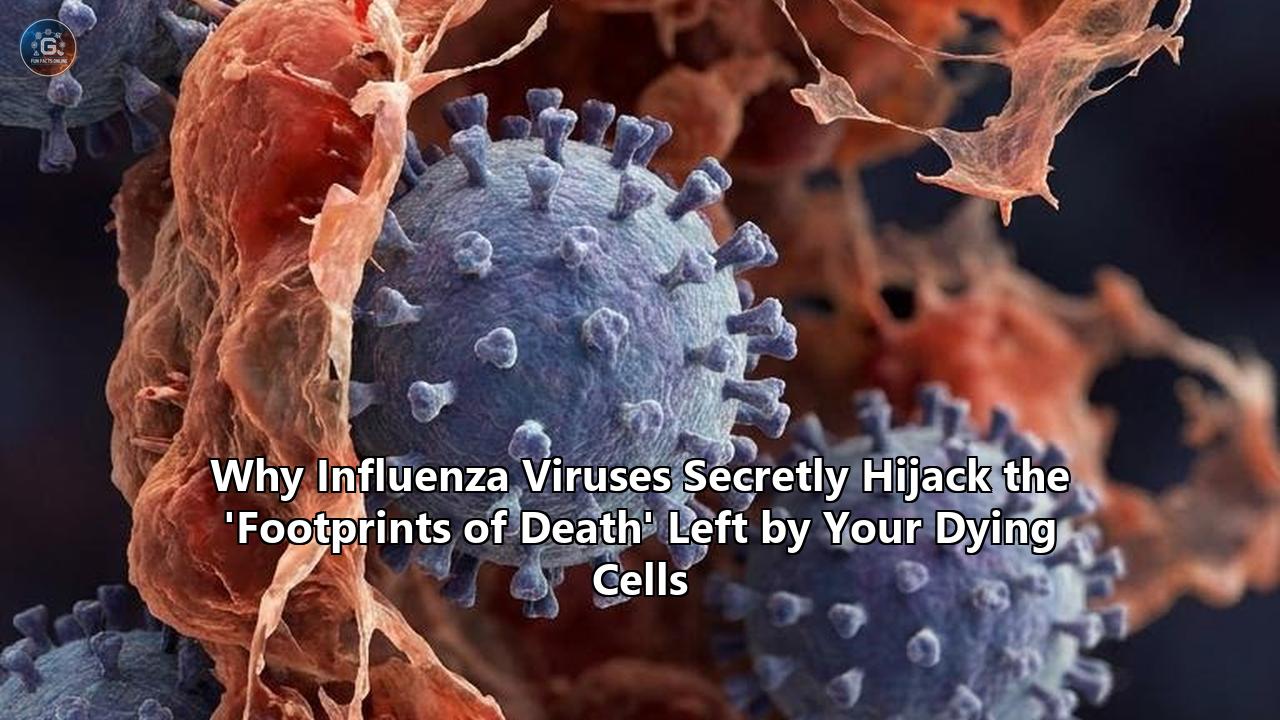
For decades, the scientific consensus held that when cells lining our airways are infected with the influenza virus, they undergo programmed cell death (apoptosis), wither away, and are swept into cellular oblivion by the immune system. However, a major discovery led by researchers at La Trobe University, the Walter and Eliza Hall Institute of Medical Research (WEHI) in Australia, and Toronto Metropolitan University in Canada, has revealed that dying cells do not simply vanish. Instead, they leave behind an organized, highly structured physical residue on the surface they once occupied—a microscopic "crime scene" dubbed the "footprint of death".
Published in the journal Nature Communications, the research was spearheaded by PhD candidate Stephanie F. Rutter alongside senior author Professor Ivan K. H. Poon, Director of the Research Centre for Extracellular Vesicles (RCEV) at the La Trobe Institute for Molecular Science (LIMS). Together with co-leader Dr. Georgia K. Atkin-Smith from WEHI, the team demonstrated that as cells self-destruct, they detach and retract, leaving behind specialized, large extracellular vesicles containing biochemical breadcrumbs to guide the immune system.
Crucially, the researchers made a disturbing discovery: the Influenza A virus (IAV) actively hijacks these cellular footprints, packing its own viral particles and genetic material into these newly discovered vesicles to use them as "Trojan Horses" to infect adjacent healthy cells.
This biological judo—turning the host's tidy cleanup mechanism into a stealth delivery vehicle—unveils a previously unrecognized dimension of viral transmission. By utilizing these "footprints," the virus is able to replicate and disseminate under the radar of the host’s immune system, bypassing antibodies and chemical defenses in the extracellular fluid. The finding changes our conceptual framework of viral exit and entry, paving the way for a new generation of host-targeted therapeutics.
Mapping the Ghost Town: The Molecular Architecture of the FOOD
To understand how the virus exploits this process, one must first examine what happens during the terminal stages of an adherent cell's life. Historically, apoptosis—the highly regulated process of programmed cell death—was seen as a somewhat inward-facing event. A cell marked for death would shrink, its chromatin would condense, its membrane would "bleb," and it would eventually split into small apoptotic bodies that are quietly devoured by patrolling phagocytes. This clean packaging prevents cellular contents from spilling out into surrounding tissues, which would otherwise trigger catastrophic inflammation or autoimmune diseases such as Systemic Lupus Erythematosus (SLE).
However, the La Trobe team used advanced live-cell imaging, including confocal laser scanning microscopy (CLSM) and lattice light-sheet microscopy, to show that apoptosis is far more structured and communicative than previously realized. When an adherent cell (such as an epithelial cell in the lungs) receives a signal to die, it begins to retract from its neighbors and the underlying extracellular matrix (ECM). But as it pulls away, it does not leave a clean slate. It leaves behind a thin, flat, membrane-encased, filamentous structure tightly anchored to the substrate. The team coined this structure the "FOotprint Of Death," or FOOD.
[Healthy Adherent Cell] (Anchored to Extracellular Matrix)
        │
        ▼ (Apoptosis Triggered by Influenza Infection)
[Cell Retraction & Blebbing] (ROCK1 Pathway Activated)
        │
        ▼ (Cell pulls away, leaving anchored membrane)
[The FOotprint Of Death (FOOD)] (F-actin & Phosphatidylserine rich)
        │
        ▼ (Membrane rounds up/vesicularizes)
[F-ApoEVs (Vesicles)] ───► Contains Hidden Influenza Virions (Trojan Horse)
        │
        ▼ (Efferocytosis)
[Neighboring Cell Ingestion] ───► Stealth Spread & Fusion (Endosomal Escape)
Rather than being random debris, the FOOD is a highly organized, active signaling hub. It is heavily enriched with filamentous actin (F-actin), adhesion-associated proteins, and—most importantly—phosphatidylserine (PtdSer). Under normal physiological conditions, PtdSer is kept strictly on the inner leaflet of the cell membrane. However, during apoptosis, scramblase enzymes flip PtdSer to the outer membrane surface. This acts as a universal, highly potent "eat me" signal to patrolling phagocytes, such as macrophages.
Over time, this flat membrane footprint does not simply dissolve. Instead, driven by the mechanical activity of the protein kinase ROCK1 (Rho-associated coiled-coil containing protein kinase 1), the membrane remnants "round up" and vesicularize into a newly discovered class of large, substrate-bound extracellular vesicles. The researchers named these structures FOOD-derived Apoptotic cell-derived Extracellular Vesicles, or F-ApoEVs.
These F-ApoEVs act like breadcrumb trails. Because they remain anchored to the exact site where the cell lived and died, they serve as highly localized beacons. Patrolling macrophages do not have to wander aimlessly looking for cellular waste; they are guided directly to the F-ApoEVs, ensuring that the dead cell is cleared swiftly, neatly, and without inducing inflammatory damage to the tissue.
The Trojan Cargo: How Influenza Recruits F-ApoEVs
Influenza A is an enveloped, single-stranded, negative-sense RNA virus that targets the respiratory epithelium. Once inside a host cell, the virus hijacks the cellular machinery to produce viral proteins and copy its eight genomic RNA segments (vRNPs). Eventually, the sheer metabolic strain and viral signaling trigger the cell's self-destruction pathway, initiating apoptosis.
For decades, virologists believed that the primary way influenza viruses multiplied was through classic viral budding. In this classical model, viral hemagglutinin (HA) and neuraminidase (NA) insert themselves into the host cell's plasma membrane, recruiting viral matrix proteins (M1) and the vRNPs. The host membrane then pinches off, releasing a free-floating, independent virus particle (virion) into the extracellular fluid to find a new cell.
But the work of Rutter, Poon, and their colleagues reveals that influenza has a backup plan—one that is far more insidious. When an infected cell undergoes apoptosis and generates the FOOD, the influenza virus does not get left behind. Instead, viral particles, structural proteins, and fully functional virions are sequestered inside and on the forming F-ApoEVs.
Using high-resolution scanning electron microscopy (SEM) and fluorescence imaging, the researchers captured images of influenza virions physically packed within these dying cell footprints. The virus exploits the host cell's caspase-cleaved ROCK1 pathway—which governs the physical contraction of the cell—to ensure that viral components are funneled directly into the membrane tethers that form the FOOD. As the FOOD vesicularizes into F-ApoEVs, these vesicles become tightly wrapped, highly concentrated packages of viral infection.
This process essentially transforms a defensive host mechanism (the creation of an immunological clean-up beacon) into an offensive viral strategy. By hiding inside the F-ApoEVs, the virus creates a specialized, protected microenvironment. This altered mechanism fundamentally expands our understanding of how influenza viruses spread within a host, revealing that viral exit is not always a chaotic release of independent particles, but often a highly coordinated transfer of mass cargo through cellular remnants.
Stealth Mechanics: A New Paradigm of Viral Dissemination
The discovery of this pathway challenges classical assumptions about virology. When influenza virions bud into the extracellular space, they face a hostile environment. The respiratory tract is flooded with neutralizing antibodies, mucins (thick glycoproteins designed to trap pathogens), and physical currents of mucus driven by ciliated cells. Furthermore, free-floating virions are easily recognized by patrolling dendritic cells and macrophages, which engulf and neutralize them before they can attach to a new host cell.
By contrast, the F-ApoEV pathway bypasses these defenses almost entirely. Because F-ApoEVs are stationary, tightly anchored to the extracellular matrix, and masked by the host's own cellular membranes, they are practically invisible to neutralizing antibodies. The viral particles are shielded inside a bilayer envelope of host origin. The antibodies circulating in the extracellular fluids cannot bind to the viral hemagglutinin (HA) or neuraminidase (NA) because these proteins are either hidden inside the vesicle lumen or structurally shielded by host membrane proteins.
So, how do these virus-laden footprints infect new cells? The answer lies in the host’s own defense mechanism: efferocytosis.
When a patrolling macrophage or neighboring healthy epithelial cell detects the "eat me" signals (the exposed phosphatidylserine) on the surface of the F-ApoEVs, it attempts to clean up the debris. The receptor-mediated uptake of apoptotic fragments is a highly efficient process. When the neighboring cell or phagocyte engulfs the F-ApoEV, it inadvertently swallows a concentrated dose of active viral particles.
Once inside the recipient cell, the vesicle is processed through the endosomal pathway. Normally, this would lead to degradation in a lysosome. However, influenza is an evolutionary master of endosomal escape. The acidic environment of the endosome triggers a conformational change in the viral hemagglutinin protein, causing the viral envelope to fuse with the endosomal membrane. This releases the viral genome directly into the cytoplasm of the new host cell, initiating a fresh cycle of replication.
       [Host Neutralizing Antibodies] & [Mucociliary Escalator]
                                  │
      ┌───────────────────────────┴───────────────────────────┐
      ▼                                                       ▼
[Budding Virions (Classical Path)]         [F-ApoEV Trojan Horses (FOOD Path)]
  - Exposed to antibodies                    - Shielded inside host membranes
  - Trapped in mucus                         - Anchored to extracellular matrix
  - Neutralized by immune cells              - Ingested safely via efferocytosis
  - High clearance rate                      - High-efficiency stealth infection
This localized propagation represents a highly sophisticated mechanism of how influenza viruses spread directly between adjacent cells without ever exposing themselves to the open extracellular environment. It is a highly efficient, low-risk strategy for the pathogen. A single F-ApoEV can carry multiple virions, ensuring that the recipient cell is hit with a high "multiplicity of infection" (MOI), which dramatically increases the likelihood of a successful, rapid takeover of the new cell.
Historically, textbook descriptions of how influenza viruses spread have focused almost exclusively on the classic budding process. This newly identified pathway forces a complete rewrite of those models. Rather than relying solely on the random diffusion of free-floating particles, the virus exploits the physical geography of the tissue, using the structural "skeletons" left behind by its victims to leapfrog to its next target.
The Viral Toolkit: From 'Dances of Death' to Tunneling Nanotubes
This discovery does not stand in isolation. It is part of an emerging, revolutionary wave of research showing that influenza and other viruses are far more active in manipulating cell morphology during death than anyone realized.
Just months prior to the Nature Communications paper on F-ApoEVs, Dr. Georgia Atkin-Smith led a separate study published in Communications Biology (July 2025). Her team discovered that influenza A virus can target and kill key white blood cells—specifically monocytes, which serve as the "reserve forces" of the immune system. When the virus infects these monocytes, it triggers a highly dynamic, programmed fragmentation process that the researchers called the "dance of death".
Instead of quietly dying, infected monocytes undergo a violent, highly coordinated mechanical breakdown, splitting into tiny fragments. The researchers captured this process in real-time using high-resolution microscopy and biochemical assays, revealing that these fragments act as "Trojan Horse" vesicles. They carry viral proteins and active viral genomes to other areas of the body, allowing the virus to travel via the bloodstream and lymphatic system, while also—paradoxically—triggering a localized antiviral immune response that can lead to severe tissue damage and hyper-inflammation.
Furthermore, in June 2025, researcher Daniel Weir and his colleagues at the University of Glasgow's Center for Virus Research (CVR) published another groundbreaking study in PLOS Pathogens. They observed, for the first time in actual infected lung tissue, that influenza viruses can trigger the formation of "tunneling nanotubes" (TNTs) as cells begin to undergo apoptosis.
These TNTs are long, thin, membrane-bound cellular bridges that project outward from an infected cell and physically connect to neighboring uninfected cells. Weir’s team proved that these nanotubes are not passive structures; instead, the virus uses them as physical conduits, literally injecting copies of the viral genome directly into the cytoplasm of neighboring cells. This direct cell-to-cell transfer was shown to occur even in the presence of neutralizing antibodies and potent antiviral drugs that block standard viral release, such as neuraminidase inhibitors (e.g., oseltamivir/Tamiflu).
When we synthesize these discoveries, a detailed picture of influenza’s mechanical versatility emerges. The virus has developed a highly sophisticated, multi-pronged physical toolkit to manipulate host cell structure during life and death:
- Tunneling Nanotubes (TNTs): Active, direct highways created by dying cells to inject viral genomes into distant, healthy neighbors.
- The "Dance of Death" (Monocyte Fragmentation): A violent disassembly of mobile immune cells into migratory, virus-carrying vesicles that distribute the infection systematically.
- The "Footprint of Death" (F-ApoEVs): A localized, substrate-anchored breadcrumb trail left by airway epithelial cells, packing viral cargo into immune-attracting cleanup beacons.
These non-canonical dissemination pathways help explain why influenza can be so incredibly difficult for the immune system to clear, and why some patients experience rapid, devastating lung damage despite having high levels of circulating antibodies.
Targeting the Dismantling Line: Haloperidol, ROCK1, and Therapeutic Futures
The identification of the "footprint of death" as a major driver of viral spread changes the playing field for drug developers. Historically, antiviral research has focused on targeting viral enzymes, such as the viral RNA polymerase (inhibited by baloxavir marboxil) or neuraminidase (inhibited by oseltamivir). However, because these viruses mutate at an astronomical rate, resistance to these direct-acting antivirals is a constant and looming threat.
By shifting our focus to host-directed therapies—specifically targeting the host cellular machinery that the virus hijacks to spread—we can potentially develop drugs that are far more resilient to viral mutation. If a virus relies on a host cell process (like apoptosis-driven cell fragmentation) to spread, it cannot easily mutate to bypass a drug that blocks that process, because the underlying cellular machinery remains unchanged.
One of the most exciting leads in this area comes from Dr. Atkin-Smith's 2025 work on monocyte fragmentation. During drug screening, her team discovered a highly unexpected candidate: Haloperidol. This drug is a commonly prescribed, FDA-approved classical antipsychotic that has been used for decades to treat schizophrenia and acute psychosis.
At a molecular level, Haloperidol was found to inhibit the violent fragmentation of dying cells. By blocking the mechanical forces that cause infected cells to break apart into "Trojan Horse" vesicles, Haloperidol successfully limited the spread of the Influenza A virus in laboratory cell cultures and preliminary mouse models.
To stop the formation of the "footprint of death" (FOOD) and F-ApoEVs specifically, researchers are looking closely at the ROCK1 (Rho-associated coiled-coil containing protein kinase 1) pathway. The Nature Communications study proved that the formation of the FOOD and its subsequent vesicularization into F-ApoEVs is entirely dependent on caspase-cleaved, active ROCK1.
           [Caspase-3 Activation (Apoptosis)]
                           │
                           ▼
          [Cleavage of ROCK1 C-Terminus]
                           │
                           ▼ (Hyperactive Actomyosin Contractility)
          [Strong Cell Body Retraction]
             ├───────────────────────┤
             ▼                       ▼
   [Cellular Retraction]   [Membrane Remnants Anchored to ECM]
             │                       │
             ▼                       ▼ (The FOOD)
     [Cell Detachment]       [F-ApoEV Vesicle Formation]
                                     │ (Targeted by Haloperidol / ROCK Inhibitors)
                                     ▼
                        [Trojan Horse Viral Release]
ROCK1 is a kinase that regulates the cell's cytoskeleton, particularly actin-myosin contractility. During apoptosis, executioner caspases (specifically caspase-3) cleave ROCK1, removing its auto-inhibitory C-terminus and making it constitutively active. This hyperactive ROCK1 drives the forceful retraction of the cell body, leaving behind the F-actin-rich FOOD.
If scientists can selectively inhibit this caspase-mediated activation of ROCK1 or use specific ROCK inhibitors (such as fasudil or Y-27632) in the respiratory tract during the early stages of an infection, they might be able to prevent the formation of the "footprint of death" entirely. Without the FOOD, the virus cannot generate the F-ApoEV "Trojan Horses," forcing the pathogen to rely on standard budding, where it is highly vulnerable to the host's natural antibodies and immune defenses.
However, developing these host-directed therapies requires a delicate balancing act. Programmed cell death and the subsequent clearance of debris are vital for maintaining tissue homeostasis. If we completely block cell disassembly, we risk causing a buildup of dead, necrotic material in the lungs. This accumulation could lead to secondary necrosis, where cells rupture and spill their highly toxic, pro-inflammatory contents (such as damage-associated molecular patterns, or DAMPs) into the tissue, triggering a hyper-inflammatory cytokine storm or triggering autoimmune conditions.
Indeed, Stephanie Rutter noted that under normal circumstances, F-ApoEVs are essential for preventing autoimmune diseases like Systemic Lupus Erythematosus (SLE). In lupus, the body's immune system fails to clear apoptotic debris efficiently, leading to chronic exposure of intracellular self-antigens (like DNA and histones) to patrolling B and T cells, which eventually generate autoantibodies.
Therefore, any therapy targeting F-ApoEV formation must be highly localized—perhaps administered via an inhaler directly to the lungs—and carefully timed to inhibit viral spread without permanently disrupting the body’s systemic garbage-disposal systems.
Rethinking the Aftermath of Viral Infection
The implications of the "footprint of death" reach far beyond influenza. It is highly probable that other enveloped and non-enveloped viruses also exploit this pathway.
For instance, respiratory syncytial virus (RSV) and Coronaviruses (including SARS-CoV-2) are notorious for causing extensive epithelial cell death in the respiratory tract. They, too, induce apoptosis and manipulate host membranes to escape immune surveillance. If SARS-CoV-2 similarly hijacks F-ApoEVs, it could explain some of the persistent inflammatory and systemic symptoms seen in patients with severe COVID-19 or "Long COVID." If viral reservoirs remain protected within these substrate-bound, antibody-shielded extracellular vesicles long after the acute infection has passed, they could serve as slow-burning, localized triggers for chronic inflammation.
Furthermore, this research forces a fundamental shift in how we view the extracellular matrix (ECM) during an infection. The ECM is not just a passive physical scaffold that holds tissues together. It is an active biological landscape. The discovery that dying cells leave behind functional, long-lasting, virus-laden "footprints" permanently anchored to the ECM means that the tissue microenvironment remains highly infectious even after the primary infected cell has detached and died.
This could explain why some viral infections are so incredibly difficult to clear from specific tissue niches. Unmasking these hidden vectors could redefine our baseline knowledge of how influenza viruses spread and persist during severe respiratory tract infections, prompting researchers to view the extracellular matrix not just as structural support, but as a potential reservoir of infectious "landmines."
Future Frontiers: What to Watch Next
As the global scientific community processes these findings, several critical questions and upcoming milestones stand at the frontier of cell biology and virology:
- In Vivo Validation and Human Clinical Relevance: While the Nature Communications study used a variety of advanced cell models (including mouse models and human epithelial cell lines), the next step is to confirm the presence and abundance of F-ApoEVs in human clinical lung biopsies from patients who have succumbed to severe influenza.
- Developing Targeted Nebulized Therapies: Researchers will need to design highly specific, localized delivery mechanisms (such as targeted dry-powder inhalers) for ROCK1 inhibitors or cell-fragmentation blockers like Haloperidol, ensuring they act strictly within the respiratory epithelium to avoid systemic side effects.
- Mapping Other Pathogens: Teams around the world are already beginning to investigate whether other deadly pathogens—ranging from bacterial toxins to other enveloped viruses like HIV and Ebola—similarly utilize the FOOD pathway to cross tissue barriers and evade phagocytosis.
- Autoimmune Intersections: Understanding whether viral hijacking of F-ApoEVs directly contributes to post-viral autoimmune syndromes is an urgent area of study. If the virus alters the biochemical signature of these "breadcrumbs," does it cause the immune system to misidentify host self-antigens, triggering chronic autoimmune conditions like lupus or rheumatoid arthritis?
What is clear is that the boundary between life, death, and viral infection is far more fluid and structurally complex than we ever imagined. The dying cells in our lungs are not going quietly into the night; instead, they are leaving behind a detailed physical legacy. Unfortunately, the influenza virus has learned to read this legacy, transforming the "footprints of death" into a map for its next conquest. Unlocking the secrets of this post-mortem communication may hold the key to finally defeating one of humanity’s oldest and most adaptable viral foes.
Reference:
- https://www.newsweek.com/footprint-of-death-scientists-discover-new-way-viruses-spread-flu-10896313
- https://www.sciencedaily.com/releases/2026/06/260623014028.htm
- https://pmc.ncbi.nlm.nih.gov/articles/PMC6156503/
- https://www.labroots.com/trending/microbiology/29724/cells-create-dangerous-footprint-death
- https://www.eurekalert.org/news-releases/1102175
- https://www.sciencedaily.com/releases/2026/06/260623014028.htm
- https://scitechdaily.com/scientists-discover-hidden-footprint-of-death-that-could-transform-how-we-fight-disease/
- https://www.newsweek.com/footprint-of-death-scientists-discover-new-way-viruses-spread-flu-10896313
- https://www.reddit.com/r/Nutraceuticalscience/comments/1ukuf29/viruses_in_the_breadcrumbs_how_cell_footprints/
- https://www.reddit.com/r/InterstellarKinetics/comments/1uev94h/exclusive_scientists_have_discovered_a_hidden/
- https://www.latrobe.edu.au/news/articles/2020/release/how-influenza-virus-hijacks-our-defence
- https://pubmed.ncbi.nlm.nih.gov/42276712/
- https://guidesplane.com/scientists-discover-hidden-footprints-of-death-that-may-help-viruses-spread/
- https://milkywaynews.com/article/scientists-just-discovered-a-hidden-footprint-of-death
- https://minerva-access.unimelb.edu.au/items/35743de0-56d3-4569-b209-036c9e840e77
- https://english.news.cn/20251017/5a9dfb2f49534e4d86ea53eb28e4f17a/c.html
- https://www.researchgate.net/figure/Apoptotic-adherent-cells-generate-a-membranous-footprint-of-death-during-cell_fig1_396517162
- https://english.news.cn/20251017/5a9dfb2f49534e4d86ea53eb28e4f17a/c.html
- https://scitechdaily.com/scientists-discover-hidden-footprint-of-death-that-could-transform-how-we-fight-disease/
- https://www.youtube.com/watch?v=Opfpwy9yUew
- https://scholars.latrobe.edu.au/s2rutter
- https://pmc.ncbi.nlm.nih.gov/articles/PMC11934494/