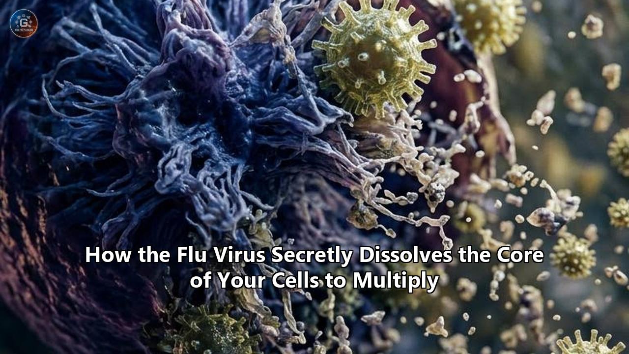
Scientists at the European Molecular Biology Laboratory (EMBL) Hamburg, working alongside researchers at the Leibniz Research Institute for Molecular Pharmacology (FMP) in Berlin and Charité – Universitätsmedizin Berlin, have mapped the intracellular architecture of influenza A infection in unprecedented detail. Published in Nature Microbiology, the study reveals a sophisticated viral sabotage mechanism: rather than merely commandeering cellular ribosomes in the cytoplasm, the influenza virus invades the innermost compartment of human host cells—the cell nucleus—and actively dissolves specialized sub-nuclear structures to multiply.
Using an advanced cross-linking mass spectrometry workflow paired with AI-driven structural modeling, the international team captured direct protein-to-protein interactions inside intact, living cells. The findings demonstrate that influenza virus infection causes the progressive breakdown and dissolution of paraspeckles—droplet-like, membraneless organelles located inside the host cell nucleus. Upon melting these structures, the virus liberates key host RNA-binding proteins trapped inside them, re-engineering those proteins to accelerate viral genome synthesis while simultaneously dismantling the cell's frontline antiviral stress responses.
The observation occurred across every human cell line and influenza strain evaluated during the project. "What surprised us most was the paraspeckles," said Iuliia Kotova, first author of the study, formerly of EMBL Hamburg and now a researcher at ETH Zurich. "Watching these tiny organelles in the nucleus dissolve, consistently across every cell line and every flu strain we tested, told us this isn't a side effect of infection—it might be a strategy."
This molecular discovery provides a clear framework for understanding the cellular mechanisms that dictate how flu virus spreads through human respiratory tissue. By neutralizing intracellular alarm systems at their nuclear source, the virus maximizes its replication rate, leading to massive viral shedding in infected upper respiratory tracts.
Anatomy of Nuclear Sabotage: How Influenza Dismantles the Cell Core
Most RNA viruses—such as coronaviruses, flaviviruses, and picornaviruses—replicate entirely within the host cell cytoplasm, avoiding the heavily fortified central repository where human DNA resides. Influenza A represents a notable exception. To copy its segmented negative-sense RNA genome, the virus must transport its viral ribonucleoprotein complexes (vRNPs) across the nuclear envelope through nuclear pore complexes, establishing a command center inside the host nucleus.
Once inside the nuclear matrix, the virus encounters paraspeckles: dynamic, liquid-liquid phase-separated condensate droplets. Lacking a physical lipid membrane, paraspeckles are maintained through delicate physical interactions between a long non-coding RNA known as NEAT1 (Nuclear Paraspeckle Assembly Transcript 1) and a dedicated suite of core proteins, including NONO (p54nrb), SFPQ (PSF), and PSPC1. Under normal physiological conditions, paraspeckles act as nuclear storage vaults and regulatory switches. They sequester specific host messenger RNAs and regulatory proteins, releasing them only when the cell experiences environmental stress, viral invasion, or mechanical damage.
+-----------------------------------------------------------------------+
|                       NORMAL HOST CELL NUCLEUS                        |
|                                                                       |
|   +---------------------------------------------------------------+   |
|   |                    Intact Paraspeckles                        |   |
|   |  - NEAT1 RNA backbone binds NONO, SFPQ, and PSPC1 proteins    |   |
|   |  - Antiviral signaling molecules sequestered & ready          |   |
|   +---------------------------------------------------------------+   |
+-----------------------------------------------------------------------+
                                   │
                                   │  Influenza A vRNP Nuclear Entry
                                   ▼
+-----------------------------------------------------------------------+
|                    INFLUENZA-INFECTED NUCLEUS                         |
|                                                                       |
|   +---------------------------------------------------------------+   |
|   |                 Dissolved Paraspeckle Core                    |   |
|   |  - Structural destabilization releases stored proteins        |   |
|   |  - Host RNA-binding proteins stolen for viral replication     |   |
|   |  - Antiviral stress response suppressed at the source         |   |
|   +---------------------------------------------------------------+   |
+-----------------------------------------------------------------------+
The EMBL-FMP research team discovered that influenza A systematically targets this phase balance. Shortly after nuclear entry, viral factors destabilize the NEAT1-protein scaffolding. The droplet-like structures dissolve into the surrounding nucleoplasm, discharging their concentrated reservoirs of RNA-binding proteins.
This dissolution delivers two distinct advantages to the invading pathogen:
- Resource Harvest: The virus recruits the liberated host RNA-binding proteins (including SFPQ and NONO) directly into its own transcription machinery, utilizing their RNA-chaperone properties to process, fold, and transcribe viral RNA molecules.
- Immune Silence: Paraspeckles regulate the expression of key cytokines and type I interferons. By melting these nuclear bodies, influenza suppresses the cell's ability to transcribe defense genes, effectively cutting the emergency alarm wires before the host can alert neighboring uninfected cells.
"There may also be a second benefit for the virus: some evidence suggests paraspeckles contribute to cellular stress responses and antiviral gene regulation, so disrupting them could also weaken parts of the cell's defense response," explained Jan Kosinski, group leader at EMBL Hamburg and the Centre for Structural Systems Biology (CSSB), who co-led the study.
Technical Precision: Mapping Interactions Inside Intact Cells
To capture this nuclear dissolution event, the research team had to overcome a fundamental barrier in structural virology. Historically, identifying interactions between viral proteins and host machinery required breaking infected cells open through physical lysis or detergent extraction.
Lysis destroys delicate, non-membrane-bound sub-cellular compartments like paraspeckles. Once compartmentalization is lost, proteins that never interact inside a living cell can artificially collide in a test tube, while brief, spatial-specific contacts dissipate unnoticed.
TRADITIONAL CELL LYSIS vs. IN-CELL CROSS-LINKING (XL-MS)

1. Traditional Lysis Workflow:
   Infected Cell  ──►  Detergent Lysis  ──►  Membraneless Organelles Destroyed  ──►  Artificial Protein Collisions / False Signals

2. In-Cell XL-MS Workflow (EMBL/FMP):
   Infected Cell  ──►  Chemical Cross-Linker  ──►  Spatial Contacts Frozen  ──►  Mass Spectrometry + AI Structural Modeling
To preserve the true intracellular architecture, the researchers deployed cross-linking mass spectrometry (XL-MS) optimized specifically for intact infected cells by collaborators Boris Bogdanow and Fan Liu at FMP Berlin.
- *Chemical Fixation in situ: A specialized chemical cross-linker penetrates live, infected human cells, forming covalent chemical bonds between proteins that are physically touching or positioned within nanometers of one another.
- Digestion & Spectrometry: The cell is subsequently digested, and mass spectrometers analyze the cross-linked peptide pairs, providing spatial coordinates of exact contact points.
- AI Structural Integration: The physical contact constraints gathered from mass spectrometry are fed directly into a modified implementation of AlphaFold. The algorithm predicts three-dimensional molecular models of host-virus protein complexes as they exist inside the intact nuclear matrix.
This integrative strategy revealed two distinct axes of host cellular re-engineering:
Axis 1: Maturation Network Hijacking
In the cell cytoplasm and endoplasmic reticulum, the viral surface glycoprotein haemagglutinin (HA) relies on a hitherto unidentified set of host chaperone proteins to fold into its functional tertiary structure. These host enzymes modify HA with precise sugar chains (glycosylation), preparing the viral envelope to attach to fresh host tissues.
Axis 2: Nuclear Core Demolition
Inside the nucleus, viral proteins engage host nuclear factors, inducing the phase-transition collapse of paraspeckles and freeing nuclear RNA-binding factors.
The Broader Arsenal: How Paraspeckle Melting Fits Into Host Shutoff
The breakdown of nuclear paraspeckles represents one phase of a broader viral strategy termed "host shutoff." Influenza viruses do not merely compete with host cellular processes; they forcibly terminate host gene expression to monopolize the cell's translational machinery.
+-----------------------------------------------------------------------------------+
|                        INFLUENZA HOST SHUTOFF MECHANISMS                          |
+---------------------------------------+-------------------------------------------+
| Viral Factor / Strategy               | Primary Subcellular Target & Effect       |
+---------------------------------------+-------------------------------------------+
| Paraspeckle Phase Dissolution         | Melts nuclear condensates; frees SFPQ/     |
|                                       | NONO proteins; degrades stress response.  |
+---------------------------------------+-------------------------------------------+
| Cap-Snatching (PB1/PB2/PA Complex)    | Cleaves 5' caps from host pre-mRNAs in     |
|                                       | nucleus; primes viral mRNA synthesis.     |
+---------------------------------------+-------------------------------------------+
| PA-X Endonuclease Activity            | Cleaves host RNA Polymerase II            |
|                                       | transcripts; triggers Xrn1 degradation.   |
+---------------------------------------+-------------------------------------------+
| NS1 Factor Blockade                   | Binds CPSF30; halts host mRNA 3'-end      |
|                                       | polyadenylation and nuclear export.       |
+---------------------------------------+-------------------------------------------+
Cap-Snatching
Because influenza A mRNA molecules lack the inherent ability to synthesize their own 5' cap structures—which are necessary for host ribosomes to recognize and translate an RNA strand into protein—the viral heterotrimeric RNA polymerase complex (composed of PB1, PB2, and PA subunits) performs "cap-snatching."
Positioned adjacent to host RNA Polymerase II inside the nucleus, the viral PB2 subunit binds to newly synthesized host pre-mRNAs. The PA subunit's endonuclease domain then cleaves the host RNA 10 to 14 nucleotides downstream from the 5' cap. The virus steals this capped leader sequence and uses it as a primer to synthesize its own viral messenger RNAs.
PA-X Mediated Transcript Destruction
Beyond cap-snatching, influenza expresses a specialized frameshift protein called PA-X. PA-X functions as a focused ribonuclease, preferentially targeting host messenger RNAs produced by RNA Polymerase II while sparing viral genomic segments.
Operating heavily within the nuclear compartment, PA-X cuts host transcripts, leaving uncapped RNA fragments that are rapidly degraded by host exonucleases like Xrn1. This selective destruction prevents host mRNAs from ever leaving the nucleus, eliminating competition for cytoplasmic ribosomes.
NS1 Blockade
Simultaneously, the viral non-structural protein 1 (NS1) binds to host cleavage and polyadenylation specificity factor 30 (CPSF30). This interaction halts the 3'-end processing of cellular pre-mRNAs, trapping host transcripts inside the nucleus where they are degraded.
When integrated with the EMBL team's discovery of paraspeckle dissolution, a clear picture emerges. Influenza A systematically dismantles the nuclear infrastructure from every angle: it stops host RNA maturation via NS1, degrades existing host transcripts via PA-X, steals host mRNA caps via its polymerase complex, and melts host nuclear organelles (paraspeckles) to scavenge essential proteins while extinguishing cellular distress beacons.
From Cell Core to Transmission: The Link to Viral Spread
Understanding the microscopic breakdown of nuclear structures provides direct insight into macro-level disease dynamics and how flu virus spreads through populations.
The primary vector for human-to-human influenza transmission is the generation of infectious fine-particle aerosols expelled during coughing, sneezing, talking, or breathing. The volume of infectious virions emitted into the surrounding air by an infected individual is dictated by the viral load present in the epithelial cells lining the upper respiratory tract.
+-----------------------------------------------------------------------------------+
|                   MICRO-TO-MACRO TRANSMISSION CASCADE                             |
+-----------------------------------------------------------------------------------+
|  1. Nuclear Invasion & Paraspeckle Dissolution                                    |
|     Influenza enters host cell nucleus and dissolves paraspeckles, harvesting     |
|     RNA-binding proteins and suppressing local interferon activation.              |
+-----------------------------------------------------------------------------------+
                                         │
                                         ▼
+-----------------------------------------------------------------------------------+
|  2. Unchecked Replication Burst                                                   |
|     Host shutoff mechanisms maximize viral protein synthesis while suppressing     |
|     antiviral cytokines, allowing rapid intracellular viral assembly.              |
+-----------------------------------------------------------------------------------+
                                         │
                                         ▼
+-----------------------------------------------------------------------------------+
|  3. Exponential Viral Load in Airway Epithelium                                    |
|     High density of infectious virions accumulates across upper respiratory       |
|     tract mucosal membranes without provoking immediate interferon-mediated clearance. |
+-----------------------------------------------------------------------------------+
                                         │
                                         ▼
+-----------------------------------------------------------------------------------+
|  4. Aerosol Generation & Population Transmission                                 |
|     Infected individuals shed high concentrations of aerosolized virus during      |
|     exhalation and coughing, driving host-to-host environmental spread.           |
+-----------------------------------------------------------------------------------+
When influenza successfully dissolves host nuclear paraspeckles and blocks interferon expression, it creates a temporal window of localized immunosuppression. During the early hours of infection, host mucosal cells fail to secrete early-warning cytokines like IFN-beta and IL-6. Unchecked by host defenses, viral replication accelerates exponentially.
A single airway cell, transformed into a viral production factory through host shutoff and nuclear scavenging, can churn out thousands of progeny virions before undergoing lysis. This high replication efficiency rapidly increases the viral titer within nasal secretions and respiratory fluids.
Consequently, when an infected individual exhales or coughs, every microscopic droplet carries a high infectious payload. The suppression of localized host defenses directly inflates viral shedding metrics, amplifying the probability that casual proximity will transmit an infectious dose to a susceptible host.
Principles of Pathogen Architecture: Condensate Exploitation
The revelation that influenza A systematically melts paraspeckles illustrates a broader theme emerging across cellular virology: biomolecular condensates formed through phase separation serve as central control hubs in host-pathogen interactions.
+-----------------------------------------------------------------------------------+
|               KEY LESSONS FROM INFLUENZA'S CONDENSATE STRATEGY                    |
+-----------------------------------------------------------------------------------+
| Principle 1: Membraneless Organelles are Primary Targets                          |
| Liquid-liquid phase-separated structures (paraspeckles, nucleoli, stress granules)|
| lack protective physical membranes, making them vulnerable to viral enzymes.      |
+-----------------------------------------------------------------------------------+
| Principle 2: Phase Dissolution Serves a Dual Purpose                              |
| Dissolving host condensates simultaneously liberates needed host machinery while  |
| dismantling host stress-signaling hubs.                                           |
+-----------------------------------------------------------------------------------+
| Principle 3: Spatial Integrity Dictates Cell Immunity                             |
| Disrupting the structural organization of host proteins is just as effective as   |
| degrading or mutating those proteins directly.                                    |
+-----------------------------------------------------------------------------------+
Principle 1: Condensates as Target-Rich Environments
Membraneless organelles—including paraspeckles, processing bodies (P-bodies), stress granules, and the nucleolus—are dense aggregates of proteins and nucleic acids created through liquid-liquid phase separation. Because they lack lipid bilayer membranes, their assembly and disassembly depend on delicate concentration thresholds and phosphorylation states. Viruses have evolved molecular triggers that disturb these thresholds, turning structural vulnerabilities into accessible reservoirs of cellular components.
Principle 2: Dual-Action Efficiency
Viruses operate under severe genomic constraints, encoding only a handful of essential proteins. To survive, viral factors must perform multiple roles simultaneously. Influenza's destruction of paraspeckles provides a clear example: a single structural disruption simultaneously harvests host RNA-processing machinery and neutralizes the host's innate immune alarm systems.
Principle 3: Spatial Disruption as Functional Suppression
Traditionally, virologists evaluated host shutoff by measuring overall protein degradation or changes in transcript levels. The EMBL findings highlight that functional suppression can occur purely through structural spatial reorganization. By altering the physical phase of host sub-nuclear compartments, the virus inactivates cellular pathways without needing to destroy every individual host enzyme.
Therapeutic Countermeasures: Protecting the Cellular Core
The discovery that influenza depends on paraspeckle dissolution and specific host-protein chaperones creates new opportunities for antiviral drug development.
+-----------------------------------------------------------------------------------+
|                        ANTIVIRAL STRATEGY COMPARISON                              |
+-----------------------------------+-----------------------------------------------+
| Traditional Antivirals            | Host-Targeted / Condensate Therapeutics       |
+-----------------------------------+-----------------------------------------------+
| Target: Viral Active Sites        | Target: Host-Pathogen Physical Interfaces     |
| (e.g., Neuraminidase, PA-Endonuclease) | (e.g., Paraspeckle Stabilizers, Chaperones) |
+-----------------------------------+-----------------------------------------------+
| High Risk of Rapid Mutation &     | Low Risk of Resistance; Host Interfaces       |
| Drug Resistance (e.g., H275Y)     | Are Genetically Stable Across Mutations       |
+-----------------------------------+-----------------------------------------------+
| Strain-Specific Efficacy          | Broad-Spectrum Efficacy Against               |
|                                   | Multiple Influenza A Strains                  |
+-----------------------------------+-----------------------------------------------+
Currently approved influenza antivirals target viral proteins directly:
- Neuraminidase Inhibitors (Oseltamivir, Zanamivir): Block the viral enzyme responsible for cleaving sialic acid residues, trapping newly assembled virions on the outer membrane of host cells.
- M2 Ion Channel Blockers (Amantadine, Rimantadine): Prevent viral uncoating inside endosomes (though widespread resistance has largely rendered this class obsolete).
- Cap-Dependent Endonuclease Inhibitors (Baloxavir Marboxil): Target the PA subunit of the viral polymerase complex to prevent cap-snatching.
While effective when administered early, these single-target drugs remain vulnerable to point mutations in the viral genome. For instance, a single amino acid substitution (such as the H275Y mutation in neuraminidase) can confer resistance to oseltamivir without compromising viral replication.
The Host-Targeted Alternative
Because host cell structures do not mutate under viral selective pressure, targeting the host-virus interface provides a more resilient therapeutic strategy. The detailed contact maps generated by the EMBL-FMP team highlight promising alternative targets:
- Condensate Stabilizers: Small molecules engineered to stabilize the phase boundaries of host paraspeckles could render these sub-nuclear structures resistant to viral dissolution. Protecting paraspeckles preserves host innate antiviral responses and prevents influenza from harvesting essential RNA-binding factors.
- Interface Disruptors: By utilizing AlphaFold structural predictions of viral-host complexes, pharmacologists can design peptidomimetics or small molecules that bind to host chaperone proteins, preventing them from assisting in the folding of viral haemagglutinin.
- Combination Regimens to Curb Spread: Combining direct-acting antivirals (like baloxavir) with host-targeted condensate stabilizers could suppress viral replication inside the nucleus while preventing the rapid viral shedding responsible for how flu virus spreads through communities.
Pandemic Preparedness: Assessing High-Consequence Strains
While the foundational experiments published by the EMBL-FMP team were conducted using established human influenza A strains, the methodology offers immediate applications for monitoring emerging zoonotic pathogens.
Pan-zootic avian influenza strains—most notably highly pathogenic avian influenza (HPAI) H5N1 clades—continue to spill over into mammalian livestock and human hosts. Assessing whether novel animal strains possess the immediate ability to dissolve human paraspeckles and hijack mammalian chaperones provides a valuable metric for evaluating potential pandemic threats.
+-----------------------------------------------------------------------------------+
|               ZOONOTIC PREPAREDNESS WORKFLOW (H5N1 / EMERGING STRAINS)            |
+-----------------------------------------------------------------------------------+
|  Isolate Emerging Zoonotic Strain (e.g., HPAI H5N1 Mammalian Spillover)           |
+-----------------------------------------------------------------------------------+
                                         │
                                         ▼
+-----------------------------------------------------------------------------------+
|  Apply In-Cell XL-MS + AlphaFold Workflow in Human Airway Epithelial Models       |
+-----------------------------------------------------------------------------------+
                                         │
                                         ▼
+-----------------------------------------------------------------------------------+
|  Evaluate Nuclear Sabotage Profile:                                               |
|  - Does viral polymerase trigger human paraspeckle dissolution?                   |
|  - Does HA utilize human ER chaperones efficiently?                               |
+-----------------------------------------------------------------------------------+
                                         │
                                         ▼
+-----------------------------------------------------------------------------------+
|  High Nuclear Efficiency  ──►  Elevated Risk Metric for Airborne Human Spread     |
|  Low Nuclear Efficiency   ──►  Barriers to Efficient Human Transmission Remain     |
+-----------------------------------------------------------------------------------+
If a bird-derived influenza strain can dissolve human nuclear paraspeckles, it indicates that the virus has already overcome critical species barriers inside the host cell core. This capacity would allow the virus to suppress human interferon responses and replicate efficiently, increasing the likelihood of human-to-human airborne transmission.
Applying this cross-linking mass spectrometry and structural modeling pipeline to patient isolates allows public health organizations to evaluate the human-adaptation risk profile of emerging virus strains in real time.
What to Watch Next in Nuclear Virology
The discovery of paraspeckle disassembly by influenza A opens several active avenues of investigation across molecular virology, biophysics, and structural biology:
- Identifying the Molecular Trigger: Researchers are working to determine precisely which viral factor initiates paraspeckle collapse. Identifying whether this process is driven by direct physical interaction between a viral protein and NEAT1* RNA, or through viral modulation of host kinase signaling, will reveal the primary enzymatic target.
- Screening Other Nuclear RNA Viruses: Structural virologists are applying the in-cell XL-MS workflow to other nuclear-replicating viruses—including respiratory syncytial virus (RSV), herpesviruses, and adenoviruses—to establish whether nuclear condensate dissolution is a universal viral strategy.
- In-Vivo Validation: Researchers are advancing from cell culture models to complex organoid models of human respiratory epithelium and animal models. These studies will examine how paraspeckle stabilization impacts viral titers, tissue pathology, and overall aerosol transmission kinetics in living organisms.
- Translating Condensate Biology to Antiviral Design: Medicinal chemists are testing high-throughput screening assays designed to identify small molecules that preserve nuclear phase boundaries during viral infection, opening a new frontier in antiviral drug discovery.
By mapping the physical contacts between host and viral factors inside intact cells, scientists have shown that the influenza virus is not merely an intracellular parasite. It operates as an architect of nuclear destruction, selectively dissolving sub-cellular structures to repurpose human proteins and suppress host defenses.
Understanding these mechanisms provides a new foundation for developing therapies designed to protect the integrity of the host cell nucleus, stop viral replication at its source, and break the cycles that dictate how flu virus spreads.
Reference:
- https://lifesciencehistory.com/scientists-map-how-the-flu-virus-rewires-the-human-cell-from-the-inside/
- https://www.youtube.com/watch?v=zew5HITFaAM
- https://en.clickpetroleoegas.com.br/flu-virus-hijacks-human-cells-dismantles-nucleus-structures-and-turns-the-organism-into-a-factory-for-new-copies-caes/
- https://www.labcompare.com/617-News/626979-How-the-Flu-Virus-Rewires-the-Human-Cell-from-the-Inside/
- https://www.medindia.net/news/scientists-reveal-how-influenza-a-reprograms-human-cells-from-within-224252-1.htm
- https://www.sciencedaily.com/releases/2026/07/260721000816.htm
- https://www.tribuneindia.com/news/health/new-study-reveals-how-flu-virus-hijacks-human-cells-to-multiply/
- https://journals.plos.org/plospathogens/article?id=10.1371/journal.ppat.1005427
- https://pmc.ncbi.nlm.nih.gov/articles/PMC4984632/
- https://biokb.lcsb.uni.lu/publications/02efaa2a-7f70-11ea-8187-001a4a160175
- https://journals.plos.org/plospathogens/article?id=10.1371/journal.ppat.1007465
- https://www.miragenews.com/flu-virus-rewiring-of-human-cells-mapped-by-1712791/