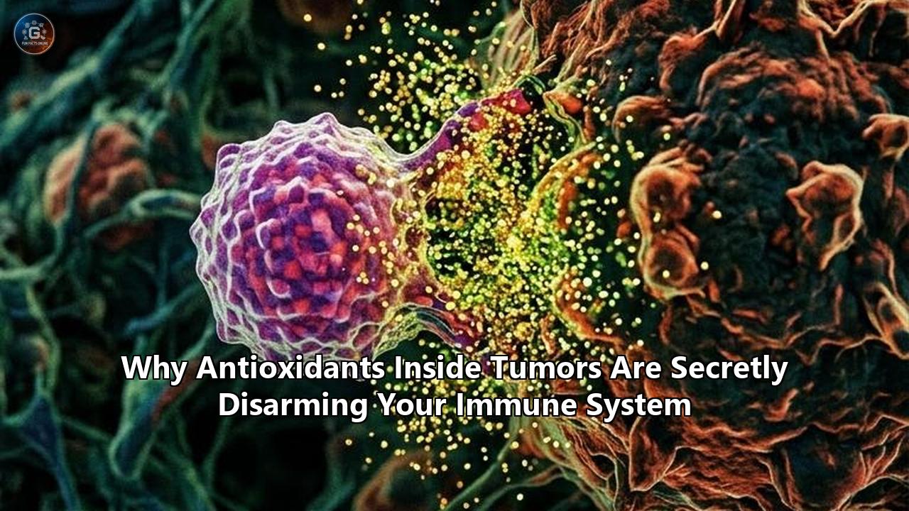
An investigation published in Science has exposed a counter-intuitive survival mechanism engineered by malignant tumors: cancer cells secrete high concentrations of natural antioxidant enzymes directly into their surrounding fluid to chemically smother and disarm attacking immune cells.
The joint investigation, led by teams at the University of Cambridge and Oregon Health & Science University (OHSU), demonstrates that cytotoxic CD8+ T lymphocytes—the immune system’s primary cellular assassins—rely on minute, tightly regulated pulses of reactive oxygen species (ROS) to trigger their tumor-killing machinery. By saturating the extracellular space with antioxidant proteins such as peroxiredoxin 1 (PRDX1), tumors strip the microenvironment of these essential oxidative signals. Deprived of ROS, T cells entering the tumor are functionally paralyzed, unable to sustain the receptor signaling required to release cytotoxic granules or synthesize interferon-gamma (IFN-γ).
This discovery resolves a medical paradox that has puzzled oncologists for over thirty years: why antioxidant compounds, widely celebrated for protecting healthy cells from DNA damage, repeatedly accelerate tumor progression and cause immunotherapies to fail in clinical trials.
THE ESCALATING REDOX TIMELINE IN ONCOLOGY
═══════════════════════════════════════════════════════════════════════════
1956–1970s │ Free Radical Theory: ROS framed as pure damage; antioxidants
           │ enter popular culture as universal protective agents.
───────────────────────────────────────────────────────────────────────────
1994–2011  │ Clinical Anomalies: ATBC, CARET, and SELECT trials show that
           │ antioxidant supplements significantly increase cancer risk.
───────────────────────────────────────────────────────────────────────────
2014–2019  │ Intracellular Shields: Research confirms cancer cells use
           │ NRF2 and GPX4 to escape apoptosis and ferroptosis.
───────────────────────────────────────────────────────────────────────────
2020–2025  │ Metabolic Checkpoints: NRF2 linked to CD8+ T cell exhaustion
           │ and immune evasion across solid tumors.
───────────────────────────────────────────────────────────────────────────
2026       │ The Extracellular Smothering Discovery: Science paper reveals
           │ tumors secrete PRDX1 to strip ROS and shut down T cell attack.
═══════════════════════════════════════════════════════════════════════════
The 1970s–1990s: The Free Radical Dogma and the Rise of the Antioxidant Myth
To understand how oncology arrived at this breaking discovery, one must trace the fifty-year trajectory of redox biology. The foundational narrative began in 1956 when Denham Harman introduced the Free Radical Theory of Aging. Harman proposed that endogenous reactive oxygen species—unstable, oxygen-bearing molecules with unpaired electrons, such as superoxide ($O_2^{\bullet-}$) and hydroxyl radicals ($\bullet OH$)—inflicted cumulative, irreversible damage upon cellular lipids, proteins, and nucleic acids.
In the 1970s, two-time Nobel laureate Linus Pauling pushed this concept into clinical and cultural ubiquity. Pauling argued that megadoses of exogenous antioxidants, particularly ascorbic acid (vitamin C) and alpha-tocopherol (vitamin E), could neutralize these free radicals, halt somatic mutagenesis, and prevent or cure malignancies.
Under Pauling’s influence, biomedical thinking adopted a rigid dichotomy:
- Reactive Oxygen Species (ROS): Harmful, mutagenic byproducts of aerobic respiration that degrade cellular components and initiate oncogenesis.
- Antioxidants: Cytoprotective molecules that neutralize ROS, maintain genomic integrity, and preserve tissue health.
This framework launched a global consumer industry and shaped clinical oncology hypotheses for decades. If free radicals drove the transition of healthy tissue into cancer, flooding human physiology with radical-scavenging antioxidants was presumed to be an effective preventive and therapeutic strategy.
Yet throughout the late 1980s, basic biochemists began detecting subtle anomalies in this paradigm. Experiments revealed that hydrogen peroxide ($H_2O_2$) was not merely an unavoidable toxic byproduct of oxidative phosphorylation. Intracellular enzymes, including the family of NADPH oxidases (NOX), deliberately synthesized ROS in response to growth factor stimulation. Rather than acting strictly as agents of decay, low concentrations of reactive oxygen species functioned as essential secondary messengers. These early physiological observations went largely unheeded by translational medicine, setting the stage for major clinical surprises.
The 1990s–2000s: Clinical Disasters and the First Major Anomalies
By the early 1990s, large-scale, randomized, double-blind, placebo-controlled clinical trials were launched to validate the cancer-preventive efficacy of high-dose antioxidant supplementation. The results produced unexpected outcomes that unsettled the field of preventative oncology.
CLINICAL TRIAL ANOMALIES: ANTIOXIDANTS IN HUMAN CANCER COHORTS
┌───────────────────────────┬───────────────────────┬───────────────────────────────┐
│ Clinical Trial            │ Intervention Target   │ Observed Clinical Outcome     │
├───────────────────────────┼───────────────────────┼───────────────────────────────┤
│ ATBC Trial                │ 29,133 male smokers;  │ 18% increase in lung cancer   │
│ (Finland, 1994)           │ Beta-carotene (20mg)  │ incidence; 8% rise in overall │
│                           │ ± Vitamin E (50mg)    │ mortality with beta-carotene. │
├───────────────────────────┼───────────────────────┼───────────────────────────────┤
│ CARET Trial               │ 18,314 high-risk      │ Halted 21 months early; 28%   │
│ (United States, 1996)     │ smokers/asbestos exp; │ more lung cancers; 17% higher │
│                           │ Beta-carotene + Vit A │ all-cause mortality.          │
├───────────────────────────┼───────────────────────┼───────────────────────────────┤
│ SELECT Trial              │ 35,533 healthy men;   │ 17% statistically significant │
│ (North America, 2011)     │ Vitamin E (400 IU)    │ increase in prostate cancer   │
│                           │ ± Selenium (200 mcg)  │ risk with Vitamin E alone.    │
└───────────────────────────┴───────────────────────┴───────────────────────────────┘
The first fracture in the dogma occurred in 1994 with the publication of the Alpha-Tocopherol, Beta-Carotene Cancer Prevention (ATBC) Study in The New England Journal of Medicine. Enrolling 29,133 male Finnish smokers aged 50 to 69, the trial tested whether daily dietary supplementation with alpha-tocopherol (50 mg), beta-carotene (20 mg), or both would decrease the incidence of lung cancer. After a median follow-up of 6.1 years, the researchers documented an 18% higher incidence of lung cancer in the cohort receiving beta-carotene, accompanied by an 8% increase in total mortality, primarily driven by lung cancer and ischemic heart disease.
Two years later, in 1996, the Beta-Carotene and Retinol Efficacy Trial (CARET), conducted across the United States with 18,314 participants at high risk for lung cancer due to heavy smoking histories or occupational asbestos exposure, ended in an emergency halt. Independent safety monitors terminated the trial 21 months ahead of schedule after determining that participants assigned to the active combination regimen (30 mg beta-carotene and 25,000 IU retinyl palmitate daily) suffered a 28% increase in lung cancer incidence and a 17% higher overall death rate compared to the placebo arm.
A third major trial, the Selenium and Vitamin E Cancer Prevention Trial (SELECT), randomized 35,533 men across 427 sites in the United States, Canada, and Puerto Rico starting in 2001 to evaluate whether selenium (200 $\mu$g/day) and/or vitamin E (400 IU/day) could prevent prostate cancer. By 2011, long-term follow-up published in JAMA revealed that dietary supplementation with vitamin E significantly increased the risk of developing prostate cancer by 17% in healthy men compared to placebo.
These clinical disasters created a deep divide in the oncology community:
- Public health advocates and alternative medicine practitioners treated the trials as isolated statistical outliers, attributing the negative results to synthetic formulations, specific chemical enantiomers, or late-stage participant intervention.
- Cancer biologists recognized a darker, mechanistic reality: advanced or pre-neoplastic lesions were somehow thriving on supplemental antioxidants, exploiting them to accelerate malignant growth.
The cellular mechanisms explaining why antioxidants favored the malignant state remained entirely unknown.
2009–2015: The Intracellular Shield — Cancer Hijacks Redox Homeostasis
The molecular explanation for these clinical trial anomalies began to take shape between 2009 and 2015, as scientists turned their focus from systemic blood markers to the internal biochemistry of the cancer cell.
THE INTRACELLULAR ESCALATION
─────────────────────────────────────────────────────────────────────────────
1. Oncogenic Activation (KRAS/BRAF) ───► Massive ROS generation
2. Intracellular Crisis             ───► Normal cells trigger p53 senescence
3. KEAP1-NRF2 Mutation              ───► Cancer hyperactivates antioxidant genes
4. System xCT & GPX4 Upregulated    ───► Intracellular ROS neutralized
5. Result                           ───► Cancer survives and metastasizes
─────────────────────────────────────────────────────────────────────────────
In 2009, a team led by Joan Brugge at Harvard Medical School published an investigation in Nature demonstrating that normal epithelial cells detached from the extracellular matrix underwent anoikis (detachment-induced apoptosis) driven by an acute loss of glucose transport and an overwhelming surge in intracellular ROS. Brugge's laboratory proved that treatment with exogenous antioxidants restored fatty acid oxidation, rescued ATP generation, and allowed detached cells to survive in suspension—a prerequisite for tumor metastasis.
Five years later, in 2014, a research team led by Martin Bergö at the University of Gothenburg in Sweden conducted experiments using genetically engineered mouse models of KRAS- and BRAF-driven lung adenocarcinoma. Supplementing the diets of these mice with clinically relevant doses of N-acetylcysteine (NAC) or vitamin E led to a threefold increase in tumor proliferation and a marked reduction in survival.
Bergö's group uncovered the underlying mechanism: oncogenic mutations in KRAS naturally drive heightened metabolic rates and generate massive amounts of endogenous ROS. In healthy cells, this severe oxidative stress triggers the tumor suppressor protein p53, forcing the cell into senescence or apoptosis. The administration of antioxidants cleared away the ROS, effectively disarming the p53 alarm system and allowing aggressive, DNA-damaged tumor cells to proliferate unchecked.
                ONCOGENIC STRESS & THE ANTIOXIDANT RESCUE

   [KRAS / BRAF Oncogene] ──► Hyper-metabolism ──► High Intracellular ROS
                                                         │
                     ┌───────────────────────────────────┴────────────────┐
                     ▼                                                    ▼
             [No Antioxidants]                                  [Antioxidant Exposure]
                     │                                                    │
           p53 Pathway Activated                                ROS Neutralized by Scavenger
                     │                                                    │
                     ▼                                                    ▼
       Cellular Senescence / Apoptosis                      p53 Checkpoint Remains Silent
                     │                                                    │
                     ▼                                                    ▼
             TUMOR ARRESTED                                      AGGRESSIVE METASTASIS
Follow-up studies in 2015 by the same team, alongside parallel findings from Sean Morrison’s laboratory at UT Southwestern, proved that antioxidants also accelerated distant melanoma metastasis. Circulating tumor cells undergoing the physical stress of hematogenous and lymphatic transit experience high oxidative stress that normally destroys them. Exogenous antioxidants protected these circulating cells from oxidative destruction, directly boosting their ability to colonize distant visceral organs.
Simultaneously, genomic sequencing initiatives, including The Cancer Genome Atlas (TCGA), began detailing recurrent somatic mutations in the KEAP1-NRF2 pathway across human non-small cell lung cancer (NSCLC), head and neck squamous cell carcinomas, and esophageal cancers. Under normal baseline conditions, Kelch-like ECH-associated protein 1 (KEAP1) acts as an adaptor for a Cullin-3-based ubiquitin ligase, constantly tagging the transcription factor NRF2 (nuclear factor erythroid 2-related factor 2) for rapid proteasomal destruction. Under conditions of oxidative stress, cysteine residues on KEAP1 are covalently modified, releasing NRF2 to translocate into the nucleus.
In malignant tumors, mutations in KEAP1 or NFE2L2 (the gene encoding NRF2) disrupt this control loop, locking NRF2 in a permanently active state. Once hyperactivated, NRF2 drives the continuous transcription of a broad antioxidant network, including:
- Glutathione Synthesizing Enzymes (GCLC/GCLM): Catalyzing the production of reduced glutathione (GSH), the primary intracellular antioxidant buffer.
- SLC7A11 (System xCT): An amino acid antiporter that imports cystine in exchange for glutamate, supplying the raw material for glutathione synthesis.
- Thioredoxin Reductase (TXNRD1): Maintaining cellular redox potential.
- Glutathione Peroxidase 4 (GPX4): An essential enzyme that prevents toxic lipid peroxidation.
By 2015, the oncology community recognized that cancer cells are not passive victims of oxidative stress. Instead, they weaponize internal antioxidant defense systems to resist therapy and maintain high proliferation rates. Yet, this insight remained focused inside the cancer cell itself. The broader implications for the surrounding immune microenvironment remained unexplored.
2016–2022: The Immune Dimension — Ferroptosis, Immunometabolism, and T-Cell Checkpoints
Between 2016 and 2022, the field of tumor immunology entered a new phase with the clinical expansion of immune checkpoint blockade therapies, particularly anti-PD-1, anti-PD-L1, and anti-CTLA-4 monoclonal antibodies. While these therapies produced long-term remissions in some patients, the majority of solid tumors showed primary or acquired resistance.
Immunologists began mapping the harsh biochemical landscape of the tumor microenvironment (TME), characterized by hypoxia, severe glucose depletion, lactic acidosis, and extracellular adenosine accumulation. As researchers analyzed the interactions between antioxidants and cancer immunity, they uncovered a deeper layer of metabolic cross-talk.
THE EVOLVING CONCEPT: ANTIOXIDANTS AND CANCER IMMUNITY
══════════════════════════════════════════════════════════════════════════════════
• Canonical View: ROS is purely destructive to both tumors and immune cells;
  antioxidants protect healthy tissue.
• Intermediate Discovery (2019): Intracellular GPX4 protects tumors from T-cell-
  mediated ferroptosis; targeting tumor antioxidants restores immunity.
• Current Model (2026): Tumors export antioxidant enzymes (PRDX1) to strip ROS
  from the microenvironment, neutralizing T-cell receptor activation.
══════════════════════════════════════════════════════════════════════════════════
In 2019, a study led by Brent Stockwell at Columbia University and Weiping Zou at the University of Michigan connected tumor antioxidant defenses directly to immune destruction in Nature. The researchers discovered that cytotoxic CD8+ T cells, upon recognizing tumor antigens, secrete interferon-gamma (IFN-γ). This cytokine acts on tumor cells to downregulate the expression of the two subunits of system xCT, SLC7A11 and SLC3A2.
By shutting down cystine uptake, T-cell-derived IFN-γ depletes the tumor's intracellular glutathione stores, crippling the antioxidant enzyme glutathione peroxidase 4 (GPX4). Without GPX4 to clear lipid peroxides, the tumor cell's membrane lipids undergo rapid, iron-catalyzed oxidation, culminating in ferroptosis—a form of non-apoptotic, immunogenic cell death.
                     HOW T CELLS KILL VIA FERROPTOSIS

   Activated CD8+ T Cell ──► Secretes Interferon-gamma (IFN-γ)
                                        │
                                        ▼
                      Tumor Surface: Downregulates SLC7A11
                                        │
                                        ▼
                      Cystine Uptake Inhibited in Cancer Cell
                                        │
                                        ▼
                         Intracellular Glutathione (GSH) Collapses
                                        │
                                        ▼
                           GPX4 Enzyme Inactivated
                                        │
                                        ▼
                     Uncontrolled Toxic Lipid Peroxidation
                                        │
                                        ▼
                         TUMOR CELL DEATH (FERROPTOSIS)
This discovery highlighted a critical dynamic in antioxidants and cancer immunity: tumor cells with hyperactivated antioxidant machinery could resist T-cell-mediated ferroptotic clearance. Tumors that upregulated their antioxidant pathways were effectively building an armored shield against the immune system's primary offensive weapon.
Simultaneously, researchers investigating the immunometabolism of tumor-infiltrating lymphocytes (TILs), such as Greg Delgoffe and Dayana Rivadeneira at the University of Pittsburgh's UPMC Hillman Cancer Center, discovered that T cells experience severe metabolic strain within solid tumors. Continuous antigen stimulation alongside hypoxia forces T-cell mitochondria to produce excessive internal ROS. This localized, internal mitochondrial stress damages T-cell telomeres and drives the lymphocytes into a state of exhaustion.
These concurrent discoveries established that the balance of antioxidants and cancer immunity is a precise biochemical tightrope:
- Cancer cells use intracellular antioxidants to survive high metabolic rates and prevent ferroptotic death.
- CD8+ T cells require protection against unmitigated, chronic mitochondrial oxidative damage to avoid terminal exhaustion.
The question remained: How did cancer cells directly manipulate the oxidative chemistry of the extracellular space to disarm infiltrating immune cells?
2023–2025: Decrypting the NRF2–T-Cell Exhaustion Network
Between 2023 and 2025, experimental evidence emerged showing that tumor antioxidant signaling was actively suppressing immune responses in patients receiving immunotherapy.
CHRONOLOGICAL DISCOVERY TRAJECTORY
┌────────────────────────────────────────────────────────────────────────────┐
│ 2023: Clinical genomics reveals KEAP1/NRF2-mutant tumors show universal    │
│       resistance to anti-PD-(L)1 immune checkpoint inhibitors.             │
├────────────────────────────────────────────────────────────────────────────┤
│ 2024: Identification of the NRF2-PTGIR axis; hyperactive antioxidant       │
│       signaling in CD8+ T cells directly drives terminal exhaustion.       │
├────────────────────────────────────────────────────────────────────────────┤
│ 2025: Discovery of NRF2-SLC2A3-DHA metabolic scavenging in esophageal      │
│       tumors, which drains extracellular redox currency.                   │
├────────────────────────────────────────────────────────────────────────────┤
│ 2026: Identification of PRDX1 secretion in tumor interstitial fluid as     │
│       an extracellular "redox checkpoint" that smothers T-cell signaling.  │
└────────────────────────────────────────────────────────────────────────────┘
In 2024, immunologists studying tumor-infiltrating lymphocytes in melanoma and colon cancer models identified an unexpected role for NRF2 within CD8+ T cells. While NRF2 activation was presumed to protect T cells from oxidative stress, researchers found that sustained, high-level NRF2 activation drove the lymphocytes into terminal functional exhaustion.
Single-cell RNA sequencing revealed that NRF2 directly controls the transcription of the prostacyclin receptor PTGIR, a G-protein-coupled receptor. When PTGIR is expressed on CD8+ T cells in response to persistent NRF2 signaling, it binds tumor-derived prostanoids, represses T-bet transcription factor activity, and downregulates the expression of Granzyme B and IFN-γ. Genetically ablating NRF2 or PTGIR in T cells restored their cytotoxic capacity, enabling sustained control of tumor growth in animal models.
Shortly thereafter, a translational oncology team at Zhongshan Hospital, Fudan University, investigated why patients with esophageal squamous cell carcinoma (ESCC) frequently experienced resistance to immune checkpoint blockade. Paired transcriptomic and metabolomic analyses of patient biopsies before and after immunotherapy revealed marked NRF2 pathway activation in non-responders.
The Fudan team traced this resistance to a metabolic mechanism: NRF2 directly upregulated SLC2A3, a high-affinity transporter for dehydroascorbic acid (DHA), the oxidized form of vitamin C. Malignant cells expressing high levels of SLC2A3 systematically pulled DHA out of the extracellular microenvironment, recycling it internally into reduced ascorbic acid and disrupting the local extracellular redox balance. Co-culture assays confirmed that this metabolic shift suppressed CD8+ T-cell proliferation, blocked IFN-γ secretion, and neutralized cytotoxic activity.
These findings confirmed that tumors manipulate redox biology to evade immune attacks. However, the most direct mechanism—how tumors physically shut down the activation of T cells via extracellular antioxidants—was published in Science.
2026: The Extracellular Redox Checkpoint — Tumors Smother Immunity
The study published in Science was led by Dr. Rahul Roychoudhuri of the University of Cambridge’s Department of Pathology and Dr. Robert L. Eil, an associate professor of surgery at the Oregon Health & Science University (OHSU) School of Medicine and Knight Cancer Institute. Their work revealed that tumors do not merely defend their own cytoplasm; they export an antioxidant shield into the extracellular fluid to disarm the immune system.
                  THE EXTRACELLULAR SMOTHERING MECHANISM

                     TUMOR MICROENVIRONMENT (INTERSTITIAL FLUID)
 ┌─────────────────────────────────────────────────────────────────────────────┐
 │                                                                             │
 │    TUMOR CELL                                            CD8+ T CELL        │
 │  ┌──────────────┐                                      ┌──────────────┐     │
 │  │ Secretes:    │                                      │ TCR Engaged  │     │
 │  │ PRDX1        ├─────────┐                            │ (Antigen)    │     │
 │  │ (Antioxidant)│         │                            └──────┬───────┘     │
 │  └──────────────┘         ▼                                   │             │
 │                     ┌────────────┐                            │             │
 │                     │ PRDX1 Mops │                            ▼             │
 │                     │ Up Free    │                     Requires Local       │
 │                     │ Extracell. │                     H2O2 Signaling       │
 │                     │ H2O2 / ROS │                            │             │
 │                     └─────┬──────┘                            │             │
 │                           │                                   │             │
 │                           │   Deprived of H2O2 Oxidative      │             │
 │                           └─── Inactivation Pulse ◄───────────┘             │
 │                                           │                                 │
 │                                           ▼                                 │
 │                              Protein Tyrosine Phosphatases                  │
 │                              (SHP-1 / CD45) Remain ACTIVE                   │
 │                                           │                                 │
 │                                           ▼                                 │
 │                              Dephosphorylates ZAP-70 / Lck                  │
 │                                           │                                 │
 │                                           ▼                                 │
 │                              T-CELL SIGNALING SHUT DOWN                     │
 │                              • Zero Granzyme B Release                      │
 │                              • Zero IFN-γ Production                        │
 │                              • Immune Attack Neutralized                    │
 │                                                                             │
 └─────────────────────────────────────────────────────────────────────────────┘
The Discovery in Tumor Interstitial Fluid
The Cambridge and OHSU teams analyzed tumor interstitial fluid—the liquid that directly bathes malignant cells, stroma, and infiltrating immune cells. Using biochemical assays and high-resolution mass spectrometry, the researchers discovered that tumor interstitial fluid possesses potent, continuous antioxidant activity, creating an environment chemically distinct from healthy peripheral tissues.
Proteomic profiling identified high concentrations of peroxiredoxin 1 (PRDX1), a natural antioxidant enzyme normally studied for its role in reducing hydrogen peroxide inside the cytoplasm. In diverse solid malignancies, however, cancer cells export large quantities of PRDX1 into the extracellular space.
"One of the surprising findings is that antioxidants aren't always beneficial in the context of cancer," said co-senior author Dr. Robert L. Eil. "While reactive oxygen species sound threatening, T cells actually need them to perform their tumor-fighting job. What we found is that tumors can exploit the T cell's dependency by removing the reactive oxygen species the immune system depends on".
Dr. Rahul Roychoudhuri of the University of Cambridge described the dynamic: "We've found a new way by which cancers shut down the immune system. Tumors do this by releasing large amounts of a protein that is a natural antioxidant, Peroxiredoxin 1 (PRDX1), which mops up reactive oxygen species and deprives T cells of the activating signals they need to perform cancer killing".
The Biochemical Mechanism: How T Cells Depend on ROS to Kill
To explain why this antioxidant secretion paralyzes the immune response, the researchers mapped the earliest nanoseconds of T-cell receptor (TCR) engagement.
When a CD8+ cytotoxic T cell encounters a cancer cell, its T-cell receptor binds to a tumor antigen presented on a major histocompatibility complex class I (MHC-I) molecule. This physical interaction triggers an immediate, localized production of reactive oxygen species—specifically hydrogen peroxide ($H_2O_2$)—generated at the immunological synapse by NADPH oxidase 2 (NOX2) complexes and mitochondrial respiratory chain complexes.
                  THE BIOCHEMICAL OFF-SWITCH IN CYTOTOXIC T CELLS

           NORMAL IMMUNE ACTIVATION             PRDX1 SMOTHERING (IN TUMORS)
           ────────────────────────             ────────────────────────────
           TCR Binds Tumor Antigen              TCR Binds Tumor Antigen
                      │                                    │
                      ▼                                    ▼
           Local Pulse of H2O2 Produced         Local H2O2 Produced
                      │                                    │
                      ▼                                    ▼
           H2O2 Oxidizes PTP Thiol:             Extracellular PRDX1 Scavenges H2O2;
           [-SH] ──► [-SOH] (Sulfenic Acid)     Zero Peroxide Reaches Synapse
                      │                                    │
                      ▼                                    ▼
           Phosphatases (SHP-1) INACTIVATED     Phosphatases (SHP-1) REMAIN ACTIVE
                      │                                    │
                      ▼                                    ▼
           Kinases (Lck/ZAP-70) Remain          Phosphatases Dephosphorylate
           Phosphorylated and Active            ZAP-70, LAT, and SLP-76
                      │                                    │
                      ▼                                    ▼
           FULL CYTOTOXIC ATTACK                IMMUNE ATTACK EXTINGUISHED
           (Granzyme, Perforin, IFN-γ)          (T-Cell Paralyzed)
This transient pulse of $H_2O_2$ serves a specific biochemical role:
- The Phosphatase Brake: Under resting conditions, cytotoxic kinase cascades driven by Lck, Fyn, and ZAP-70 are held in check by Protein Tyrosine Phosphatases (PTPs), including SHP-1 (PTPN6) and CD45. These phosphatases continuously remove activating phosphate groups from downstream signaling proteins, preventing accidental immune activation.
- The Oxidative Inactivation: When the T cell detects an antigen, the localized burst of $H_2O_2$ oxidizes the catalytic cysteine residues inside the active site of these PTPs. The nucleophilic thiol group ($-SH$) on the cysteine is converted into a sulfenic acid intermediate ($-SOH$).
- Sustained Kinase Activation: This reversible oxidative modification temporarily inactivates the phosphatases. With the phosphatase brake removed, ZAP-70, LAT, and SLP-76 remain phosphorylated, allowing downstream calcium influx, MAP kinase activation, cytolytic granule polarization (perforin and granzyme B), and transcriptional activation of cytokine genes.
When a tumor floods its interstitial fluid with PRDX1, this regulatory system is short-circuited. The extracellular PRDX1 acts as an enzymatic vacuum, consuming every stray molecule of $H_2O_2$ before it can oxidize the regulatory phosphatases. As a result, the PTPs remain un-oxidized and fully active. The moment the T-cell receptor binds a tumor antigen, the active phosphatases dephosphorylate ZAP-70 and LAT, cutting off downstream signaling. The T cell is functionally paralyzed before it can launch an attack.
ENZYMATIC OXIDATION DYNAMICS AT THE IMMUNOLOGICAL SYNAPSE

Target Molecule: Protein Tyrosine Phosphatase Catalytic Site (SHP-1 / PTPN6)
Normal Activation:
   Cys-SH  +  H2O2  ──►  Cys-SOH (Sulfenic Acid) + H2O  [Enzyme Inactivated]
                                   │
                                   ▼
                Kinase cascades continue uninterrupted

Tumor Suppression (PRDX1 Present):
   2 PRDX1-Cys-SH  +  H2O2  ──►  PRDX1-Cys-S-S-Cys + 2 H2O  [Peroxide Eliminated]
                                   │
                                   ▼
   Target PTP remains Cys-SH (Fully Active) ──► Dephosphorylates ZAP-70/LAT
Preclinical Reversal and Human Cross-Cohort Validation
To confirm whether this mechanism shields tumors from immune destruction, the research team used CRISPR-Cas9 genome editing to delete the Prdx1 gene in mouse cancer cell lines.
- When PRDX1-deficient cancer cells were implanted into immunocompetent mice, the tumors showed a marked increase in infiltrating, activated CD8+ T cells and sustained substantial tumor shrinkage.
- When PRDX1-deficient tumors were implanted into immunodeficient mice lacking functional T cells, the therapeutic benefit disappeared, confirming that PRDX1 acts specifically as an immune checkpoint.
- Combining PRDX1 genetic ablation with anti-PD-1 checkpoint inhibitors caused established, therapy-resistant tumors to shrink and, in many cases, cleared the malignancy entirely.
The team then expanded their investigation to human clinical datasets, analyzing:
- Secretome profiling from hundreds of human cancer cell lines.
- Bulk and single-cell RNA-sequencing data spanning thousands of human primary tumors across TCGA datasets.
- Direct mass spectrometry analysis of interstitial fluid collected from primary human tumors removed during surgery.
All three independent approaches confirmed the preclinical findings: human malignancies consistently upregulate and secrete PRDX1 into the extracellular space. High PRDX1 expression correlates with T-cell exclusion ("cold" immune deserts), decreased expression of cytotoxic effector genes, and primary resistance to standard immune checkpoint inhibitors.
The study demonstrated that cancer cells upregulate PRDX1 during immunoediting—the evolutionary selection process wherein tumors adapt under pressure from an active immune system to survive. PRDX1 secretion operates as a dedicated redox checkpoint, allowing cancer cells to escape immune detection.
The Dual Nature of ROS in Cancer Immunology
The discovery of the extracellular PRDX1 redox checkpoint highlights the dual, context-dependent nature of reactive oxygen species in oncology. The biological effects of ROS are determined entirely by subcellular compartmentalization, concentration, and timing.
THE REDOX COMPARTMENTALIZATION MATRIX
┌──────────────────────────────┬──────────────────────────────┬──────────────────────────────┐
│ Anatomical / Cellular Zone   │ Oxidative State (ROS/Antiox) │ Impact on Cancer Immunity    │
├──────────────────────────────┼──────────────────────────────┼──────────────────────────────┤
│ T-Cell Receptor Synapse      │ High localized H2O2          │ BENEFICIAL: Inactivates      │
│ (Subcellular micro-domain)   │ (Micromolar burst)           │ PTPs to allow T-cell killing │
├──────────────────────────────┼──────────────────────────────┼──────────────────────────────┤
│ Tumor Interstitial Fluid     │ High PRDX1 / High Antiox     │ HARMFUL: Smothers T cells,   │
│ (Extracellular Matrix)       │ (Low extracellular ROS)      │ blocks immune attack         │
├──────────────────────────────┼──────────────────────────────┼──────────────────────────────┤
│ Cancer Cell Cytoplasm        │ High NRF2 / GPX4 / GSH       │ HARMFUL: Prevents tumor cell │
│ (Intracellular)              │ (Low intracellular ROS)      │ death and ferroptosis        │
├──────────────────────────────┼──────────────────────────────┼──────────────────────────────┤
│ T-Cell Mitochondria          │ Chronic Overproduction       │ HARMFUL: Drives telomere     │
│ (Intracellular / Exhaustion) │ of Mitochondrial ROS         │ damage and T-cell exhaustion │
└──────────────────────────────┴──────────────────────────────┴──────────────────────────────┘
The dual roles of ROS in tumor immunology can be broken down across these compartments:
1. The Immunological Synapse (Signaling Hub)
- Status: Requires localized, short bursts of $H_2O_2$.
- Function: Oxidatively inactivates inhibitory protein tyrosine phosphatases (SHP-1, DEP-1, CD45).
- Immune Impact: Enables TCR-driven kinase signaling, cytokine synthesis (IFN-γ, IL-2), and cytotoxic degranulation against target tumor cells.
- Disruption: Tumor-derived PRDX1 scavenges this localized ROS pool, terminating T-cell activation.
2. The Extracellular Tumor Matrix (Chemical Buffer)
- Status: Kept in a reduced, antioxidant-rich state by secretable tumor enzymes.
- Function: Neutralizes oxidative signaling molecules generated by innate and adaptive immune cells.
- Immune Impact: Creates an immune-excluded microenvironment, preventing T-cell activation and blunting the efficacy of checkpoint inhibitors.
3. The Malignant Cytoplasm (Ferroptosis Barrier)
- Status: Maintained by NRF2, SLC7A11, and GPX4.
- Function: Scavenges lipid hydroperoxides generated by cellular metabolism and immune-derived cytokines.
- Immune Impact: Prevents T-cell-induced ferroptotic cell death, driving therapy resistance and metastasis.
4. The Lymphocyte Mitochondria (Exhaustion Driver)
- Status: Overloaded with electron-leak ROS under conditions of chronic hypoxia and sustained stimulation.
- Function: Inflicts oxidative damage on mitochondrial DNA and nuclear telomeres.
- Immune Impact: Triggers a DNA damage response that drives T cells into terminal metabolic exhaustion and senescence.
                     THE REDOX LOCALIZATION PARADOX

     T-CELL ACTIVATION BURST                     T-CELL EXHAUSTION OVERLOAD

        Immunological Synapse                       T-Cell Mitochondrion
     ┌────────────────────────┐                  ┌────────────────────────┐
     │  NOX2 / Complex I/III  │                  │  Chronic Hypoxia / TCR │
     │           │            │                  │           │            │
     │           ▼            │                  │           ▼            │
     │     PULSED H2O2        │                  │      CHRONIC ROS       │
     │           │            │                  │           │            │
     │           ▼            │                  │           ▼            │
     │  Reversible PTP Shift  │                  │  Telomeric DNA Damage  │
     │     (SH ──► SOH)       │                  │           │            │
     │           │            │                  │           ▼            │
     │           ▼            │                  │    Terminal T-Cell     │
     │     CYTOTOXICITY       │                  │       Exhaustion       │
     └────────────────────────┘                  └────────────────────────┘
            [DESIRED]                                   [UNDESIRED]
This spatial compartmentalization explains why dietary antioxidant supplements repeatedly failed in clinical trials. Systemic administration of un-targeted antioxidants (such as high-dose vitamin C, vitamin E, or N-acetylcysteine) indiscriminately floods all compartments. Instead of protecting T cells from internal mitochondrial exhaustion, systemic supplements help tumors maintain their extracellular PRDX1 shield and support the intracellular GPX4 barrier, preventing both immune recognition and ferroptotic death.
Translating the Redox Checkpoint into Cancer Therapeutics
The discovery of extracellular antioxidant-mediated immune suppression points to several new therapeutic avenues. Rather than using broad-spectrum antioxidants or non-selective pro-oxidant chemotherapies, researchers are developing precision tools to selectively dismantle the tumor's antioxidant shield while preserving necessary immune functions.
NEXT-GENERATION REDOX IMMUNOTHERAPY STRATEGIES
══════════════════════════════════════════════════════════════════════════════
1. PRDX1-Neutralizing Monoclonal Antibodies
   • Target extracellular PRDX1 directly in the tumor interstitial fluid.
   • Prevent the mop-up of H2O2, restoring the T-cell activation synapse.
──────────────────────────────────────────────────────────────────────────────
2. Tumor-Selective NRF2 / GPX4 PROTAC Degraders
   • Proteolysis-Targeting Chimeras tagged for tumor-specific surface antigens.
   • Selectively downregulate the NRF2 antioxidant program inside tumor cells.
──────────────────────────────────────────────────────────────────────────────
3. Synthetic Biology: Redox-Armored CAR-T Cells
   • Engineer CAR-T / TCR-T cells to carry phosphatase mutants resistant
     to dephosphorylation or autonomous local ROS generation cassettes.
──────────────────────────────────────────────────────────────────────────────
4. Dual Immunotherapy Combinations
   • Combine extracellular PRDX1 blockade with anti-PD-1/PD-L1 antibodies.
   • Simultaneously release the PD-1 brake and dismantle the PRDX1 shield.
══════════════════════════════════════════════════════════════════════════════
1. Monoclonal Antibodies Targeting Extracellular PRDX1
Because PRDX1 acts in the extracellular tumor interstitial fluid to disarm T cells, it is directly accessible to systemically administered monoclonal antibodies (mAbs).
Therapeutic antibodies designed to bind and neutralize extracellular PRDX1 prevent the enzyme from scavenging localized hydrogen peroxide at the immunological synapse. This approach leaves normal intracellular peroxiredoxins unaffected inside healthy tissues, minimizing off-target toxicities while restoring T-cell activation specifically within the tumor microenvironment.
2. Targeted NRF2 and GPX4 Degraders
Small-molecule inhibitors and PROTACs (proteolysis-targeting chimeras) that selectively target the NRF2 transcription factor or GPX4 are advancing through early-stage drug pipelines. Conjugating these molecules to tumor-homing vehicles—such as antibody-drug conjugates (ADCs) or N-acetylgalactosamine (GalNAc) moieties—enables the selective shutdown of intracellular antioxidant machinery inside malignant cells without disrupting normal tissues. This leaves the tumor vulnerable to T-cell-induced ferroptosis.
3. Engineering Redox-Resistant T-Cell Therapies
Cellular immunotherapies, including chimeric antigen receptor (CAR) T-cell and engineered TCR-T cell platforms, can be modified to bypass the tumor's antioxidant defenses:
- Synaptic Catalytic Engines: Engineering CAR-T cells to carry membrane-anchored enzymes that generate localized $H_2O_2$ upon antigen binding, overpowering extracellular PRDX1.
- Phosphatase Modification: CRISPR editing of specific cysteine residues in PTPs (such as SHP-1) to reduce their ability to dephosphorylate ZAP-70 in low-ROS environments.
- Mitochondrial Protection: Selectively targeting antioxidants to T-cell mitochondria using triphenylphosphonium (TPP) conjugates to prevent internal telomeric damage and avoid exhaustion, while leaving synaptic ROS signaling active.
                PRECISION REDOX ENGINEERING IN CAR-T CELLS

               ┌──────────────────────────────────────────────┐
               │         ENGINEERED CAR-T LYMPHOCYTE          │
               │                                              │
               │   [Synaptic ROS Module]                      │
               │   Membrane-anchored NOX2 domain creates      │
               │   concentrated H2O2 pulse directly at tumor  │
               │   junction, overwhelming extracellular PRDX1 │
               │                      │                       │
               │                      ▼                       │
               │   [Engineered SHP-1]                         │
               │   Phosphatase modified to prevent aberrant   │
               │   inactivation of ZAP-70 and LAT kinases     │
               │                      │                       │
               │                      ▼                       │
               │   [Mitochondrial TPP-Antioxidant]            │
               │   Internal scavenger prevents telomeric      │
               │   damage and blocks terminal exhaustion      │
               │                                              │
               └──────────────────────┬───────────────────────┘
                                      │
                                      ▼
                      SUSTAINED, LONG-TERM TUMOR CLEARANCE
4. Strategic Combination with Checkpoint Inhibitors
Preclinical data from the Cambridge–OHSU study confirm that removing the PRDX1 antioxidant shield sensitizes unresponsive tumors to anti-PD-1 and anti-CTLA-4 immunotherapies.
Checkpoint inhibitors remove the inhibitory surface signals (such as PD-1/PD-L1 binding) that tell T cells to stand down. Neutralizing PRDX1 removes the chemical shield that extinguishes T-cell receptor activation. Combining these two approaches targets both the surface receptors and the metabolic environment that tumors use to evade destruction.
Upcoming Milestones and Clinical Questions
The revelation that tumors actively deploy antioxidant enzymes as an immune evasion strategy opens several key areas for ongoing translational research:
1. Clinical Stratification Biomarkers
Will interstitial PRDX1 levels, combined with KEAP1/NRF2 mutation status, serve as predictive clinical biomarkers to identify which patients will experience resistance to standard PD-1 blockade? Clinical trials will need to incorporate tumor fluid secretome profiling and spatial proteomic assays into patient screening protocols.
2. Patient Dietary Guidance
How should oncologists counsel patients regarding high-dose, over-the-counter antioxidant supplements (such as vitamins C and E, NAC, and selenium) during active immunotherapy?
While the Cambridge and OHSU teams emphasize that cancer patients should not alter their diets or clinical regimens without consulting their oncologists, preclinical and clinical trial data continue to demonstrate that blanket, high-dose antioxidant supplementation can inadvertently protect malignant tumors and blunt immune efficacy.
3. Therapeutic Window and Systemic Safety
Can pharmacological agents selectively disable extracellular PRDX1 in the tumor microenvironment without interfering with the essential antioxidant systems required by healthy tissues, such as erythrocyte membrane stabilization and cardiac protection? Early-phase clinical trials will need to establish the therapeutic safety window for targeted redox-modulating compounds.
THE EVOLUTION OF ANTIOXIDANT UNDERSTANDING IN ONCOLOGY
═══════════════════════════════════════════════════════════════════════════════
ERA 1: The Protective Myth (1956–1990)
• Free radicals cause cancer; external antioxidants prevent it.
• Result: Multibillion-dollar supplement industry; broad clinical assumptions.

ERA 2: The Clinical Reality (1994–2011)
• Large trials (ATBC, CARET, SELECT) show antioxidants increase cancer risk.
• Result: Confusion in public health; clinical hesitation.

ERA 3: The Intracellular Defense (2014–2022)
• Cancer cells mutate NRF2/KEAP1 to build internal antioxidant shields.
• Intracellular GPX4 protects tumors from T-cell-mediated ferroptosis.
• Result: Recognition of antioxidant defenses as drivers of therapy resistance.

ERA 4: The Extracellular Immune Checkpoint (2026–Present)
• Tumors export PRDX1 into interstitial fluid to strip ROS from the environment.
• Deprived of ROS, cytotoxic T cells suffer immediate signaling failure.
• Result: Development of PRDX1-targeted therapies to restore immune clearance.
═══════════════════════════════════════════════════════════════════════════════
The discovery published in Science shifts our understanding of antioxidants and cancer immunity. What was long viewed as a simple battle between damaging free radicals and protective nutrients is now recognized as a complex network of compartmentalized, receptor-level redox signaling.
By uncovering how malignancies use extracellular antioxidant enzymes to chemically disarm attacking lymphocytes, researchers have identified a previously unrecognized vulnerability in the tumor microenvironment. The therapeutic objective is no longer to flood human physiology with non-selective antioxidants, but to dismantle the tumor's antioxidant shield—restoring the immune system's natural ability to recognize and clear malignant cells.
Reference:
- https://www.cam.ac.uk/research/news/cancer-cells-release-antioxidants-to-prevent-immune-cells-from-destroying-them
- https://pubmed.ncbi.nlm.nih.gov/42691162/
- https://pmc.ncbi.nlm.nih.gov/articles/PMC11230227/
- https://pmc.ncbi.nlm.nih.gov/articles/PMC12339922/
- https://www.the-scientist.com/antioxidants-help-cancer-cells-43949
- https://pmc.ncbi.nlm.nih.gov/articles/PMC10812565/
- https://pubmed.ncbi.nlm.nih.gov/33205526/
- https://www.explorationpub.com/Journals/em/Article/1001367
- https://www.musc.edu/content-hub/News/2019/07/10/antioxidant-therapies
- https://aacrjournals.org/cancerres/article/79/13_Supplement/884/637501/Abstract-884-Interplay-between-NRF2-and-GPX4-is
- https://www.sciopen.com/article/10.32604/or.2024.054201
- https://www.drugtargetreview.com/tumour-stress-drives-t-cell-exhaustion-antioxidants-may-reverse-it/677336.article
- https://www.aats.org/resources/abstract.pdf?abstract=3022998
- https://www.researchgate.net/figure/NRF2-activation-promotes-CD8-T-cell-dysfunction-in-tumors-a-Weighted-nearest-neighbor_fig2_393095432
- https://news.ohsu.edu/2026/09/03/cancer-cells-use-antioxidant-protein-to-suppress-t-cells-resist-immunotherapy-study-finds