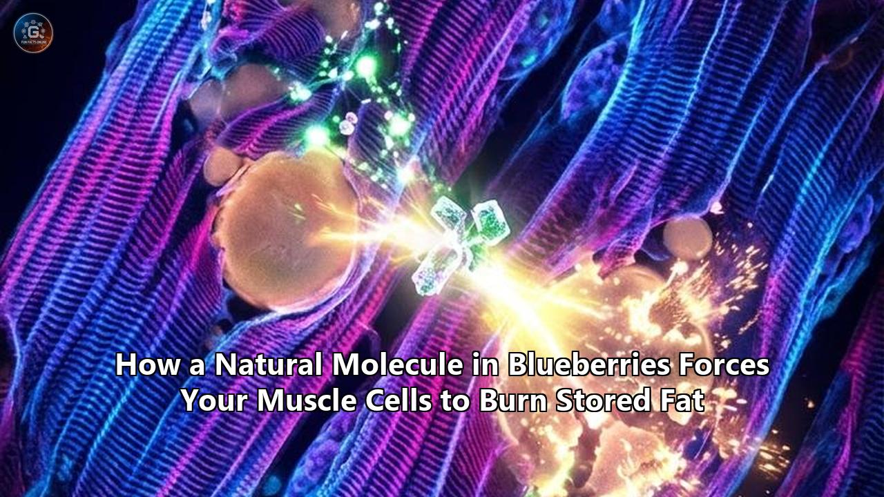
A molecular discovery from researchers at Shinshu University in Japan has pinpointed the precise cellular mechanism through which a specific compound found in blueberries forces skeletal muscle cells to dismantle stored intracellular fat. The study, led by Associate Professor Takakazu Mitani and published in peer-reviewed findings, reveals that pterostilbene—a naturally occurring methylated stilbenoid concentrated in berries—prevents the proteasomal degradation of Peroxisome Proliferator-Activated Receptor delta (PPARδ). By stabilizing this master nuclear receptor inside muscle cells, the compound accelerates lipid droplet clearance, upregulates fatty acid oxidation machinery, and triggers substantial lipolysis without disrupting normal muscle development.
                     +---------------------------------------+
                     | Pterostilbene (Blueberry Stilbenoid)  |
                     +-------------------+-------------------+
                                         |
                                         v
                     +---------------------------------------+
                     | Blocks 26S Proteasome Degradation     |
                     | of Nuclear Receptor PPARδ             |
                     +-------------------+-------------------+
                                         |
                                         v
                     +---------------------------------------+
                     | PPARδ Heterodimerizes with RXR        |
                     | Binds PPRE Genomic Response Elements  |
                     +-------------------+-------------------+
                                         |
          +------------------------------+------------------------------+
          |                                                             |
          v                                                             v
+-----------------------------+                               +-----------------------------+
| Intracellular Lipolysis     |                               | Mitochondrial Beta-Oxidation|
| • ATGL / HSL activation     |                               | • CPT1 / FAT/CD36 induction |
| • IMTG -> DAG -> Glycerol   |                               | • Acyl-CoA matrix transport |
| • Reduces toxic ceramides   |                               | • PDK4 / UCP3 upregulation  |
+-----------------------------+                               +-----------------------------+
          |                                                             |
          +------------------------------+------------------------------+
                                         |
                                         v
                     +---------------------------------------+
                     | Elevated Muscle Fat Oxidation &       |
                     | Restored Sarcolemmal GLUT4 Insertion  |
                     +---------------------------------------+
The Shinshu finding provides the missing mechanistic bridge for recent clinical data gathered across sports science and metabolic research. In human trials conducted at the Cal Poly Humboldt Human Performance Lab, researchers observed that daily intake of freeze-dried wild blueberry powder produced a 43.2% increase in fat oxidation rates during exercise, simultaneously decreasing reliance on carbohydrate reserves and lowering circulating lactate levels.
For years, dietary polyphenols were dismissed by mainstream pharmacology as generic, low-potency antioxidants whose biological effects were too diffuse to drive targeted clinical changes. The revelation that blueberry molecules operate as direct molecular stabilizers of genetic metabolic switches rewrites the understanding of nutritional bioactives, providing a non-synthetic blueprint for clearing muscle steatosis and enhancing systemic metabolic flexibility.
The Hidden Pathology: Ectopic Intramuscular Lipids and Metabolic Paralysis
To understand why stabilizing a single nuclear receptor matters, one must look at how the body mishandles energy when physical activity drops or caloric intake surges. When excess energy circulates through the bloodstream, adipose tissue eventually reaches its functional storage ceiling. Lipids then spill over into non-adipose organs—the liver, the heart, and skeletal muscle tissue. This ectopic storage creates intramyocellular lipids (IMCL), visible under electron microscopy as dense lipid droplets packed adjacent to the myofibrils and mitochondria.
Intramuscular fat is not inherently destructive; in elite endurance athletes, high IMCL levels serve as a readily available, harmless fuel source, a phenomenon known in exercise physiology as the "athlete’s paradox." In sedentary, aging, or metabolically impaired individuals, however, intramyocellular lipid pools become toxic.
Sedentary / Overfed State:
Unoxidized Fatty Acids ---> sn-1,2-Diacylglycerol (DAG) & Ceramides
                                         |
                                         v
                         Membrane Translocation of PKCθ / PKCε
                                         |
                                         v
                    Inhibitory Serine Phosphorylation of IRS-1
                                         |
                                         v
                        Blockade of PI3K / Akt Activation
                                         |
                                         v
                       Arrest of GLUT4 Translocation
                                         |
                                         v
                    Metabolic Inflexibility & Muscle Insulin Resistance
When muscle cells fail to balance lipid influx with mitochondrial combustion, triacylglycerols break down incompletely into reactive lipid intermediates, most notably:
- sn-1,2-diacylglycerols (DAGs)
- Ceramides
- Long-chain acyl-CoAs
These intermediates act as destructive signaling molecules. DAG accumulation recruits novel isoforms of protein kinase C, specifically PKCθ and PKCε, from the cytosol to the plasma membrane. Once anchored to the membrane, PKCθ phosphorylates Insulin Receptor Substrate 1 (IRS-1) on inhibitory serine residues (such as Ser307 and Ser1101) instead of allowing normal tyrosine phosphorylation.
This aberrant phosphorylation shuts down downstream phosphatidylinositol 3-kinase (PI3K) and protein kinase B (Akt) signaling. As a result, glucose transporter type 4 (GLUT4) storage vesicles remain locked within the intracellular matrix, unable to fuse with the sarcolemma.
The outcome is muscle insulin resistance and metabolic inflexibility. Skeletal muscle accounts for approximately 70% to 80% of all postprandial glucose disposal in the human body. When intramyocellular lipotoxicity paralyzes GLUT4 translocation, blood glucose surges, pancreatic beta cells face compensatory stress, and the muscle cell loses its ability to switch between carbohydrates and fatty acids for fuel.
Restoring metabolic health requires clearing these toxic lipid intermediates from inside the muscle fiber. Until recently, sustained low-intensity exercise was considered the primary means of clearing intramyocellular lipids; the Shinshu University data proves that specific bioactive molecules from blueberries force muscle cells to burn these stores directly.
Molecular Architecture: Why Pterostilbene Outperforms Resveratrol
The primary compound at the center of the Shinshu University breakthrough is pterostilbene (trans-3,5-dimethoxy-4'-hydroxystilbene). Belonging to the stilbenoid class of natural phytoalexins, pterostilbene shares an underlying chemical backbone with resveratrol (trans-3,5,4'-trihydroxystilbene), a molecule that received widespread scientific attention for its interactions with sirtuins and longevity pathways. Resveratrol’s therapeutic application, however, has consistently hit an insurmountable biological barrier: low systemic bioavailability.
       Resveratrol (3,5,4'-trihydroxy)          Pterostilbene (3,5-dimethoxy-4'-hydroxy)
              OH                                           OCH3
             /                                            /
      HO--< O >--CH=CH--< O >--OH                 H3CO--< O >--CH=CH--< O >--OH

      • 3 Hydroxyl (-OH) Groups                    • 2 Methoxy (-OCH3) + 1 Hydroxyl (-OH)
      • Rapid Phase II Glucuronidation             • High Resistance to First-Pass Metabolism
      • Bioavailability: < 1-2%                    • Bioavailability: ~80%
      • Lipophilicity: LogP ~3.1                   • Lipophilicity: LogP ~3.7 (Cell-Permeable)
Resveratrol contains three hydroxyl (-OH) functional groups. When ingested, intestinal enterocytes and hepatic phase II enzymes recognize these exposed hydroxyl sites, rapidly conjugating them via glucuronidation and sulfation into inactive metabolites. Less than 1% to 2% of ingested resveratrol ever reaches peripheral circulation in its intact, biologically active aglycone form.
Pterostilbene circumvents this pharmacodynamic trap through a structural evolution:
Structural Methylation
In pterostilbene, two of the hydroxyl groups (at carbon positions 3 and 5 of the A-ring) are substituted with methoxy (-OCH3) groups. Only a single hydroxyl group remains exposed at position 4' on the B-ring.
Resistance to Conjugation
The methyl capping of the 3- and 5-carbon positions sterically blocks the access of UDP-glucuronosyltransferases (UGTs) and sulfotransferases (SULTs). As a consequence, pterostilbene slips past first-pass hepatic metabolism with a substantial portion of its parent molecule intact.
Enhanced Lipophilicity and Membrane Permeation
The two methoxy groups lower polarity and increase the compound's octanol-water partition coefficient (logP of ~3.7 for pterostilbene versus ~3.1 for resveratrol). This elevated lipophilicity allows pterostilbene to pass directly through the lipid bilayer of skeletal muscle cells without requiring active peptide transporter channels.
Extended Plasma Half-Life
Pharmacokinetic tracking in mammalian models demonstrates that pterostilbene achieves an oral bioavailability approaching 80%, exhibiting a biological half-life nearly seven times longer than that of non-methylated stilbenes.
Because of this prolonged half-life, when someone consumes wild blueberries, pterostilbene achieves biologically relevant micromolar concentrations in the deep interstitial fluid bathing skeletal myofibers.
The Shinshu Breakthrough: The Ubiquitin-Proteasome Bypass of PPARδ
The critical advance from the Shinshu University investigation, led by Takakazu Mitani, lies in identifying how pterostilbene controls Peroxisome Proliferator-Activated Receptor delta (PPARδ).
                                  +-----------------------------+
                                  | Native PPARδ Protein Pool   |
                                  +--------------+--------------+
                                                 |
                       +-------------------------+-------------------------+
                       |                                                   |
       [Sedentary / Unstimulated]                                [Pterostilbene Present]
                       |                                                   |
                       v                                                   v
         E3 Ubiquitin Ligase Attachment                          Allosteric Stabilization /
                       |                                         Blockade of Ubiquitin Sites
                       v                                                   |
           Polyubiquitination Chain                                        v
                       |                                            Protection Against
                       v                                          Proteasomal Recognition
         26S Proteasome Degradation                                        |
                       |                                                   v
                       v                                       +-----------------------+
          +------------------------+                           | Prolonged Intranuclear|
          | Depleted PPARδ Levels  |                           | Receptor Half-Life    |
          | Arrested Fat Oxidation |                           +-----------+-----------+
          +------------------------+                                       |
                                                                           v
                                                               Direct Transcription of:
                                                               • CPT1A / CPT1B
                                                               • PDK4
                                                               • FAT/CD36
                                                               • ACO
PPARδ is a ligand-activated transcription factor belonging to the nuclear receptor superfamily. Unlike its sister isoforms—PPARα, which operates predominantly in the liver to control systemic ketogenesis, and PPARγ, which directs adipogenesis in adipose tissue—PPARδ is the dominant isoform expressed in mammalian skeletal muscle, present at concentrations up to 50 times higher than PPARα.
When activated, PPARδ drives the transcription of genes responsible for fatty acid import, transport, and mitochondrial beta-oxidation. Under basal conditions, however, the muscle cell strictly limits PPARδ activity. The receptor has a rapid turnover rate; it is continuously tagged with ubiquitin molecules by specific E3 ubiquitin ligases and ferried to the 26S proteasome for proteolytic destruction.
This rapid proteasomal clearance acts as a physiological brake, preventing the skeletal muscle from combusting lipids uncontrollably when rapid glycolytic energy is required.
Mitani’s team exposed cultured C2C12 skeletal muscle cells to high lipid loads to replicate the intracellular lipid clogging observed in metabolic disorders. They then screened multiple dietary compounds to observe their effects on lipid droplet clearance. Pterostilbene emerged as the most potent agent, clearing intracellular fat accumulations without reducing cell viability or halting differentiation.
Through Western blotting, immunoprecipitation, and pulse-chase degradation assays, the researchers mapped the underlying mechanism:
- Non-Competitive Structural Protection: Rather than serving merely as an agonist that transiently fits into the ligand-binding pocket of PPARδ, pterostilbene induces conformational stability across the protein's tertiary structure.
- Ubiquitination Blockade: This structural stabilization prevents E3 ubiquitin ligases from attaching polyubiquitin chains to target lysine residues on the PPARδ protein.
- Proteasome Shielding: Shielded from the 26S proteasome, the half-life of intact PPARδ inside the nucleus increases significantly, allowing the basal pool of the receptor to remain functional for extended periods.
- Enhanced Gene Transcription: The stabilized PPARδ binds with high affinity to its obligate partner, the Retinoid X Receptor (RXR). The resulting heterodimer binds directly to Peroxisome Proliferator Response Elements (PPRE) across the genome.
- Direct Lipolysis: The team confirmed that this was not a matter of blocking fatty acid entry into the muscle cell; extracellular glycerol levels surged in the culture media. Because glycerol is the chemical backbone of triacylglycerols released only when intracellular lipid droplets are cleaved, the elevated glycerol proved that the muscle cells were actively breaking down their own stored fat stores.
The Enzymatic Engine: The CPT-1 Gate and Beta-Oxidation Spiral
Once the pterostilbene-stabilized PPARδ-RXR complex occupies the PPRE consensus sequences in skeletal muscle DNA, it initiates a coordinated transcriptional cascade. This program systematically upregulates every major rate-limiting enzyme required for cellular fat disposal.
EXTRACELLULAR SPACE / SARCOLEMMA
  Fatty Acids  ===[ FAT/CD36 Translocase ]===>  Cytosolic Long-Chain Acyl-CoA
                                                          |
OUTER MITOCHONDRIAL MEMBRANE                              |
  Acyl-CoA + Carnitine ===[ CPT1 (Uninhibited) ]===> Acylcarnitine + CoASH
                                                          |
INTERMEMBRANE SPACE                                       |
                                             [ CACT Translocase ]
                                                          |
INNER MITOCHONDRIAL MEMBRANE                              |
  Acylcarnitine + CoASH ===[ CPT2 Enzyme ]=========> Matrix Acyl-CoA + Carnitine
                                                          |
MITOCHONDRIAL MATRIX                                      |
  +-------------------------------------------------------+
  |
  +---> [ BETA-OXIDATION SPIRAL ]
        1. VLCAD / LCAD (Acyl-CoA Dehydrogenase)   ---> Generates FADH2
        2. EHHADH (Enoyl-CoA Hydratase)
        3. HADHA (3-Hydroxyacyl-CoA Dehydrogenase) ---> Generates NADH
        4. ACAA2 (Beta-Ketothiolase)
        |
        v
  Cleaves 2-Carbon Units into Acetyl-CoA ===> Enters Citric Acid Cycle / ATP Production
1. Sarcolemmal Fatty Acid Uptake: FAT/CD36
PPARδ upregulates Fatty Acid Translocase (FAT/CD36). This glycoprotein localizes to the muscle cell membrane, intercepting circulating non-esterified fatty acids (NEFAs) from the bloodstream and transporting them across the sarcolemma into the cytoplasm.
2. The Mitochondrial Gatekeeper: Carnitine Palmitoyltransferase-1 (CPT-1)
Once inside the cytosol, fatty acids are activated by long-chain acyl-CoA synthetases into acyl-CoA molecules. Long-chain acyl-CoAs, however, cannot pass through the impermeable inner mitochondrial membrane on their own. They must pass through the carnitine shuttle, where Carnitine Palmitoyltransferase-1 (CPT1A/CPT1B) is the absolute rate-limiting gatekeeper.
CPT-1 sits anchored in the outer mitochondrial membrane, where it strips the CoA group from the fatty acid and attaches a carnitine molecule, forming acylcarnitine. The acylcarnitine is shuttled across the inner membrane by Carnitine-Acylcarnitine Translocase (CACT), after which Carnitine Palmitoyltransferase-2 (CPT2) reconstitutes the fatty acyl-CoA inside the mitochondrial matrix, recycling the carnitine back to the cytosol.
Under normal conditions, CPT-1 is strongly inhibited by malonyl-CoA, a metabolic intermediate formed during carbohydrate surplus. The gene transcription driven by stabilized PPARδ floods the mitochondrial membrane with newly synthesized CPT-1 enzymes, overwhelming baseline malonyl-CoA inhibition and opening the transport gate for fatty acids to enter the mitochondrial core.
3. The Beta-Oxidation Spiral
Inside the mitochondrial matrix, the fatty acyl-CoA enters the beta-oxidation spiral, a four-step enzymatic pathway:
- Dehydrogenation: Very-long-chain or long-chain acyl-CoA dehydrogenases (VLCAD/LCAD) oxidize the acyl-CoA, transferring electrons to flavin adenine dinucleotide to create $\text{FADH}_2$.
- Hydration: Enoyl-CoA hydratase adds a water molecule across the newly formed double bond.
- Oxidation: 3-hydroxyacyl-CoA dehydrogenase oxidizes the hydroxyl group to a keto group, reducing $\text{NAD}^+$ to $\text{NADH}$.
- Thiolysis: Beta-ketothiolase cleaves off a terminal two-carbon fragment in the presence of free Coenzyme A, yielding one molecule of Acetyl-CoA and a fatty acyl-CoA chain shortened by two carbons.
This cycle repeats until the entire fatty acid chain is broken down. The resulting Acetyl-CoA enters the tricarboxylic acid (TCA) cycle, while the accumulated $\text{FADH}_2$ and $\text{NADH}$ deliver high-energy electrons directly to Complexes I and II of the electron transport chain, generating ATP through oxidative phosphorylation.
4. Pyruvate Dehydrogenase Kinase 4 (PDK4) Induction
Simultaneously, PPARδ transcriptionally activates PDK4. This enzyme phosphorylates and inactivates the Pyruvate Dehydrogenase Complex (PDC), the primary gateway through which carbohydrate-derived pyruvate enters the mitochondria.
By deactivating the PDC, the muscle cell shuts down glucose combustion, prioritizing fatty acids as its primary substrate.
The Anthocyanin Synergy: The AMPK/Malonyl-CoA Axis
While pterostilbene acts via the nuclear PPARδ pathway, it does not operate in isolation inside the blueberry matrix. Blueberries are among the densest dietary sources of anthocyanins—water-soluble flavonoid pigments that include cyanidin, delphinidin, malvidin, peonidin, and petunidin glycosides.
Research shows that these anthocyanins work through an independent, complementary mechanism: the 5'-AMP-activated protein kinase (AMPK) cascade.
                     +---------------------------------------+
                     | Blueberry Anthocyanins & Metabolites  |
                     | (Cyanidin-3-Glucoside, Protocatechuate)|
                     +-------------------+-------------------+
                                         |
                                         v
                     +---------------------------------------+
                     | Activation of Upstream Kinases        |
                     | (LKB1 & CaMKKβ)                       |
                     +-------------------+-------------------+
                                         |
                                         v
                     +---------------------------------------+
                     | Phosphorylation of AMPK at Thr172      |
                     +-------------------+-------------------+
                                         |
                                         v
                     +---------------------------------------+
                     | Phosphorylation of ACC1 / ACC2        |
                     | at Ser79 (INACTIVATION)               |
                     +-------------------+-------------------+
                                         |
                                         v
                     +---------------------------------------+
                     | Sharp Depletion of Malonyl-CoA Pool   |
                     +-------------------+-------------------+
                                         |
                                         v
                     +---------------------------------------+
                     | Relief of Allosteric Inhibition       |
                     | on CPT-1 Transporter                  |
                     +-------------------+-------------------+
                                         |
                                         v
                     +---------------------------------------+
                     | Accelerated Mitochondrial Influx      |
                     | of Long-Chain Fatty Acids             |
                     +---------------------------------------+
AMPK is the master cellular energy gauge, responding to shifts in the AMP-to-ATP and ADP-to-ATP ratios. When energy stress occurs, AMPK is phosphorylated at its Thr172 activation loop by upstream kinases, primarily Liver Kinase B1 (LKB1) and Calcium/Calmodulin-Dependent Protein Kinase Kinase Beta (CaMKKβ).
Anthocyanin metabolites, particularly cyanidin-3-glucoside (C3G) and its primary gut breakdown product, protocatechuic acid (PCA), trigger this phosphorylation event without requiring severe cellular energy depletion.
Once activated, phosphorylated AMPK targets Acetyl-CoA Carboxylase (ACC), specifically the ACC1 and ACC2 isoforms located near the outer mitochondrial membrane.
- ACC normally synthesizes malonyl-CoA from acetyl-CoA.
- Phosphorylation by AMPK at serine residue 79 (Ser79) completely inactivates ACC.
- This enzymatic shutdown rapidly depletes the cellular pool of malonyl-CoA.
Because malonyl-CoA is the primary allosteric inhibitor of CPT-1, its depletion removes the physical barrier that normally limits fatty acid transport into the mitochondria.
When examining whole-food interactions, this dual mechanism explains the effectiveness of blueberries: pterostilbene stabilizes PPARδ to increase the total number of CPT-1 and beta-oxidation enzymes, while anthocyanins activate AMPK to remove malonyl-CoA inhibition from those same enzymes.
The result is a coordinated biochemical drive that channels cytosolic lipids directly into the mitochondrial combustion pathway.
Clinical Validation: Inside the Human Performance Laboratory
The clinical application of these cellular mechanisms was confirmed in a controlled human trial conducted by Dr. Taylor Bloedon and her research team at Cal Poly Humboldt, published in the journal Nutrients.
While previous investigations had focused on sedentary or rodent models, the Cal Poly Humboldt trial examined how dietary blueberry supplementation alters real-time substrate oxidation kinetics during exercise in healthy, aerobically trained subjects.
FAT OXIDATION SURGE (Cal Poly Humboldt Human Trial Data)
  Baseline (Control) vs. 14-Day Wild Blueberry Powder (25g/day = 1 Cup Fresh Equivalent)

  Fat Oxidation Rate Increases:
  Minute 20 of Steady-State Cycling:  [+19.7% Increase]
  Minute 30 of Steady-State Cycling:  [+43.2% Increase]  <--- Peak Metabolic Shift
  Minute 40 of Steady-State Cycling:  [+31.1% Increase]

  Carbohydrate Oxidation Reductions:
  Minute 20 of Steady-State Cycling:  [-10.1% Decrease]
  Minute 30 of Steady-State Cycling:  [-19.2% Decrease]
  Minute 40 of Steady-State Cycling:  [-14.8% Decrease]

  Blood Lactate Accumulation:
  Minute 20: 2.6 mmol/L (WB) vs 3.0 mmol/L (Control)
  Minute 30: 2.2 mmol/L (WB) vs 2.9 mmol/L (Control)  [-24.1% Lower Lactate Accumulation]
  Minute 40: 1.9 mmol/L (WB) vs 2.5 mmol/L (Control)  [-24.0% Lower Lactate Accumulation]
Protocol Design
The trial enrolled aerobically trained male athletes ($26.0 \pm 7.5$ years of age) to eliminate the confounding variable of sedentary deconditioning. Participants completed an initial two-week nutritional washout period, strictly avoiding all exogenous dietary sources of anthocyanins and stilbenes.
Following baseline testing, participants consumed 25 grams of freeze-dried wild blueberry powder daily for 14 consecutive days—a dose equivalent to approximately one cup of fresh, raw wild blueberries containing roughly 375 mg of total anthocyanins.
Substrate oxidation rates were quantified using breath-by-breath indirect calorimetry inside the Human Performance Lab during a 40-minute stationary cycling protocol fixed at a moderate intensity of $65\%\ \text{VO}_{2\text{peak}}$. Serial blood draws and respiratory exchange ratio (RER) metrics were tracked at continuous 10-minute intervals.
Respiratory Exchange Ratio (RER) Comparison:
RER = VCO2 / VO2
• Pure Carbohydrate Oxidation: RER = 1.00
• Pure Fatty Acid Oxidation:   RER = 0.70

Control Protocol Mean RER:       ~0.88 - 0.91 (Predominantly Glycolytic)
Blueberry Intervention Mean RER: ~0.82 - 0.84 (Substantial Shift Toward Lipid Combustion)
The physiological results demonstrated a clear shift in whole-body bioenergetics:
- Surge in Fat Combustion: In the control condition, fat oxidation rates plateaued and then declined as glycogen pathways took over. Following blueberry supplementation, whole-body fat oxidation rates increased by 19.7% at 20 minutes, peaked at a 43.2% increase at 30 minutes, and remained elevated at 31.1% at 40 minutes.
- Carbohydrate Sparing: Concurrently, carbohydrate oxidation rates dropped significantly: down 10.1% at 20 minutes, 19.2% at 30 minutes, and 14.8% at 40 minutes.
- Lactate Blunting: Blood lactate accumulation dropped from $2.9\ \text{mmol/L}$ to $2.2\ \text{mmol/L}$ at 30 minutes and from $2.5\ \text{mmol/L}$ to $1.9\ \text{mmol/L}$ at 40 minutes. Lower lactate accumulation at identical power outputs confirms that mitochondrial oxidative phosphorylation was handling a larger share of the ATP generation workload, reducing the need for anaerobic glycolysis.
This metabolic profile highlights a key advantage of the intervention. Under standard nutritional protocols, increasing the body's rate of lipid oxidation requires weeks of extreme carbohydrate restriction (such as ketogenic adaptation) or prolonged fasting, both of which can compromise high-intensity exercise capacity and cause thyroid or hormonal down-regulation.
The Cal Poly Humboldt data demonstrated that introducing wild blueberries fat burning signaling molecules induced a substantial shift toward lipid utilization during steady-state exertion while preserving carbohydrate reserves.
The Shadow Industry: Synthetic Agonists, Cardarine, and the Safety Pivot
The discovery that natural dietary molecules can stabilize PPARδ addresses a major pharmacological challenge that has persisted for more than two decades.
+------------------------------------+------------------------------------+
| Synthetic Super-Agonists (GW501516)| Natural Stilbenes (Pterostilbene)  |
+------------------------------------+------------------------------------+
| • Supramaximal affinity (Kd ~1 nM) | • Modulates protein turnover via   |
| • Hyper-activates transcription    |   proteasomal stabilization        |
| • Bypasses endogenous cellular     | • Retains normal physiological     |
|   checkpoint controls              |   transcription limits             |
| • Triggered rapid multi-organ      | • Extensively studied safe food    |
|   carcinogenesis in animal models  |   compound with zero oncogenic     |
| • Abandoned by pharma; black-      |   toxicity flags across human and  |
|   market athletic doping risk      |   animal literature                |
+------------------------------------+------------------------------------+
In the early 2000s, pharmaceutical companies identified PPARδ as a target for treating dyslipidemia, obesity, and type 2 diabetes. GlaxoSmithKline (GSK), in partnership with Ligand Pharmaceuticals, developed GW501516 (known as Cardarine or Endurobol).
GW501516 is a synthetic, high-affinity ligand designed to bind directly into the ligand-binding pocket of PPARδ with an equilibrium dissociation constant ($\text{K}_d$) in the sub-nanomolar range.
The compound produced striking physiological changes. Sedentary rodents given GW501516 doubled their running endurance, remodeled their fast-twitch glycolytic muscle fibers into oxidative slow-twitch fibers, and rapidly burned visceral and intramyocellular fat without requiring physical training. It quickly became one of the most widely used performance-enhancing drugs on the athletic black market, leading to a total ban by the World Anti-Doping Agency (WADA) in 2009.
Behind the scenes, however, pharmaceutical development had hit a fatal obstacle:
Pharmaceutical Development Timeline:
2000-2006: GSK advances GW501516 through Phase I and Phase II metabolic trials.
2006-2007: Long-term rodent safety studies reveal rapid, multi-organ neoplasia.
           Tumors developed across the liver, thyroid, stomach, tongue, and colon.
2007:      GSK halts all human clinical trials and completely abandons the molecule.
2009:      WADA adds GW501516 to the prohibited list, issuing warnings about toxicity.
The failure of GW501516 stemmed from its pharmacological mechanism: supramaximal, unrelenting ligand activation. By forcibly locking the PPARδ receptor into an active state across all tissues, synthetic super-agonists trigger uncontrolled cell-cycle proliferation, driving oncogenesis in vulnerable epithelial tissues.
This history underscores the significance of the Shinshu University findings on pterostilbene. Pterostilbene does not act as a high-affinity synthetic agonist that overrides natural transcriptional controls. Instead, it functions as a proteasomal stabilizer that protects existing PPARδ pools within skeletal muscle from premature degradation.
By prolonging the receptor's active half-life without triggering excessive transcription, it supports healthy fat oxidation while operating safely within the body's natural regulatory boundaries.
The Anthocyanin Bioavailability Paradox and the Gut Microbiome
A central point of discussion among nutritional biochemists is the "Anthocyanin Bioavailability Paradox." If you analyze human blood plasma two hours after someone eats a bowl of fresh blueberries, intact anthocyanin molecules are present in trace amounts, reflecting a systemic bioavailability of roughly 0.1% to 1.5%.
Historically, this led critics to argue that in vitro cellular findings could never translate to living human physiology.
Modern metabolomics has resolved this question by tracking the biotransformation of these compounds through the gastrointestinal tract:
INGESTION: Whole Blueberries (Anthocyanin Glycosides: Delphinidin, Malvidin, Cyanidin)
                               |
                               v
STOMACH & PROXIMAL JEJUNUM: Low Absorption (~1-2% via SGLT1 / GLUT2 enterocyte transport)
                               |
                               v
DISTAL ILEUM & COLONIC MICROBIOME: Extensive Microbial Catabolism
  • Microbes: Bifidobacterium, Akkermansia muciniphila, Faecalibacterium prausnitzii
  • Cleavage of sugar moieties via microbial beta-glucosidases
  • Ring fission of the flavonoid C-ring
                               |
                               v
GENERATION OF BIOACTIVE PHENOLIC METABOLITES:
  1. Protocatechuic Acid (PCA)
  2. Gallic Acid
  3. Vanillic Acid
  4. Hippuric Acid & Hydroxyhippuric Acids
  5. 4-Hydroxybenzoic Acid
                               |
                               v
COLONIC ENTEROCYTE ABSORPTION & ENTEROHEPATIC CIRCULATION
  • Peak plasma concentrations achieved at 4 to 24 hours post-ingestion
  • Plasma concentrations of bioactive metabolites reach 100x to 1000x that of parent molecules
  • Circulate systemically to penetrate peripheral skeletal muscle tissue
The gut microbiome converts high-molecular-weight parent anthocyanins into biologically active phenolic acids. While parent anthocyanins (such as cyanidin-3-glucoside) are largely cleared within two hours, their microbial breakdown products—such as protocatechuic acid (PCA) and hippuric acid—achieve micromolar concentrations in the bloodstream between 4 and 24 hours after ingestion.
These low-molecular-weight metabolites cross the sarcolemma of skeletal muscle cells, where they:
- Activate LKB1 to stimulate AMPK phosphorylation.
- Act as free-radical scavengers at the mitochondrial outer membrane, neutralizing reactive oxygen species (ROS) produced by electron leak during accelerated beta-oxidation.
- Promote the growth of beneficial gut bacteria (including Akkermansia muciniphila and Faecalibacterium prausnitzii), which produce short-chain fatty acids (SCFAs) like acetate, propionate, and butyrate.
These circulating SCFAs enter systemic circulation, bind to free fatty acid receptors (FFAR2/GPR43) on muscle cells, and enhance insulin sensitivity and mitochondrial biogenesis.
Nutritional Engineering: Cultivar Chemistry, Dosing Thresholds, and Interference
Translating this biochemistry into everyday practice requires understanding that not all blueberries share the same molecular profile. The concentration of pterostilbene and anthocyanins varies widely across different species, agricultural growing conditions, and post-harvest processing methods.
+------------------------------------+------------------------------------+
| Cultivated Highbush Blueberries    | Lowbush Wild Blueberries           |
| (Vaccinium corymbosum)             | (Vaccinium angustifolium)          |
+------------------------------------+------------------------------------+
| • Larger berry size                | • Smaller berry size               |
| • Lower skin-to-pulp ratio         | • High skin-to-pulp surface ratio  |
| • Moderate anthocyanin density     | • Up to 2x-3x higher anthocyanins  |
| • Lower concentrations of          | • Significantly higher levels of   |
|   pterostilbene and chlorogenic    |   pterostilbene and complex        |
|   acid                             |   polyphenols                      |
+------------------------------------+------------------------------------+
Cultivar Differences: Wild vs. Cultivated
Wild lowbush blueberries (Vaccinium angustifolium) grow in challenging, high-stress sub-arctic and boreal climates, such as those found in Maine and Eastern Canada. To protect their tissues from cold shock and intense UV exposure, wild lowbush berries produce significantly higher concentrations of defensive polyphenols than commercial cultivated highbush berries (Vaccinium corymbosum).
Because anthocyanins and pterostilbene are concentrated in the skin rather than the watery pulp, the higher skin-to-volume ratio of wild blueberries delivers a far greater payload of bioactive compounds per gram.
Effective Dosing Protocols
The clinical data from Cal Poly Humboldt and related metabolic trials establish a clear minimum threshold for activating these lipid-clearing pathways:
- Freeze-Dried Wild Blueberry Powder: 25 grams daily (yielding $\sim 375\ \text{mg}$ of total anthocyanins alongside natural pterostilbene fractions).
- Fresh or Frozen Whole Wild Blueberries: Approximately 1 cup (150 grams) daily.
- Duration: The structural stabilization of PPARδ and subsequent gene transcription require an accumulation period of 7 to 14 consecutive days to establish elevated baseline fat oxidation rates.
                +-----------------------------------------------+
                | Daily Wild Blueberry Intake (1 Cup / 25g Pwd) |
                +-----------------------+-----------------------+
                                        |
                 [ Avoid Dairy Matrix for 60-90 Minutes ]
                                        |
                                        v
  +-------------------------------------+-------------------------------------+
  |                                                                           |
  v                                                                           v
+-----------------------------------+                       +-----------------------------------+
| Pterostilbene Molecule            |                       | Anthocyanins & Metabolites        |
| • Crosses sarcolemma directly     |                       | • Catabolized by gut microbiome   |
| • Shields PPARδ from 26S          |                       | • Phosphorylates AMPK via LKB1    |
|   proteasome degradation          |                       | • Inactivates ACC (lowers         |
| • Increases CPT1 & PDK4 genes     |                       |   malonyl-CoA barrier)            |
+-----------------+-----------------+                       +-----------------+-----------------+
                  |                                                           |
                  +-----------------------------+-----------------------------+
                                                |
                                                v
                              +-----------------------------------+
                              | Sustained Intramyocellular        |
                              | Lipid Clearance & Muscle Fat      |
                              | Combustion                        |
                              +-----------------------------------+
The Matrix Interference Problem: Dairy Proteins
A common dietary mistake is consuming blueberries alongside milk, yogurt, or whey protein. Dairy proteins—specifically alpha-casein and beta-casein—contain hydrophobic proline-rich domains that rapidly bind to polyphenolic rings.
This creates insoluble polyphenol-protein complexes that resist enzymatic cleavage in the small intestine, preventing the absorption of anthocyanins and stilbenes.
To ensure optimal uptake and fully support the muscle-level signaling that drives blueberries fat burning mechanisms, blueberries or blueberry extracts should be consumed with water or plant-based matrices, keeping a 60-to-90-minute window away from intact casein-containing dairy products.
Metabolic Impact and Emerging Research Frontiers
The discovery of the pterostilbene-PPARδ stabilization pathway opens several key areas for metabolic and clinical research:
                            EMERGING RESEARCH FRONTIERS
                                         |
     +-----------------------------------+-----------------------------------+
     |                                   |                                   |
     v                                   v                                   v
+-----------------------+   +-----------------------+   +-----------------------+
| Sarcopenic Obesity &  |   | GLP-1 Agonist Adjunct |   | Epigenetic Remodeling |
| Healthy Aging Trials  |   | Co-Therapy Protocols  |   | of Muscle Chromatin   |
|                       |   |                       |   |                       |
| • Reverses muscle     |   | • Mitigates loss of   |   | • Investigating how   |
|   steatosis           |   |   lean mass during    |   |   PPARδ stabilization |
| • Restores baseline   |   |   rapid weight loss   |   |   alters long-term    |
|   insulin sensitivity |   | • Clears residual     |   |   histone acetylation |
|   in older adults     |   |   intramuscular fat   |   |   and DNA methylation |
+-----------------------+   +-----------------------+   +-----------------------+
1. Sarcopenic Obesity and Healthy Aging
As the body ages, skeletal muscle mass declines while intramuscular fat deposition increases. This combination—sarcopenic obesity—is a primary cause of physical frailty and metabolic decline in older adults.
Clinical teams are now testing whether daily wild blueberry supplementation can safely clear intramuscular fat, improve muscle quality, and restore insulin sensitivity in older populations without relying on high-volume, strenuous exercise.
2. Combination Therapies Alongside GLP-1 Receptor Agonists
With the widespread clinical adoption of GLP-1 receptor agonists (such as semaglutide and tirzepatide), clinicians have noted that rapid weight loss is often accompanied by a loss of lean muscle mass alongside adipose tissue.
Research programs are beginning to evaluate whether combining GLP-1 therapy with pterostilbene-rich whole-food matrices can protect muscle metabolic health, directing fuel consumption toward fat oxidation within remaining myofibers and preserving overall metabolic rate.
3. Epigenetic Remodeling and Chromatin Accessibility
Beyond immediate transcription, researchers are investigating whether sustained PPARδ stabilization modifies the muscle cell's long-term epigenetic landscape.
By recruiting histone acetyltransferases (HATs) and clearing corepressor complexes from chromatin, regular exposure to blueberry stilbenoids may permanently enhance the accessibility of mitochondrial promoter regions, reinforcing long-term metabolic flexibility.
Key Takeaways
The findings from Shinshu University and recent clinical trials clarify how wild blueberries support metabolic health:
- Direct Molecular Target: Pterostilbene, a naturally methylated stilbene in blueberries, prevents the 26S proteasome from degrading PPARδ, stabilizing this master nuclear receptor inside skeletal muscle cells.
- Lipid Clearance Mechanism: Stabilized PPARδ upregulates key metabolic genes—including CPT-1, PDK4, and FAT/CD36—driving intracellular lipolysis and burning intramyocellular fat stores.
- Complementary Synergies: Dietary anthocyanins and their gut-derived metabolites activate AMPK, inactivating ACC and depleting malonyl-CoA to eliminate the physiological barrier to mitochondrial fat entry.
- Demonstrated Clinical Outcomes: Human trials confirm that consuming the equivalent of one cup of wild blueberries daily yields significant increases in fat oxidation (up to 43.2%) during physical activity, accompanied by lower blood lactate accumulation and carbohydrate sparing.
- A Safe Alternative to Synthetic Agonists: Unlike high-potency synthetic agonists (such as GW501516) that were abandoned due to severe toxicity, the natural stabilization of PPARδ by food-derived molecules operates safely within endogenous cellular limits.
These discoveries demonstrate that specific molecules in blueberries function as targeted metabolic modulators, showing how nutritional bioactives can directly influence cellular machinery to clear stored fats and support metabolic resilience.
Reference:
- https://www.sciencedaily.com/releases/2026/09/260903064219.htm
- https://www.eurekalert.org/news-releases/1142364
- https://www.researchgate.net/publication/23144151_AMPK_and_PPARdelta_agonists_are_exercise_mimetics
- https://www.news-medical.net/news/20230327/Wild-blueberries-may-help-accelerate-fat-oxidation-during-exercise.aspx
- https://pmc.ncbi.nlm.nih.gov/articles/PMC10058338/
- https://www.researchgate.net/publication/8444519_Resveratrol_Pterostilbene_and_Piceatannol_in_Vaccinium_Berries
- https://www.jayfeldmanwellness.com/ep-64-the-true-cause-of-fatty-liver-nafld-part-2/
- https://www.researchgate.net/publication/41422925_Purified_Blueberry_Anthocyanins_and_Blueberry_Juice_Alter_Development_of_Obesity_in_Mice_Fed_an_Obesogenic_High-Fat_Diet
- https://www.mdpi.com/2076-3921/15/1/61
- https://digitalcommons.humboldt.edu/cgi/viewcontent.cgi?article=1311&context=etd
- https://pmc.ncbi.nlm.nih.gov/articles/PMC7699863/
- https://www.mdpi.com/2673-8430/2/2/19
- https://pmc.ncbi.nlm.nih.gov/articles/PMC11817741/
- https://www.frontiersin.org/journals/pharmacology/articles/10.3389/fphar.2020.01300/full
- https://www.mdpi.com/2072-6643/17/20/3303