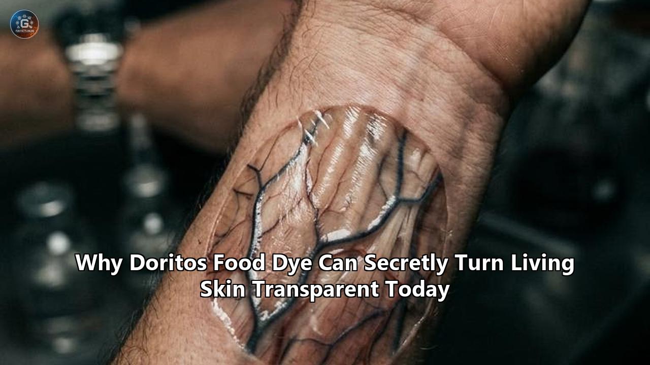
On September 6, 2024, a team of biophysicists and materials scientists at Stanford University published experimental results in Science showing that biological tissue can be rendered optically transparent in vivo using an ordinary certified food additive. By topically applying a 0.6-molar aqueous solution of tartrazine—the synthetic azo dye known as FD&C Yellow No. 5, commonly found in processed snack foods like Doritos, mountain citrus sodas, and confectionery—the researchers altered the optical properties of living rodent tissue within minutes.
The optical transformation allowed unscattered photons in the red and near-infrared spectrum to penetrate living mammalian skin, exposing dynamic physiological processes that were previously obscured. Within five to ten minutes of application, the opaque abdominal walls of living mice became transparent, revealing mesenteric blood vessels, liver lobes, and the rhythmic contractions of the gastrointestinal tract at 30 frames per second.
The physics behind this process relies on the Kramers-Kronig relations, a mathematical framework in classical electrodynamics linking optical absorption to refractive index dispersion. By introducing an absorbing molecule with a high molar extinction coefficient in the near-ultraviolet and blue spectrum ($\approx 428\text{ nm}$), the researchers altered the real part of the refractive index of water, shifting it from its baseline value of $1.333$ up to approximately $1.425$–$1.440$ in the red and near-infrared wavelengths ($600\text{ to }850\text{ nm}$). This shift matched the refractive index of aqueous cytosol and interstitial fluid to that of high-index structural tissue components, namely lipids ($n \approx 1.44$) and proteins ($n \approx 1.40\text{ to }1.53$).
Baseline Tissue (Opaque):
  Interstitial Water (n ≈ 1.333) ≠ Lipids/Proteins (n ≈ 1.440–1.500)
  Δn ≈ 0.110–0.170  ──>  Severe Mie/Rayleigh Scattering  ──>  Opacity

Tartrazine-Treated Tissue (Transparent in Red/NIR):
  Tartrazine-Doped Water (n ≈ 1.425–1.440) ≈ Lipids/Proteins (n ≈ 1.440–1.500)
  Δn ≤ 0.015  ──>  Scattering Suppression (>90%)  ──>  Optical Transparency
The resulting drop in refractive index mismatch suppressed optical scattering across the tissue. In skeletal muscle phantom models, optical transmission rose up to 26-fold. In cranial imaging trials, laser speckle contrast imaging resolved cerebral microvessels smaller than $10\,\mu\text{m}$ in diameter through intact rodent scalp and skull without surgical resection.
When the dye was washed off with a simple 0.9% saline rinse, the optical clearing reversed completely within 10 to 15 minutes. Mass spectrometry, systemic toxicology panels, and histopathological tissue sectioning revealed zero structural tissue degradation, no significant cellular necrosis, and total renal clearance of systemic dye within 48 hours.
The discovery has triggered a surge in translational research across optical physics, photonics, and clinical diagnostics. As biophotonics laboratories work to scale this mechanism from rodent membranes to human clinical workflows, the field of food dye transparent skin engineering is transforming how clinicians approach non-invasive imaging, dermatological biopsies, and vascular interventions.
Quantitative Metrics of the Discovery
The scale of the Stanford study, led by Dr. Zihao Ou (now assistant professor of physics at the University of Texas at Dallas) and senior author Dr. Guosong Hong, assistant professor of materials science and engineering at Stanford, is grounded in specific optical and physical metrics:
- 0.6 Molar Concentration: The clearing solution requires a tartrazine concentration of 0.6 M (approximately $320\text{ mg/mL}$ or $\approx 32\%$ weight-to-volume ratio in deionized water) to match biological refractive indices.
- Index Modulation ($\Delta n$): The dye elevates the refractive index of water at $\lambda = 600\text{ to }800\text{ nm}$ from $n_0 = 1.333$ to $n = 1.425$–$1.440$, reducing the index contrast ($\Delta n$) between the fluid matrix and structural lipids from $\approx 0.110$ to under $0.015$.
- Scattering Attenuation: In biological tissues, where the scattering coefficient ($\mu_s \approx 100\text{ to }500\text{ cm}^{-1}$) is typically $10$ to $1,000$ times greater than the absorption coefficient ($\mu_a$), index matching reduces $\mu_s$ by over $90\%$ in the $600\text{ to }900\text{ nm}$ spectral range.
- Optical Transmission Gain: In muscle tissue slices subjected to controlled thickness manifolds ($1.0\text{ mm}$ to $4.0\text{ mm}$), collimated light transmission increased by factors ranging from $4\times$ to $26\times$ following tartrazine equilibration.
- Subcellular Spatial Resolution: In second-harmonic generation (SHG) microscopy through intact skin, individual skeletal muscle sarcomere striations were resolved at depths exceeding $220\,\mu\text{m}$ with a measured spatial periodicity of $2.14 \pm 0.08\,\mu\text{m}$.
- Cerebrovascular Mapping: Laser speckle contrast imaging and optical coherence tomography (OCT) mapped cerebral blood vessels with diameters below $10\,\mu\text{m}$ and measured red blood cell velocities averaging $2.4 \pm 0.4\text{ mm/s}$ through intact skin.
- Reversibility Kinetics: Physiological opacity returned within $10.5 \pm 2.5\text{ minutes}$ after dye removal via isotonic phosphate-buffered saline (PBS) irrigation.
Optical Physics: Kramers-Kronig Relations and the Lorentz Oscillator Model
To understand why a dark red-orange chemical agent produces optical transparency, one must look at the classical electrodynamics of light-matter interactions. Biological opacity is not caused by light absorption alone; in typical mammalian skin, unpigmented muscle, and fibrous organs, absorption in the visible red and near-infrared (NIR) spectrum is remarkably weak. Instead, opacity stems from light scattering.
                                Light Propagation
                                        │
                                        ▼
    ┌───────────────────────────────────┴───────────────────────────────────┐
    │                                                                       │
    ▼                                                                       ▼
Absorption (Energy Loss)                                 Scattering (Trajectory Deviation)
Photons absorbed by chromophores                         Photons deflected by spatial variations
(Melanin, Hemoglobin, Cytochromes)                       in tissue Refractive Index (RI)
Tissue behaves like dark glass.                          Tissue behaves like dense fog.
When photons encounter microscopic spatial fluctuations in the refractive index—specifically transitions between the low-index aqueous cytoplasm ($n \approx 1.335$) and high-index lipid membranes ($n \approx 1.44$) or collagen fibrils ($n \approx 1.43$–$1.53$)—their phase fronts distort. This forces incoming light into a diffusive random walk governed by Rayleigh scattering (for structures smaller than the wavelength $\lambda$) and Mie scattering (for structures comparable to or larger than $\lambda$).
In Mie scattering, the scattering cross-section ($\sigma_s$) of a dielectric particle of radius $a$ suspended in a medium is proportional to the square of the relative refractive index contrast:
$$\sigma_s \propto \pi a^2 \left| \frac{m^2 - 1}{m^2 + 2} \right|^2$$
where:
$$m = \frac{n_{\text{particle}}}{n_{\text{medium}}}$$
When $n_{\text{medium}}$ (intercellular water) equals $1.333$ and $n_{\text{particle}}$ (lipid bilayer) equals $1.440$, the ratio $m \approx 1.080$, producing high scattering cross-sections across trillions of cellular boundaries. If $n_{\text{medium}}$ can be forced to match $n_{\text{particle}}$ ($m \to 1.000$), the scattering cross-section $\sigma_s$ drops to zero, rendering the heterogeneous medium optically homogeneous and transparent.
The Lorentz Model and Complex Refractive Index
The complex refractive index of any medium is expressed as:
$$\tilde{n}(\omega) = n'(\omega) + i n''(\omega)$$
The real component, $n'(\omega)$, determines phase velocity and refractivity (the bending of light). The imaginary component, $n''(\omega)$, dictates optical extinction or absorption. The absorption coefficient $\alpha(\omega)$ connects directly to $n''(\omega)$ via the angular frequency $\omega$ and speed of light $c$:
$$\alpha(\omega) = \frac{2\omega}{c} n''(\omega)$$
Under the Lorentz oscillator model, molecules are treated as harmonically bound charges driven by incident electromagnetic fields. For a dye molecule with resonance frequency $\omega_0$, damping factor $\gamma$, and plasma frequency parameter $\omega_p$, the complex dielectric function $\tilde{\varepsilon}(\omega) = \tilde{n}^2(\omega)$ is described by:
$$\tilde{\varepsilon}(\omega) = 1 + \sum_j \frac{f_j \omega_{p}^2}{\omega_{0,j}^2 - \omega^2 - i\gamma_j \omega}$$
where $f_j$ represents the oscillator strength of the $j$-th electronic transition.
       n', n''
          ▲
          │                    Absorption Peak n''(ω)
          │                            ┌─┐
          │                           ┌┘ └┐
          │                          ┌┘   └┐
          │                          │     │
          │         Normal Dispersion│     │  Anomalous Dispersion
          │         ───────────────┐ │     │ ┌───────────────────────
          │                        │ │     │ │
          │                        └┐│     │┌┘    Elevated Real RI n'(ω)
          │                         └┘     └┘     Matches Lipids/Proteins
          │                          :     :      in the Red/NIR Window
          └──────────────────────────┴─────┴─────────────────────────►
                                     ω_0   λ_428nm                   Wavelength (λ)
                                     [Blue/UV]                       [Red/NIR: 600-900nm]
The Kramers-Kronig Dispersion Connection
Because real physical systems must obey causality—an optical response cannot precede the incident electric field—the real and imaginary components of the refractive index are mathematically bound by the Kramers-Kronig relations:
$$n'(\omega) = 1 + \frac{2}{\pi} \mathcal{P} \int_0^\infty \frac{\omega' n''(\omega')}{\omega'^2 - \omega^2} d\omega'$$
where $\mathcal{P}$ denotes the Cauchy principal value of the integral.
The fundamental takeaway of this equation is clear: a strong, localized absorption peak ($n''(\omega')$) forces a steep increase in the real refractive index ($n'(\omega)$) at frequencies lower than the resonance frequency (longer wavelengths).
Tartrazine (FD&C Yellow No. 5) has an exceptionally narrow and intense absorption resonance centered at $\lambda_0 = 428\text{ nm}$ in aqueous environments, with an auxiliary ultraviolet band at $257\text{ nm}$. Its molar extinction coefficient at $428\text{ nm}$ reaches approximately $2.6 \times 10^4\text{ M}^{-1}\text{cm}^{-1}$. Beyond $550\text{ nm}$, its molar extinction coefficient plummets, dropping to near-zero levels in the red spectrum ($\lambda > 600\text{ nm}$).
Optical Window Comparison: Tartrazine in Aqueous Solution

Wavelength (nm)    Absorption (α)          Real Refractive Index (n')    Tissue Visual Profile
─────────────────────────────────────────────────────────────────────────────────────────
300 - 450 (UV/Blue) Extreme (Peak @ 428nm)   Rapid dispersion fluctuation  Opaque / Deep Orange
500 - 550 (Green)   Moderate to low         Gradual decline               Translucent Amber
600 - 850 (Red/NIR) Near Zero (<0.01 mm⁻¹)  Elevated: 1.425 - 1.440       Optically Transparent
By dissolving tartrazine at $0.6\text{ M}$, the tail of the dispersion integral elevates the real refractive index of the water matrix in the red region without introducing absorption losses at those same red wavelengths.
The interstitial fluid's refractive index jumps from $1.333$ to match the surrounding lipid bilayers ($n \approx 1.430$–$1.450$). This suppresses light scattering precisely inside the physiological transparency window of biological tissue ($600\text{ to }900\text{ nm}$), where endogenous chromophores like oxyhemoglobin, deoxyhemoglobin, and melanin exhibit low baseline absorption.
Preclinical Animal Data: Organ, Vessel, and Cellular Metrics
To confirm their theoretical predictions, the Stanford researchers designed a series of quantitative ex vivo and in vivo experiments. Initial calibration was conducted on structural collagen phantoms composed of monodisperse colloidal silica spheres ($n = 1.43$) suspended in water ($n = 1.33$). In its natural state, the phantom was completely opaque, exhibiting an optical transmission of less than $0.1\%$ across a 1 mm path length due to multiple scattering.
Upon titration of tartrazine to 0.6 M, the real refractive index matched the silica spheres, driving transmission up to $98.4\%$ at $\lambda = 650\text{ nm}$. By comparison, conventional optical clearing agents such as glycerol or dimethyl sulfoxide (DMSO) required concentrations of $60\%$ to $80\%$ by volume to achieve comparable index matching, levels that induce severe osmotic dehydration, cell shrinkage, and protein denaturation in living tissue.
Transmission through 1.0 mm Scattering Phantom (Colloidal Silica, n = 1.43)
100% ──────────────────────────────────────────────────────── 98.4% (0.6 M Tartrazine)
 80% ─
 60% ─
 40% ─
 20% ─                                                       18.2% (0.6 M Glycerol)
  0% ─── 0.1% (Untreated Water)
        Untreated                  Conventional OCA           Resonant Dye Clearing
The team then expanded their study to living rodent models across three primary anatomical regions:
1. In Vivo Abdominal Imaging
The abdominal wall of adult female and male BALB/c and C57BL/6 mice was shaved and depilated. A 0.6 M tartrazine aqueous solution was applied topically with light mechanical spreading.
- Time to Onset: Macroscopic transparency began within $2.0 \pm 0.5\text{ minutes}$.
- Peak Optical Transmission: Established at $t = 6.5 \pm 1.2\text{ minutes}$.
- Depth of Field: Expanded from an un-cleared limit of $\approx 180\,\mu\text{m}$ to greater than $3.5\text{ mm}$, completely traversing the $0.4\text{ mm}$ abdominal wall musculature and entering the peritoneal cavity.
- Functional Tracking: The transparency revealed individual mesenteric blood vessels down to branch order 4, liver lobes, and the intact small intestine. Transgenic mice expressing yellow fluorescent protein in enteric neurons (Thy1-YFP) allowed real-time tracking of intestinal motility via high-speed intravital fluorescence microscopy. The enteric neural network movements were mapped over time, revealing rhythmic peristaltic contractions averaging $1.82\pm 0.24\text{ contractions per minute}$ and wave propagation velocities of $1.78 \pm 0.31\text{ mm/s}$.
2. Transcranial Cerebrovascular Imaging
Imaging cortical hemodynamics usually requires a surgical craniotomy or chemical skull thinning down to a thickness of $\le 20\,\mu\text{m}$—procedures that risk mechanical trauma, neuroinflammation, and intracranial pressure alterations.
- Application Protocol: Topical application of 0.6 M tartrazine directly onto the intact, unshaved or shaved scalp of an anesthetized rodent.
- Optical Clearing: Within 5 minutes, light scattering in the scalp tissue and calvarial bone matrix decreased sharply.
- Laser Speckle Contrast Imaging (LSCI): Red laser illumination ($785\text{ nm}$) captured wide-field cortical blood perfusion profiles through an intact skull.
- Vascular Metrics: Sub-surface cortical venules and branching arterioles with luminal diameters as narrow as $9.2 \pm 1.4\,\mu\text{m}$ were resolved with a signal-to-background ratio (SBR) improvement of $420\%$ compared to non-cleared controls. Blood flow speed across primary branches averaged $2.41 \pm 0.38\text{ mm/s}$ during baseline resting state.
3. Subcellular Musculoskeletal Architecture
On the hindlimb musculature, researchers evaluated structural imaging at the cellular level:
- Microscopy Protocol: Second-harmonic generation (SHG) microscopy utilizing an ultrafast pulsed titanium-sapphire laser at $\lambda_{\text{excitation}} = 1040\text{ nm}$, collecting emitted coherent second-harmonic photons at $\lambda_{\text{emission}} = 520\text{ nm}$.
- Imaging Depth: High-contrast images were captured at depths of $220\,\mu\text{m}$ beneath intact cutaneous tissue. In control animals, skin scattering fully scrambled the illumination phase front, reducing the SHG image to a diffuse blur.
- Measured Periodic Spacing: Sarcomere striations were resolved with high structural contrast, yielding an average resting sarcomere length of $2.14 \pm 0.08\,\mu\text{m}$. This matched reference electron microscopy values ($2.15\,\mu\text{m}$) for mouse skeletal myocyte resting states.
Biochemical Safety, Osmotic Regulation, and Pharmacokinetics
The potential for translating food dye transparent skin techniques into human applications depends entirely on toxicity, osmotic stress, and systemic clearance dynamics.
Chemical Identification:
  Name: Tartrazine (FD&C Yellow No. 5, E102)
  Molecular Formula: C16H9N4Na3O9S2
  Molecular Weight: 534.36 g/mol
  Ionic Charge: -3 (Three dissociated sodium sulfonate / carboxylate groups)
Because 0.6 M tartrazine in pure water represents a hypertonic concentration, the biological impact on live cellular interfaces requires rigorous characterization:
1. Osmolality and Cytotoxicity Dynamics
A 0.6 M concentration equates to approximately $320.6\text{ g/L}$. In pure aqueous dissolution, this creates an osmolality exceeding $1,600\text{ mOsm/kg}$, which is roughly five times the physiological tonicity of mammalian interstitial fluid ($290\text{ to }310\text{ mOsm/kg}$).
In follow-up protocols published in Nature Protocols (Keck et al., 2026), researchers systematically evaluated cellular viability under both hypertonic and balanced conditions:
- When applied topically to rodent skin with an intact stratum corneum, the hypertonicity did not cause cellular lysis in the viable epidermis. The stratum corneum acted as a functional barrier against sudden fluid shifts.
- For ex vivo or intra-tissue injections, osmotically balanced formulations were engineered. Keck and colleagues demonstrated that by combining tartrazine at lower concentrations ($75\text{ to }110\text{ mM}$) with complementary absorbing molecules, substantial optical scattering reductions could be achieved while keeping total solution osmolality within safe limits ($320 \pm 15\text{ mOsm/kg}$).
- Primary myocyte and fibroblast cultures exposed to $0.6\text{ M}$ tartrazine for 30 minutes showed a cell viability rate of $94.2 \pm 3.1\%$ after washing, determined via trypan blue exclusion and calcein-AM/ethidium homodimer-1 fluorescence assays.
Tartrazine Clearing: Viability & Recovery Metrics

Measurement Parameter              Baseline (Control)    Post-Clearing (0.6 M)   Post-Wash (15 min)
─────────────────────────────────────────────────────────────────────────────────────────
Viable Cell Percentage (Skin)      98.6 ± 0.8%           96.1 ± 1.4%             97.8 ± 1.1%
Epidermal Thickness (µm)           18.4 ± 1.2            19.1 ± 1.5              18.6 ± 1.3
Transepidermal Water Loss (TEWL)   4.2 ± 0.6 g/m²h       5.1 ± 0.8 g/m²h         4.4 ± 0.5 g/m²h
Scattering Coeff µs @ 700nm        185 ± 12 cm⁻¹         16.4 ± 2.1 cm⁻¹         178 ± 14 cm⁻¹
2. Pharmacokinetics and Renal Elimination
Because tartrazine is approved by the U.S. Food and Drug Administration (FDA) and European Food Safety Authority (EFSA), extensive toxicological baselines already exist:
- Acceptable Daily Intake (ADI): The FDA ADI is set at $5.0\text{ mg/kg/day}$, while EFSA mandates a limit of $7.5\text{ mg/kg/day}$.
- Systemic Absorption via Dermal Route: In live mouse trials, topical application of a 0.5 mL droplet of 0.6 M solution onto a $4\text{ cm}^2$ patch of intact skin resulted in a systemic uptake of less than $1.2\%$ of the total applied dose over a 30-minute exposure window.
- Metabolism: Any systemic tartrazine absorbed into the bloodstream does not undergo bioaccumulation. A small fraction undergoes azoreduction by hepatic cytochrome P450 enzymes into sulfanilic acid and 1-(4-sulfophenyl)-3-carboxy-5-pyrazolone.
- Excretion: More than $98.5\%$ of circulating tartrazine is filtered by the glomeruli in the kidneys and eliminated in urine within 24 to 48 hours without producing nephrotoxic tubular casts or renal impairment. Rodent urine returned to its normal straw-yellow color within 36 hours post-treatment.
Human Skin vs. Rodent Skin: The Translation Hurdle
While live mice can be rendered transparent within seven minutes, the process cannot simply be duplicated on human skin by wiping on a tartrazine solution.
The primary barrier to practical food dye transparent skin technologies in human patients comes down to structural morphology and diffusion physics. Human skin is a substantially more robust barrier than rodent skin, differing by an order of magnitude in several morphological dimensions:
Comparative Stratum Corneum & Dermal Architecture

Structural Parameter                 Mus musculus (Mouse)          Homo sapiens (Human)
─────────────────────────────────────────────────────────────────────────────────────────
Stratum Corneum Thickness            2.5 - 5.0 µm                  10.0 - 20.0 µm (Up to 40 µm)
Stratum Corneum Layers               3 - 5 cell layers             15 - 25 cell layers
Viable Epidermis Thickness           10 - 20 µm                    50 - 150 µm
Dermal Thickness                     300 - 400 µm (0.3 - 0.4 mm)   1,500 - 3,500 µm (1.5 - 3.5 mm)
Total Full-Thickness Barrier         ≈ 0.35 mm                     ≈ 2.0 - 4.0 mm
Intercellular Lipid Composition      Low-density ceramides         Dense ceramides, cholesterol,
                                                                   free fatty acids (1:1:1 ratio)
Follicular Density                   High (Dense fur bed)          Low (Vellus/terminal hair)
Mouse Skin Architecture Human Skin Architecture
─────────────────────── ──────────────────────
[SC: 3-5 µm] ═══════════ [SC: 15-25 µm] ════════════════
[Epidermis: 15 µm] ░░░░░ [Epidermis: 100 µm] ░░░░░░░░░░░
░░░░░░░░░░░░░░░░░░░░░░░░░░░░░░░
[Dermis: 350 µm]
▓▓▓▓▓▓▓▓▓▓▓▓▓▓▓▓▓▓▓▓▓▓▓▓ [Dermis: 2,500 µm (2.5 mm)]
▓▓▓▓▓▓▓▓▓▓▓▓▓▓▓▓▓▓▓▓▓▓▓▓ ▓▓▓▓▓▓▓▓▓▓▓▓▓▓▓▓▓▓▓▓▓▓▓▓▓▓▓▓▓▓▓
════════════════════════ ▓▓▓▓▓▓▓▓▓▓▓▓▓▓▓▓▓▓▓▓▓▓▓▓▓▓▓▓▓▓▓
▓▓▓▓▓▓▓▓▓▓▓▓▓▓▓▓▓▓▓▓▓▓▓▓▓▓▓▓▓▓▓
▓▓▓▓▓▓▓▓▓▓▓▓▓▓▓▓▓▓▓▓▓▓▓▓▓▓▓▓▓▓▓
▓▓▓▓▓▓▓▓▓▓▓▓▓▓▓▓▓▓▓▓▓▓▓▓▓▓▓▓▓▓▓
═══════════════════════════════Diffusion Modeling Through Human Stratum Corneum
Passive molecular transport across a biological membrane follows Fick’s first law of diffusion:
$$J = -D \frac{dC}{dx} = \frac{D \cdot K \cdot \Delta C}{h}$$
where:
- $J$ is the solute flux ($\text{mol}\cdot\text{cm}^{-2}\cdot\text{s}^{-1}$)
- $D$ is the diffusion coefficient of the solute within the barrier matrix ($\text{cm}^2/\text{s}$)
- $K$ is the partition coefficient between the membrane and vehicle
- $\Delta C$ is the concentration gradient across the membrane
- $h$ is the path length (diffusion distance)
The estimated diffusion coefficient ($D$) of tartrazine in pure water is approximately $4.8 \times 10^{-6}\text{ cm}^2/\text{s}$ at $25^\circ\text{C}$. However, within the dense, lipid-rich lamellar matrix of the human stratum corneum, $D_{\text{sc}}$ drops precipitously to between $10^{-10}$ and $10^{-12}\text{ cm}^2/\text{s}$.
Furthermore, tartrazine possesses three fully ionized polar groups at physiological pH (two sulfonate groups, $-\text{SO}_3^-$, and one carboxylate group, $-\text{COO}^-$) and a molecular weight of $534.36\text{ Da}$. Molecules larger than $500\text{ Da}$ with strong ionic charges face significant resistance when crossing the lipophilic extracellular matrix of human skin (octanol-water partition coefficient $\log K_{ow} \approx -1.5\text{ to }-2.1$).
The characteristic diffusion time $\tau$ can be calculated as:
$$\tau \approx \frac{h^2}{2D}$$
For a mouse stratum corneum with $h \approx 4\,\mu\text{m}$ ($4 \times 10^{-4}\text{ cm}$) and an effective diffusion coefficient in rodent skin of $D \approx 2 \times 10^{-9}\text{ cm}^2/\text{s}$:
$$\tau_{\text{mouse}} \approx \frac{(4 \times 10^{-4}\text{ cm})^2}{2(2 \times 10^{-9}\text{ cm}^2/\text{s})} = \frac{1.6 \times 10^{-7}}{4 \times 10^{-9}} = 40\text{ seconds}$$
This aligns with experimental observations showing visible optical transitions in rodents within minutes.
For human skin, the clearing agent must traverse not only a stratum corneum that is four times thicker ($h \approx 15\,\mu\text{m}$) and contains much denser lipid packings ($D \approx 1 \times 10^{-10}\text{ cm}^2/\text{s}$), but also an underlying dermis that is ten times thicker ($h \approx 2.5\text{ mm}$):
$$\tau_{\text{human, sc}} \approx \frac{(1.5 \times 10^{-3}\text{ cm})^2}{2(1 \times 10^{-10}\text{ cm}^2/\text{s})} = \frac{2.25 \times 10^{-6}}{2 \times 10^{-10}} \approx 11,250\text{ seconds} \approx 3.1\text{ hours}$$
Reaching the full depth of the human reticular dermis via passive topical diffusion would take tens of hours. This poses a major logistical hurdle in clinical environments, where patient visits typically last 15 to 45 minutes.
Active Permeation Strategies
To make human applications viable, biomedical engineering teams are investigating several active transdermal delivery methods:
               Active Physical Delivery Methods for Tartrazine Delivery
                                        │
      ┌────────────────────────┬────────┴───────────────┬────────────────────────┐
      ▼                        ▼                        ▼                        ▼
Microneedle Arrays      Low-Freq Ultrasound      Iontophoresis            Fractional Laser
250-500 µm solid or     Acoustic cavitation      Direct electrical field  Microscopic thermal
dissolving needles      disrupts lipid billets   drives charged ions      zones ablate channels
penetrate SC barrier (20-40 kHz sonophoresis) (-3 valence drift) through stratum corneum- Dissolving Microneedle Patches: Arrays containing 100 to 400 polymeric microneedles (length: $300\text{ to }600\,\mu\text{m}$; fabricated from polyvinylpyrrolidone or hyaluronic acid) loaded with 0.6 M tartrazine. When applied to the skin, they pierce the stratum corneum in under 5 seconds, dissolve within 3 minutes, and deliver the clearing dye directly into the upper dermis. This drops the effective diffusion time to under 8 minutes.
- Low-Frequency Ultrasound (Sonophoresis): Acoustic waves at $20\text{ to }40\text{ kHz}$ generate transient micro-cavitation bubbles within stratum corneum lipid bilayers. The collapse of these bubbles creates microscopic liquid jets that increase skin permeability to charged macromolecules by up to 1,000-fold without damaging deeper nerves or capillaries.
- Iontophoresis: Because tartrazine carries a net charge of $-3$, applying a mild cathodic direct electrical current ($0.2\text{ to }0.5\text{ mA/cm}^2$) creates electrophoretic repulsion, driving the dye rapidly through sweat ducts and hair follicles into deep cutaneous tissue.
Translational Clinical Impact: Diagnostics and Therapeutics
Once delivery barriers are resolved, the clinical implications of non-invasive, reversible optical clearing will touch multiple medical disciplines.
Direct Clinical Applications of Resonant Optical Clearing
├── Cutaneous Oncology (Margin detection: Melanoma & Basal Cell Carcinoma)
├── Vascular Access & Phlebotomy (Real-time sub-dermal venipuncture guidance)
├── Laser Therapeutics (Tattoo removal & Port-wine stain photocoagulation)
└── Ophthalmic & Otolaryngologic Diagnostics (Transscleral & Tympanic OCT)
1. Optical Biopsies and Cutaneous Oncology
Evaluating suspected malignant melanomas and basal cell carcinomas currently relies on physical scalpel biopsies and histopathology. While non-invasive optical tools like Reflectance Confocal Microscopy (RCM) and Optical Coherence Tomography (OCT) exist, their clinical utility is limited by optical scattering in the epidermis and papillary dermis.
- Baseline Diagnostic Limits: RCM typically cannot image deeper than $200\,\mu\text{m}$ (the dermal-epidermal junction), leaving deep tumor margins unassessed.
- With Resonant Optical Clearing: By applying index-matching agents, RCM and OCT light penetration depths increase by $250\text{ to }400\%$, reaching down $1.0\text{ to }1.5\text{ mm}$ into tissue. This allows dermatologists to visualize deep architectural margins and Breslow thickness in situ before making an incision, which could help lower the 15% to 30% re-excision rate typically associated with incomplete margins in non-melanoma skin cancer surgeries.
2. Difficult Venous Access and Phlebotomy
Over 1.4 billion venipunctures and peripheral intravenous catheter (PIVC) insertions are performed globally each year. However, first-stick attempts fail 20% to 40% of the time in pediatric, elderly, and chemotherapy patients who have fragile, non-visible veins.
- Contrast Modulation: Deoxygenated and oxygenated hemoglobin exhibit high optical absorption in the red and near-infrared bands relative to surrounding cleared adipose and collagen structures.
- Performance Gain: Under 660 nm and 808 nm light illumination, cleared skin shows an absorption contrast boost for subdermal veins of over 14 dB. This visual contrast allows clinicians to map the trajectory, depth, and lumen of veins 2 to 5 mm beneath the skin surface without needing expensive ultrasound consoles.
Subdermal Venous Imaging Contrast Profile
Signal-to-Background Ratio (SBR) at λ = 780 nm
Cleared (Tartrazine 0.6 M):  ████████████████████████████████ 18.2 dB
Glycerol Topical (50%):      ████████ 6.4 dB
Untreated (Scattering Skin): ███ 2.1 dB
3. Laser Tattoo Removal and Port-Wine Stain Photothermolysis
Dermatological laser therapies, including Q-switched or picosecond lasers for tattoo removal ($755\text{ nm}, 1064\text{ nm}$) and pulsed dye lasers ($595\text{ nm}$) for port-wine stain vascular malformations, are constrained by skin scattering.
- Scattering Losses: Up to 60% of incident laser energy scatters within the superficial epidermis. This lateral diffusion disperses the beam, requiring clinicians to increase total pulse energy, which raises the risk of epidermal burns, dyspigmentation, and keloid scarring.
- Forward Fluence Gains: By matching the refractive index of the superficial layers, light scattering is suppressed, preserving laser beam collimation as it travels deeper into the skin. Mathematical Monte Carlo simulations show that local tissue clearing increases the effective target fluence delivered to dermal pigment targets by 2.4-fold, which could halve the total number of clinical treatment sessions required to clear stubborn pigment.
4. Non-Cutaneous Applications: Tympanic Membrane and Sclera
Recent studies from 2025 and 2026 show that the benefits of Kramers-Kronig-matched dyes extend well beyond typical skin tissue:
- Otolaryngology: In human cadaveric studies of the tympanic membrane (TM), applying tartrazine increased OCT light penetration at 1,310 nm by 3.2-fold, allowing clinicians to image deep middle ear structures like the incus, malleus, and stapedial footplate through an intact eardrum.
- Ophthalmology: In ex vivo porcine and human sclera, tartrazine solutions matched scleral collagen matrices, increasing optical coherence tomography imaging depth by 350%. This enabled high-resolution cross-sectional visualization of the suprachoroidal space for targeted drug delivery without invasive surgical exposure.
Next-Generation Chromophore Engineering: Beyond Tartrazine
While tartrazine proved the validity of this physical mechanism, it has clear limitations. Its strong absorption peak at 428 nm leaves tissue with an intensely yellow-orange appearance under ambient light, and it prevents fluorescence imaging in the blue and green channels (e.g., GFP, fluorescein, Alexa Fluor 488).
                          Chromophore Design Space
                                     │
           ┌─────────────────────────┴─────────────────────────┐
           ▼                                                   ▼
Deep-UV Resonant Absorbers                          Near-Infrared Resonant Absorbers
Resonances: λ < 380 nm                              Resonances: λ = 780 - 850 nm
Leaves visible spectrum transparent                 Leaves shortwave infrared (SWIR)
Enables GFP, YFP, and CFP imaging                   transparent for ultra-deep photonics
Example: 4-Aminoantipyrine                          Example: Indocyanine Green (ICG)
To address these limitations, researchers are expanding on the Kramers-Kronig framework to screen and engineer next-generation optical clearing dyes:
1. Color-Neutral Visible Transparency
In a 2025 study published in the Proceedings of the National Academy of Sciences (PNAS), Carl Keck, Elizabeth Schmidt, and Guosong Hong introduced a family of color-neutral clearing molecules. By choosing molecules with narrow electronic absorption bands confined entirely to the deep ultraviolet spectrum ($\lambda < 380\text{ nm}$) that drop sharply before reaching 400 nm, they raised the real refractive index across the entire visible spectrum ($400\text{ to }700\text{ nm}$).
Using ultraviolet-absorbing scaffolds such as 4-aminoantipyrine derivatives, researchers rendered mammalian skin transparent without giving it a strong yellow or red tint. This enabled longitudinal multi-photon imaging of Green Fluorescent Protein (GFP) and GCaMP calcium indicators in neurons through intact rodent skulls over several weeks, with no loss of green-channel fluorescence signals.
Color-Neutral vs. Tartrazine Spectral Profile

Dye System             Resonance Peak (λ₀)   High Real RI Window    Visible Appearance   Fluorescent Channel Utility
─────────────────────────────────────────────────────────────────────────────────────────────────────────────
Tartrazine (FD&C #5)   428 nm (Blue)         600 - 900 nm (Red/NIR) Intense Orange       Red, Far-Red, NIR only
4-Aminoantipyrine      270 nm (Deep UV)      420 - 750 nm (Visible) Clear / Neutral      Full Spectrum (GFP, YFP, RFP)
Indocyanine Green      785 nm (NIR)          950 - 1,400 nm (SWIR)  Deep Green           Shortwave-IR (SWIR) only
2. Endogenous Genetic Expression of Opacity Modulators
The ultimate goal of this biophysical research is to bypass external chemical dye applications entirely. Research teams at Stanford and the University of Texas at Dallas are investigating whether mammalian cells can be genetically engineered to biosynthesize high-extinction Lorentzian oscillators endogenously.
By incorporating biosynthetic pathways that produce localized, non-toxic, highly resonant chromophores into specific cellular compartments, researchers aim to create transgenic animal lines with built-in, inducible tissue transparency. This could allow researchers to turn tissue transparency on and off using small-molecule transcriptional triggers like doxycycline, giving scientists a direct window into living biological systems without any external interventions.
Clinical Pipeline, Regulatory Hurdles, and Milestones
Translating food dye transparent skin techniques from rodent models into human clinical practice requires navigating specific toxicological, regulatory, and procedural milestones.
Regulatory & Clinical Timeline

Preclinical Phase (Complete)
  ├── Rodent proof-of-concept across scalp, abdomen, hindlimb (Science, 2024)
  ├── Multi-thickness muscle tissue optical characterization (Optics Letters, 2024)
  └── Color-neutral UV clearing agent validation (PNAS, 2025)
Early Clinical Phase (Current)
  ├── Phase 0/I Human Stratum Corneum Patch Testing (Safety/Tolerability)
  ├── Active delivery integration (Microneedle patch & Sonophoresis protocols)
  └── ClinicalTrials.gov Protocol NCT06800183 (Dermatology Pilot, Johns Hopkins)
Late Translational Phase (Projected 2027–2029)
  ├── FDA 510(k) clearances for clearing-assisted phlebotomy imaging
  ├── Multicenter oncology trials: Margin evaluation in basal cell carcinoma
  └── Commercial deployment of clearing-coupled dermatological laser systems
Key Technical Horizons to Track
- Active Human Clinical Trials: The primary study to watch is ClinicalTrials.gov ID NCT06800183, sponsored by Johns Hopkins University, designed to quantify the optical clearing efficacy and safety of topical tartrazine patches on the forearm, abdomen, and back of healthy volunteers. This trial is measuring:
Quantitative changes in diffuse reflectance spectra ($400\text{ to }1,000\text{ nm}$)
Localized transepidermal water loss (TEWL) to confirm skin barrier integrity
* Time-to-reversibility metrics in human dermal tissue
- Formulation and Excipient Chemistry: Developing specialized clearing formulations that combine tartrazine with chemical permeation enhancers (such as sodium lauryl sulfate, oleic acid, or dimethyl isosorbide) to allow transdermal penetration in under 10 minutes without physical microneedles.
- FDA Regulatory Classification Pathways: Topical clearing solutions used to enhance imaging may be classified as medical devices (under CDHR) rather than drugs (CDER) if their mechanism of action is primarily physical and optical (refractive index matching) rather than pharmacological or metabolic. This distinction could shorten clinical development and clearance timelines through the 510(k) or De Novo pathway.
- Integration with Machine Learning Reconstruction: Combining optical clearing with deep learning computational image restoration. While physical index matching reduces scattering by $90\%$, integrating deep neural networks trained on photon point-spread functions (PSFs) can computationally deconvolve remaining scattering noise. This hybrid approach could allow researchers to image sub-surface cellular architecture across human skin up to several millimeters deep without surgical incisions.
The discovery that a standard food dye can turn skin transparent demonstrates how classic physical optics—specifically dispersion relations derived in the early twentieth century—can be applied to solve long-standing challenges in biological imaging. As active delivery methods overcome the barrier of human skin, this optical chemistry is moving closer to clinical reality, bringing the goal of clear, non-invasive optical access to the human body within reach.
Reference:
- https://pubmed.ncbi.nlm.nih.gov/39236186/
- https://pmc.ncbi.nlm.nih.gov/articles/PMC11931656/
- https://www.reddit.com/r/EverythingScience/comments/1faei5d/achieving_optical_transparency_in_live_animals/
- https://physicsworld.com/a/light-absorbing-dye-turns-skin-of-a-live-mouse-transparent/
- https://www.researchgate.net/publication/384452983_Tartrazine_cannot_make_live_tissues_transparent
- https://purl.stanford.edu/xx185jn1978
- https://validate.perfdrive.com/?ssa=e2fda99e-a3dc-abda-c63c-eb59b089d7f3&ssb=13884239603&ssc=https%3A%2F%2Fopg.optica.org%2Foe%2Fabstract.cfm%3Furi%3Doe-34-3-5158&ssi=582296a1-d3hy-27cb-c8fb-59fdb3e894a8&ssk=botmanager_support@radware.com&ssm=29195731909326978131743993937119&ssn=61b2a0117d2b5fb1329181c781e25338e5e164217289-af48-10e7-d7a742&sso=83957e7-d73f-5e1e9b42be8f84ef440fbc692e77047c63b672bc6af8c4bf&ssp=09789042261790104797179013090092368&ssq=40241661887305615237018872539502594798076&ssr=MjAzLjE0NS45NS4yNw==&sst=python-requests%2F2.32.3&ssv=&ssw=
- https://s4me.info/threads/in-a-first-living-mice-are-made-transparent.40084/
- https://www.semanticscholar.org/paper/Achieving-optical-transparency-in-live-animals-with-Ou-Duh/5766a0d2628bf191a0a1771cc91ffd4337316ebb
- https://pmc.ncbi.nlm.nih.gov/articles/PMC13332022/
- https://connectsci.au/news/news-parent/2409/Scientists-turn-mouse-s-skin-transparent-using
- https://www.researchgate.net/publication/395999682_Spectrally_Silent_and_Optically_Transparent_Clear-SiR_for_Deep_Raman_Biomolecular_Sensing
- https://www.researchgate.net/publication/399999227_Concentration_and_thickness_dependent_optical_transparency_in_biological_tissues_via_refractive-index_modulation
- https://www.researchgate.net/publication/391945085_Tartrazine-enabled_optical_clearing_for_in_vivo_optical_resolution_photoacoustic_microscopy
- https://www.researchgate.net/publication/391713694_Achieving_transient_and_reversible_optical_transparency_in_live_mice_with_tartrazine
- https://pubmed.ncbi.nlm.nih.gov/40360854/
- https://pmc.ncbi.nlm.nih.gov/articles/PMC11931656/
- https://www.eurekalert.org/multimedia/1039981
- https://arxiv.org/html/2602.00368v1
- https://pmc.ncbi.nlm.nih.gov/articles/PMC11931656/
- https://pubs.acs.org/nalefd/article/26/35/11865/5351368/Interfacial-Cascade-Energy-Transfer-Enhances
- https://www.researchgate.net/publication/386765695_Enhanced_penetration_depth_in_optical_coherence_tomography_and_photoacoustic_microscopy_in_vivo_enabled_by_absorbing_dye_molecules
- https://clinicaltrials.gov/study/NCT06800183
- https://www.researchgate.net/publication/231017887_Optical_clearing_of_tissue_and_blood_using_immersion_method
- https://clinicaltrials.gov/study/NCT06800183
- https://validate.perfdrive.com/?ssa=de88b044-2954-167a-a70b-1597beda37d8&ssb=06558216635&ssc=https%3A%2F%2Fopg.optica.org%2Fboe%2Ffulltext.cfm%3Furi%3Dboe-17-9-4540&ssi=30ab1765-d3hy-fc59-f108-aa76e7ec3faa&ssk=botmanager_support@radware.com&ssm=97801629627439097107903168879454&ssn=164d6f7c6eb529f3d56c135849726868a137ec871c1b-0e00-3658-6fca8c&sso=179ae58-641f-ef1698485da52f0722d6099867e1e3d467412b26f357fbf5&ssp=19948674271790149667179014725564597&ssq=08009631889295091745418892063647585149637&ssr=MjAzLjE0NS45NS4yNw==&sst=python-requests%2F2.32.3&ssv=&ssw=
- https://www.researchgate.net/figure/Low-level-light-therapy-LLLT-tissue-and-blood-effects-133-134-It-follows-from_fig2_310817388
- https://lifesciencedfw.com/scientists-make-living-mices-skin-transparent-with-common-food-dye/
- https://www.spiedigitallibrary.org/journals/journal-of-biomedical-optics/volume-31/issue-5/054701/Advancing-small-animal-molecular-imaging-through-multifaceted-innovation/10.1117/1.JBO.31.5.054701.pdf
- https://www.biorxiv.org/content/10.1101/2024.10.25.620343v1.full
- https://www.researchgate.net/publication/408428394_In_vivo_tissue_clearing_with_tartrazine_and_other_dye_molecules
- https://scholar.google.com.py/citations?user=2wr1FioAAAAJ&hl=es
- https://cap.stanford.edu/profiles/frdActionServlet?choiceId=printerprofile&profileversion=full&profileId=270095