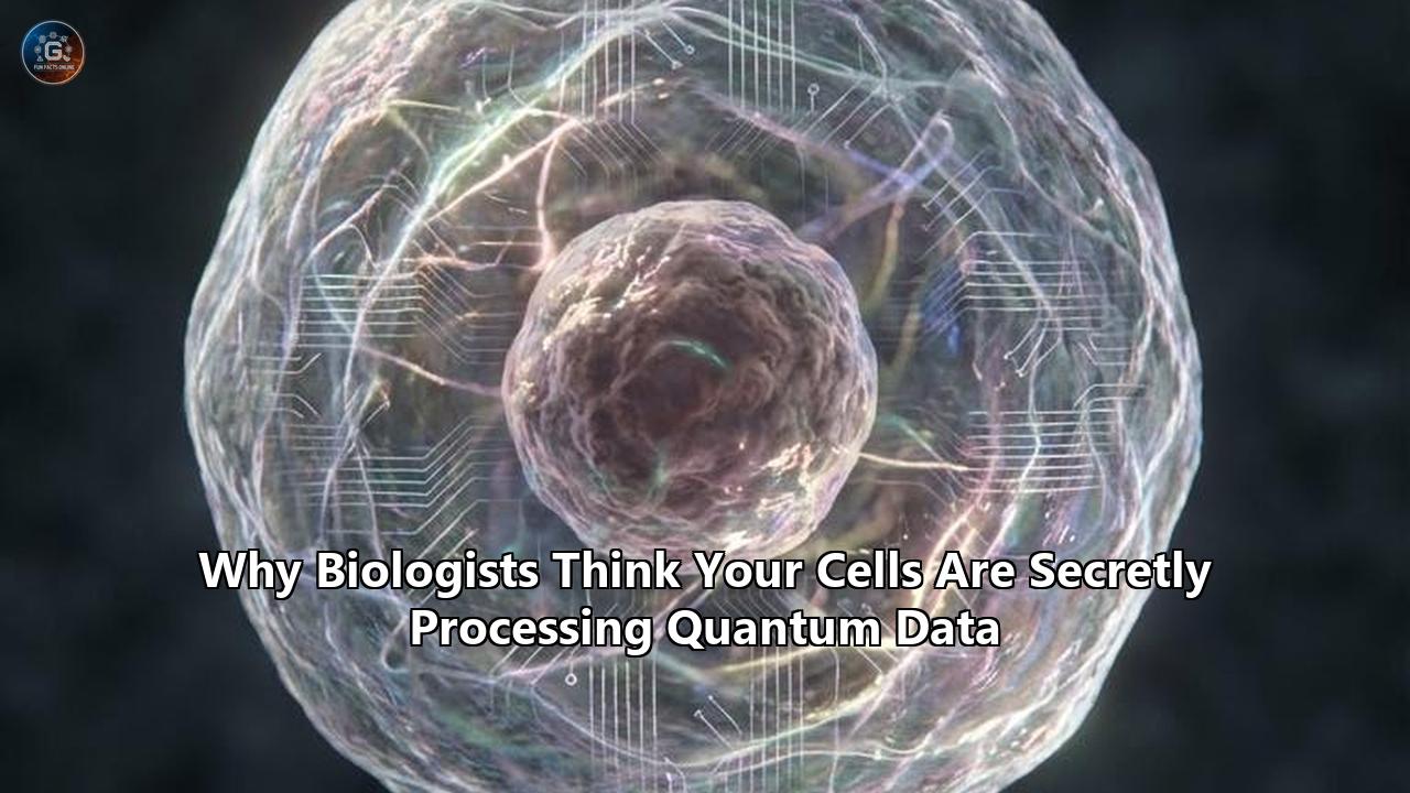
Just weeks ago, a multidisciplinary team of physicists and geneticists at the University of Chicago and the University of Oxford executed an experiment that fundamentally rewrites our understanding of organic life. Operating entirely at room temperature, they successfully turned an ordinary protein—the exact kind synthesized continuously inside your own body—into a functioning biological quantum bit, or qubit.
By encoding spin states directly into a molecular protein lattice, researchers engineered a quantum sensor that maintains its coherent state inside a living, breathing, electrically noisy cell. Simultaneous research published in early 2026 by the Quantum Biology Laboratory at Howard University revealed that the structural scaffolding of human cells, known as microtubules, actively utilizes a quantum optical phenomenon called "superradiance". Inside these cellular tubes, networks of the amino acid tryptophan absorb and emit ultraviolet light not as isolated particles, but as a single, unified quantum system operating billions of times faster than standard chemical reactions allow.
For decades, the global scientific consensus held that biological tissue was too warm, wet, and chaotic to sustain quantum mechanics. Building a commercial quantum computer currently requires cooling superconducting materials to fractions of a degree above absolute zero and shielding them inside billion-dollar vacuum chambers just to keep the qubits from collapsing. Yet, these recent discoveries prove that carbon-based life solved the exact same engineering problem billions of years ago. Your cells are not just mechanical factories governed by classical physics; they are active quantum information processors.
The implications of this extend far beyond academic physics. By reverse-engineering how proteins shield delicate quantum states from thermal interference, the technology sector gains a direct blueprint for room-temperature quantum computing. In the medical sector, biological qubits open the door to molecular-scale MRI scanners capable of detecting the earliest atomic misalignments of neurodegenerative diseases long before symptoms manifest.
To understand how a wet, 37-degree Celsius human cell manages to outperform IBM and Google’s most advanced cryogenic hardware, we have to examine the exact mechanisms keeping biological data intact.
The Decoherence Dilemma and the Biological Workaround
The central hurdle in maintaining any quantum system is a process called decoherence. In the quantum realm, particles like electrons and photons exist in a state of superposition—occupying multiple potential states or locations simultaneously. This allows a quantum processor to evaluate millions of possibilities at once, rather than sequentially. However, this state is incredibly fragile. The slightest interaction with the outside environment—a stray photon, a spike in temperature, or a slight magnetic fluctuation—causes the wave function to collapse. The particle snaps back into a single, fixed classical state, and the computational advantage evaporates instantly.
Physicists assumed that the interior of a human cell—a microscopic soup of swirling water molecules, vibrating proteins, and continuous electrical discharges—would cause instantaneous decoherence. The assumption was that biological processes had to be strictly classical, operating like a complex, predictable clockwork of chemical locks and keys.
That assumption failed to account for the unique architectural properties of biological polymers. The UChicago Pritzker School of Molecular Engineering (PME) breakthrough, developed in part by assistant professor Peter Maurer, bypasses the decoherence problem not by freezing the environment, but by utilizing the specific geometry of proteins.
Maurer’s team discovered that certain protein structures act as a natural isolation chamber for electron spin states. While traditional tech companies build synthetic qubits using superconducting circuits or trapped ions, the UChicago researchers embedded a rare-earth element, erbium, into a biological molecular host. Because proteins are incredibly complex macromolecules that fold into highly specific three-dimensional shapes, they create localized pockets that physically shield the embedded spin states from the thermal noise of the surrounding cellular fluid.
Furthermore, biological water itself behaves differently than previously understood. Research indicates that the water immediately surrounding structural proteins in the cell does not act like bulk liquid. Instead, it enters a highly ordered, "liquid crystal" phase. The dipole moments of these constrained water molecules align, restricting random thermal vibrations and essentially creating a quiet, low-noise microenvironment that allows quantum coherence to persist.
Superradiance in the Cellular Fiber-Optic Network
If localized proteins act as isolated quantum sensors, how does the cell transmit complex data across its vast internal distances? The answer lies in the cytoskeleton, specifically within microtubules.
Microtubules are the physical support beams of the cell. They are hollow cylinders constructed from alternating alpha and beta-tubulin proteins, responsible for everything from pulling chromosomes apart during cell division to transporting neurotransmitters down the long axons of nerve cells. But recent models advanced by Philip Kurian and his colleagues reveal that these structures double as biological fiber-optic cables.
Every tubulin protein contains tightly packed clusters of tryptophan. Tryptophan is highly responsive to ultraviolet light. When Kurian’s team mapped the precise geometry of these tryptophan molecules within the microtubule lattice, they found that the distances and dipole orientations were perfectly spaced to facilitate cooperative quantum effects.
In a classical system, if you shine light on a group of molecules, each one absorbs and re-emits a photon independently, scattering the energy randomly. In the microtubule networks, the tryptophan molecules engage in optical superradiance. Because of their precise geometric arrangement, the molecules couple together to form a delocalized excitonic state. When energy enters the system, the entire network absorbs it collectively and emits it in a highly synchronized, unidirectional pulse.
This is not a theoretical model; it is measurable. In March 2026, experimental fluorescence yield measurements demonstrated that the increase in quantum emission from individual tubulin units to fully assembled microtubules mathematically aligns with the emergence of superradiant states. The physical architecture of the microtubule modifies the dipole-dipole coupling matrix, fundamentally altering how light and matter interact within the neuron. The brain is actively using quantum optics to route energy and information without the massive heat loss that would accompany classical chemical diffusion.
The Anesthesia Enigma and the Roots of Consciousness
The realization that neuronal microtubules are processing quantum data has dramatically revived one of the most controversial hypotheses in neuroscience: the Orchestrated Objective Reduction (Orch OR) theory.
First proposed in the 1990s by mathematical physicist Roger Penrose and anesthesiologist Stuart Hameroff, Orch OR suggested that human consciousness is not merely the byproduct of chemical synapses firing in the brain, but the result of quantum computations occurring within the microtubules of our neurons. For decades, the broader neuroscience community dismissed the idea, citing the exact "warm and wet" decoherence arguments that recent discoveries have systematically dismantled.
The strongest experimental validation for this cellular quantum processing comes from the exact mechanism of general anesthesia. Despite using anesthetics for over a century, the medical field has never fully understood how these gases actually extinguish consciousness. They do not bind to specific receptors like typical pharmaceutical drugs. Instead, their potency perfectly correlates with their solubility in lipid environments (the Meyer-Overton correlation).
Researchers recently targeted the microtubule directly to observe its relationship with consciousness. In a mid-2025 controlled study using Sprague-Dawley rats, scientists administered Epothilone B, a drug uniquely known to cross the blood-brain barrier and structurally stabilize microtubule lattices. The rats were then exposed to isoflurane, a standard volatile anesthetic known to interact with and disrupt microtubule dynamics.
The results were stark. The rats pre-treated with the microtubule-stabilizing agent took 69 seconds longer to lose their righting reflex (the standard metric for unconsciousness) compared to the control group. The effect size was massive (Cohen's d ≈ 1.9), directly indicating that the physical stability of the microtubule lattice is intimately tied to the maintenance of the conscious state.
If consciousness relies on delocalized pi-electrons maintaining a state of quantum entanglement across the brain's microtubule networks, then volatile anesthetics work by physically wedging themselves into the tubulin structures, disrupting the dipole couplings, and forcing a premature collapse of the quantum wave function. The loss of coherence is the loss of consciousness.
Spin Chemistry: Seeing the Invisible Magnetic Triggers
While superradiance dictates how cells move energy, another quantum mechanism dictates how they sense their environment: the radical pair mechanism.
This mechanism was initially identified to explain avian magnetoreception. Birds, such as the European Robin, navigate across thousands of miles by "seeing" the Earth's weak magnetic field. This is achieved through a light-sensitive protein in their retinas called cryptochrome. When a photon strikes the cryptochrome, it knocks an electron out of place, creating a "radical pair"—two molecules, each with an unpaired electron.
Because these two electrons were created simultaneously, they are quantumly entangled. Their spins (a fundamental quantum property analogous to angular momentum) are linked. As they exist in this brief state, the Earth’s magnetic field gently nudges their spin alignment, pushing them between parallel and antiparallel states. The specific spin state dictates the chemical output of the molecule when the electrons eventually recombine. Thus, a subatomic quantum spin dictates a macro-biological chemical reaction, telling the bird which way is north.
For years, tracking this inside living systems was nearly impossible because the intermediate spin states are "dark" molecules—they do not emit light and vanish in nanoseconds. That barrier fell in April 2026. A research team led by Noboru Ikeya and Jonathan R. Woodward at the University of Tokyo successfully unveiled a pump-field-probe fluorescence microscopy platform capable of visually isolating these spin-dependent reactions.
By synchronizing nanosecond magnetic pulses with perfectly timed light pulses, the Tokyo researchers isolated the exact moment these magnetically sensitive intermediates formed and vanished. This represents a monumental leap in biological imaging. Scientists can now directly examine how weak magnetic fields alter chemical pathways in real-time.
Crucially, cryptochromes are not restricted to avian retinas; they are found throughout human cells, primarily regulating our circadian rhythms. The Tokyo microscopy platform provides the exact diagnostic tool needed to investigate how environmental electromagnetic fields impact human tissue at the quantum level, offering a clear mechanism for how external forces initiate cascading changes in human cellular chemistry.
Biomimetics and the Room-Temperature Quantum Leap
The economic implications of mapping these biological algorithms are staggering. The global race for quantum supremacy is currently bottlenecked by hardware limitations. Companies are pouring billions into massive dilution refrigerators that use rare isotopes like Helium-3 to keep quantum processors at 0.015 Kelvin. This hardware is static, incredibly fragile, and requires enormous energy inputs.
The shift toward bio-inspired quantum computing is currently reorganizing the tech sector. If directed evolution can optimize a protein to maintain coherence at 300 Kelvin (room temperature) in a chaotic liquid environment, tech conglomerates can adapt these molecular architectures to build stable, scalable, and portable quantum computers.
Gabriel Abrahams and his team at Oxford demonstrated this principle perfectly. By applying directed evolution to a protein originally found in oat plants, they engineered "Mag2", a biological quantum sensor. By steering the evolutionary process in bacteria, they forced the biological system to solve the coherence problem natively. This shifts the engineering paradigm entirely: instead of trying to isolate synthetic qubits from the environment, researchers are now designing molecular qubits that use the surrounding structural noise to their advantage, locking their spin states into protected geometric pockets.
In Japan, this intersection of biology and quantum mechanics has become a primary driver of industrial strategy. By pairing the massive simulation capabilities of the Fugaku supercomputer (capable of 442 petaflops) with biological modeling, pharmaceutical and technology conglomerates are shifting toward biocomputing paradigms. Designing synthetic materials that mimic the superradiant excitonic transport of microtubules will drastically alter the efficiency of solar panels and optical sensors.
The Medical Horizon: Molecular Diagnostics
The most immediate practical application of quantum biology lies in medical diagnostics. Current Magnetic Resonance Imaging (MRI) machines are massive, requiring superconducting magnets to align the protons in the water of your body to generate an image. They are excellent at showing macro-structures like tumors or torn ligaments, but they cannot see disease at its origin point: the individual molecule.
The biological qubits engineered at UChicago function essentially as molecular-scale MRI scanners. Because the spin states of these protein-based qubits are exquisitely sensitive to their immediate surroundings, they can be injected directly into a living cell. Once inside, they act as quantum spies, measuring microscopic fluctuations in temperature, pressure, and magnetic fields at the nanoscale.
This completely redefines predictive medicine. Alzheimer’s disease, Parkinson’s, and many cancers begin with subtle misfoldings of proteins and localized chemical imbalances long before a structural tumor forms or memory fails. By utilizing biological quantum sensors, oncologists and neurologists will be able to detect the precise magnetic signature of a single misfolded amyloid-beta protein or the aberrant electron transfer of a mutating cell in real-time. This pushes the diagnostic window back by years, potentially decades, moving medicine from reactive treatment to exact, molecular prevention.
Calculating the Biological Singularity
As we expand our diagnostic capabilities, we are also forced to reconsider our place within the computational hierarchy of the universe. The traditional view of Earth’s biological history is a timeline of gradual chemical complexity, slowly evolving from single-celled organisms to human intelligence.
Philip Kurian’s 2025 calculations, published during the International Year of Quantum Science and Technology, reframe this timeline entirely. By quantifying the quantum optical properties of cytoskeletal filaments across all carbon-based life, Kurian proposed an updated upper limit on the total information-processing capacity of the biosphere.
When every bacterial cell, plant, and animal is recognized not just as a chemical reactor, but as an active network of biological qubits processing data through superradiance and excitonic tunneling, the math changes drastically. The total computational capacity of carbon-based life throughout Earth’s history is mathematically immense, rivaling the theoretical computational bounds of all matter in the observable universe. Life is not just surviving; it is calculating.
What to Watch For Next
The era of classical biology is effectively over. Over the next 36 months, the integration of quantum mechanics into biological sciences will accelerate rapidly, driven by the new imaging and engineering tools developed in early 2026.
Watch for the expansion of the Tokyo pump-field-probe microscopy platform into complex, live-tissue human trials. The ability to track "dark" spin states in real-time will likely map the exact pathways by which human cells interpret magnetic and optical data, potentially unveiling new, non-chemical communication networks within the human nervous system.
Furthermore, keep an eye on the commercialization of biological qubits. As institutions like the Berggren Center for Quantum Biology and Medicine transition these molecular sensors from proof-of-concept to deployable medical devices, we will likely see the first FDA trials for intracellular diagnostic sensors.
We are finally shedding the mechanical view of the human body. Your cells are not mere bags of water and enzymes bumping into each other in the dark. They are highly structured, exquisitely tuned quantum arrays, harvesting light, entangling electrons, and processing the mathematics of survival at the very limits of physics.
Reference:
- https://www.youtube.com/watch?v=LU-WY4MxUmk
- https://www.youtube.com/watch?v=4JBiYzjJSh4
- https://www.spiedigitallibrary.org/conference-proceedings-of-spie/13872/1387202/Quantum-optical-effects-in-neuronal-microtubules--superradiance-under-structural/10.1117/12.3072614.full
- https://scitechdaily.com/scientists-just-discovered-quantum-signals-inside-life-itself/
- https://pme.uchicago.edu/news/world-quantum-day-2026-shaping-future-quantum-technology
- https://scienceandnonduality.com/article/quantum-coherence-and-the-hidden-secret-behind-our-bodies/
- https://www.neuroba.com/post/understanding-the-role-of-microtubules-in-quantum-consciousness-neuroba
- https://pmc.ncbi.nlm.nih.gov/articles/PMC8393322/
- https://www.reddit.com/r/consciousness/comments/1m8qih9/microtubules_and_consciousness_a_new_experimental/
- https://www.frontiersin.org/journals/neuroscience/articles/10.3389/fnins.2024.1430432/full
- https://scitechdaily.com/scientists-unveil-microscopy-breakthrough-that-reveals-invisible-molecular-states/
- https://itbusinesstoday.com/health-tech/quantum-biology-japans-role-in-the-next-medical-frontier/
- https://biotechnology-conferences.magnusgroup.org/program/scientific-sessions/quantum-biology-emerging-biotech-trends