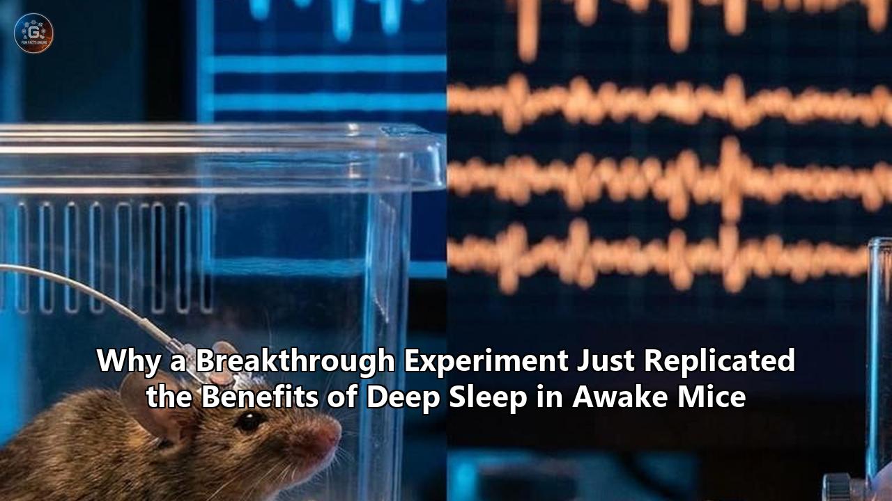
The laboratory at the Wisconsin Institute for Sleep and Consciousness is typically quiet, but the data streaming across the monitors in the summer of 2026 represents a noisy, fundamental disruption to our understanding of the mammalian brain.
On a table in the center of the room, a mouse actively moves about its enclosure. It whisks its snout, rears on its hind legs, and sniffs at a small plastic block newly introduced to its cage. By every behavioral definition, the animal is wide awake, alert, and engaged with its environment. Yet, deep within the sensory and motor regions of its cerebral cortex, a highly synchronized, rhythmic dance is taking place—one that has, until now, only ever been observed during the deepest states of unconscious slumber.
This paradoxical state is the centerpiece of a study published in Nature Neuroscience. Led by graduate researcher Kort Driessen, along with pioneering sleep neuroscientists Fabio Squarcio, Giulio Tononi, and Chiara Cirelli, the team has achieved what was long thought impossible. By using highly targeted pulses of light to orchestrate the firing of specific neural populations, they successfully replicated the physiological benefits of deep sleep in the brains of mice that remained completely awake and active.
The implications of this experiment stretch far beyond the walls of the Madison, Wisconsin laboratory. It represents a major pivot in sleep science, decoupling the physical state of being asleep from the restorative processes that sleep provides. For decades, scientists have operated under the assumption that to heal, rebuild, and organize memories, the brain must go dark—shutting out the external world entirely. The Wisconsin study suggests otherwise, raising a provocative question: can we harvest the restorative power of rest without ever losing consciousness?
The Synaptic Toll of Consciousness
To understand how the researchers pulled off this biological illusion, one must first look at why we sleep in the first place. For more than twenty years, Cirelli and Tononi have championed a framework known as the Synaptic Homeostasis Hypothesis (SHY).
When an animal is awake, it is constantly interacting with its environment. It learns, navigates obstacles, registers sensory inputs, and forms new memories. At the microscopic level, this cognitive activity requires the connections between brain cells—synapses—to grow stronger and more numerous. If you learn a new path, the synapses along that specific neural pathway are reinforced, physically packing themselves with neurotransmitter receptors to make the pathway fire more easily.
But this continuous strengthening comes at a massive cost.
"Being awake is expensive," explains Cirelli, a professor of psychiatry at the University of Wisconsin-Madison. "If your synapses constantly get stronger day after day without ever resetting, the brain quickly runs into a resource crisis. It consumes too much energy, runs out of physical space for new receptors, and eventually loses its ability to encode any new information. It becomes electrically saturated."
According to the SHY model, this is where non-rapid eye movement (NREM) sleep—the phase commonly referred to as deep sleep—steps in. NREM sleep accounts for roughly 80% of our total sleep time. During this phase, the brain undergoes a massive, systemic recalibration. It systematically evaluates the junctions between neurons, protecting the critical connections that represent important, long-term memories, while systematically pruning and weakening the less important, day-to-day noise.
These micro-adjustments represent the core deep sleep benefits, ensuring that the brain does not become physically overloaded, while restoring its capacity to learn fresh information the following morning.
For years, the gold standard indicator of this restorative process has been Slow-Wave Activity (SWA). When a mammal is sleep-deprived, their subsequent EEG readings show intense, high-amplitude slow waves during their first few hours of sleep. This SWA acts as a biological pressure gauge: the longer you stay awake, the higher your "sleep pressure" builds, and the more intense the slow waves are when you finally drift off. As you sleep, these waves gradually decay, signaling that the synapses have been successfully recalibrated and the sleep debt has been paid.
Historically, this reset was believed to be a global, whole-brain event that absolutely required behavioral sleep. But Driessen and his colleagues set out to challenge this dogma. They wanted to see if they could bypass the behavioral requirement of sleep entirely, unlocking these exact deep sleep benefits by manually driving the slow-wave machinery in an awake, conscious animal.
Engineering a Biological Mirage
To accomplish this, the Wisconsin team turned to optogenetics, an advanced neurological tool that allows researchers to control the electrical activity of specific brain cells using precise flashes of light.
The researchers genetically engineered two distinct groups of mice to express light-sensitive proteins within specific populations of cortical neurons.
- In one model, they targeted somatostatin-expressing (SOM+) interneurons—cells that act as the brain's natural "off-switches" by releasing inhibitory chemicals to quiet down neighboring neurons.
- In the other model, they targeted excitatory pyramidal neurons directly, using light to control the brain's primary communicators.
The team then surgically implanted tiny, dual-purpose fiber-optic devices, known as optrodes, into the sensorimotor cortex of the mice. These implants allowed the researchers to simultaneously record local electrical activity and deliver precise, microsecond pulses of light to the surrounding tissue. Crucially, these implants were placed on both the left and right hemispheres of the brain, allowing one side to receive the light stimulation while the other served as an internal, untreated control.
                OPTICAL FIBER (OPTRODE)
                         ||
                         ||  (Blue Light Pulses)
                         \/
              [====================]   <-- Skull
             /   SENSORIMOTOR       \
            /       CORTEX           \
           |   (SOM+ / Pyramidal)     |
           |                          |
           |   (*)  (*)  (*)  (*)     | <-- Genetically modified
           |    \    /    \    /      |     light-sensitive neurons
           |     \  /      \  /       |
           |     [ON]      [OFF]      | <-- Induced 0.5 - 1.0 Hz rhythm
           |     Burst     Silence    |
With the hardware in place, the true test began. The researchers sleep-deprived the mice for five hours. To keep the animals awake without causing undue stress, the lab staff continually introduced novel objects—colorful plastic blocks, running wheels, and textured materials—into their cages. The mice remained highly active, curious, and awake.
During the final 30 minutes of this five-hour sleep-deprivation window, the researchers turned on the blue lasers.
They did not simply flood the brain with light. Instead, they carefully programmed the pulses to mimic the exact rhythm of natural deep sleep. During NREM sleep, millions of cortical neurons fire in highly synchronized bursts, followed immediately by brief periods of absolute silence. This cycle—an "ON" state of high activity followed by an "OFF" state of quiet—repeats at a frequency of roughly 0.5 to 1 Hertz.
By pulsing the light at this exact frequency, the researchers forced a small, localized region of the sensorimotor cortex on one side of the awake mouse's brain to cycle through these ON and OFF periods. The other hemisphere was left completely untouched to experience the full, uninterrupted pressure of sleep deprivation.
Throughout the entire 30-minute stimulation period, the mice remained completely awake, moving around their cages, exploring the novel objects, and showing no behavioral signs of drowsiness or distress. But underneath the skull, a quiet revolution was taking place.
Layer One: The Vanishing Sleep Pressure
Once the 30-minute stimulation period ended, the sleep deprivation protocol was halted, and the mice were finally permitted to curl up and fall asleep naturally. This is where the researchers began to uncover their findings, layer by layer, starting with the electrophysiological evidence.
As the mice drifted into natural sleep, the team closely monitored the electrical signals coming from both the left and right hemispheres of the sensorimotor cortex.
On the control side of the brain—the hemisphere that had not received the optogenetic light flashes—the EEG showed a massive spike in Slow-Wave Activity. This was entirely expected; after five hours of forced wakefulness, the untreated cortex was under immense sleep pressure, screaming for rest.
But on the stimulated side of the brain, the picture was entirely different.
The cortex that had received the artificial, sleep-like ON/OFF pulses while the mouse was awake exhibited significantly less slow-wave activity during subsequent recovery sleep. In fact, the sleep pressure on that side of the brain had been discharged. The brain tissue behaved as though it had already received a substantial amount of deep sleep, despite the fact that the mouse had been actively walking around and exploring while the treatment was administered.
"What we're essentially doing is forcing sleep in a local region of the brain," Cirelli explains. "While that part of the cortex is solidifying memories and restoring learning capacity, other parts stay aware, vigilant, and connected to the environment. It is a highly localized discharge of the brain's biological need for sleep."
This was the first piece of hard evidence showing that sleep pressure is not a global currency that must be managed by the brain as a single, unified entity. Instead, sleep pressure is regional, modular, and can be artificially manipulated on a highly localized scale.
Layer Two: The Molecular Pruning Event
While the electrophysiological data was compelling, the team needed to prove that this artificial sleep rhythm was actually doing the biochemical work of real sleep. They had to look at the physical connections between the brain cells themselves.
To do this, the researchers ran a parallel experiment with a separate group of twenty-four mice. These animals went through the same five-hour sleep deprivation and 30-minute optogenetic stimulation protocol. However, the moment the 30-minute stimulation ended, the researchers immediately collected brain tissue from the sensorimotor cortex of both hemispheres—without allowing the mice to fall asleep.
If the Synaptic Homeostasis Hypothesis was correct, the wakeful state should have left both hemispheres cluttered with strong, high-receptor synapses. However, if the artificial ON/OFF rhythms had truly mimicked deep sleep, the stimulated side should show a marked reduction in synaptic strength.
The team measured the levels of specific neurotransmitter receptors—specifically, AMPA-type glutamate receptors, which serve as the primary chemical gateways for fast excitatory transmission in the cortex. The results were unmistakable.
        UNTREATED HEMISPHERE (Sleep Deprived)
        High density of AMPA receptors (cluttered, saturated synapses)
         [ Neuronal Terminal ]
           o   o   o   o  (Neurotransmitters)
         [====================]
           |   |   |   |   |  (AMPA Receptors - Strong/Saturated)
         [ Post-Synaptic Cell ]

        STIMULATED HEMISPHERE (Awake Sleep-Induction)
        Low density of AMPA receptors (pruned, recalibrated synapses)
         [ Neuronal Terminal ]
           o   o   o   o  (Neurotransmitters)
         [====================]
           |       |       |  (AMPA Receptors - Pruned/Renormalized)
         [ Post-Synaptic Cell ]
On the untreated side of the cortex, AMPA receptor levels remained exceptionally high, indicating that the synapses were still highly stressed and saturated. But on the stimulated side, the levels of these receptor proteins had dropped significantly, mirroring the exact, natural weakening of cellular connections that occurs during a normal, restful night of sleep.
The molecular data showed that the physical connections between brain cells had indeed been reset, capturing one of the most critical deep sleep benefits at the biochemical level. The 30-minute artificial rhythm had performed the essential maintenance work of sleep, clearing the metaphorical cognitive desk and freeing up resources for future learning, all while the animal was awake and active.
Layer Three: Cognitive Salvation
The ultimate test of any sleep intervention is functional: does it actually protect and restore cognitive performance? Can an animal whose brain has been artificially "rested" while awake actually learn and remember things as well as a well-rested animal?
To find out, the Wisconsin team designed a behavioral experiment focused on tactile memory—a cognitive capability that is heavily dependent on the somatosensory cortex and highly sensitive to sleep deprivation.
They gathered thirty mice and introduced them to an enclosed chamber on "Day 1". The floor of the chamber featured two identical, highly specific textures (for example, a rough, ridged pattern). The mice were allowed to explore the chamber for fifteen minutes, using their paws and whiskers to map out and memorize the texture of the floor.
Following this initial exploration, the thirty mice were split into three testing groups:
- The Sleepers (9 mice): These animals were returned to their cages and allowed to sleep normally.
- The Sleep-Deprived (13 mice): These animals were kept awake for an hour following the training, with no intervention.
- The Awake-Stimulated (8 mice): These animals were also kept awake for an hour, but during that time, they received the rhythmic, optogenetic ON/OFF brain stimulation to both hemispheres of their somatosensory cortex.
On "Day 2," all thirty mice were placed back into the testing chamber. This time, however, the floor of the chamber had been altered. One side of the floor featured the familiar, ridged texture from the previous day, while the other side featured an entirely new, smooth texture.
Mice are naturally drawn to novelty; if they remember an object or texture they have already experienced, they will spend the vast majority of their time investigating the new one. If they do not remember, they will divide their time equally between the two, treating both as brand new.
  BEHAVIORAL TEST RESULTS (Day 2: Texture Exploration)

  [Group 1: The Sleepers]  (Rested)
  Familiar Floor:   [====] (20%)
  Novel Floor:      [====================] (80%) <-- Remembered!

  [Group 2: Sleep-Deprived] (No Stimulation)
  Familiar Floor:   [==========] (50%)
  Novel Floor:      [==========] (50%) <-- Failed to remember

  [Group 3: Awake-Stimulated] (Rhythmic light pulses while awake)
  Familiar Floor:   [====] (20%)
  Novel Floor:      [====================] (80%) <-- Restored Memory!
The results of the behavioral test were striking.
- The mice that had slept normally easily recognized the old floor and spent nearly all of their time exploring the novel texture.
- The sleep-deprived mice failed the memory test completely, wandering aimlessly and spending an equal amount of time on both floors. Their lack of sleep had prevented their brains from consolidating the memory of the first texture.
- The awake-stimulated mice—which had been kept awake but received the rhythmic light pulses—performed just as well as the well-rested group, instantly recognizing the old floor and focusing their attention on the new texture.
These findings suggest that we can successfully harvest the deep sleep benefits of memory consolidation without requiring the brain to surrender its connection to the outside world. The memory of the floor texture had been successfully consolidated, and cognitive capacity was fully rescued, despite the mice never having closed their eyes.
The Secret Is in the Beat: Why Silence Is Not Enough
As the layers of the study began to unfold, a critical scientific question emerged: what, exactly, was driving this restorative effect? Was it simply a matter of quieting down overactive neurons, giving them a metabolic "break" from firing, or was there something unique about the deep sleep rhythm itself?
To isolate this variable, Driessen and his team designed a clever control experiment.
They took another group of sleep-deprived mice and used a different optogenetic protocol to suppress neural activity in the same region of the cortex. Instead of driving the rhythmic, alternating 0.5 to 1 Hz ON/OFF pulses, they simply used a continuous, steady pulse of light to suppress neuronal firing. The cells were kept completely quiet, calm, and resting, but without the active, synchronized slow-wave oscillation.
If the restorative nature of sleep was merely about metabolic rest—allowing tired cells to stop firing and recover—this quiet state should have produced the same benefits.
It did not.
When the researchers measured the synaptic strength and subsequent sleep pressure in these "quieted" mice, they found no restorative effects whatsoever. The molecular markers of synaptic strength remained high, and the sleep pressure was completely undischarged.
This revelation was the smoking gun of the study. It proved that the restorative benefits of sleep are not a passive consequence of inactivity. Rather, they are driven by an active, highly coordinated, rhythmic engine. The specific, alternating ON/OFF pattern of deep sleep is a mechanical necessity, acting like a cellular metronome that actively shakes loose excess receptors and reorganizes the synaptic architecture of the brain.
"The rhythm itself is the key factor here," says Driessen, emphasizing this critical point. "Just putting the brain to rest doesn't do the trick. You need that active, rhythmic oscillation to drive the molecular cleaning process."
The Dolphin Blueprint and the Modular Brain
The concept of a brain being partially awake and partially asleep is not entirely new to evolutionary biology. Nature has already designed several working prototypes.
"Dolphins do something very similar," points out Cirelli. "They, along with fur seals and many migratory bird species, sleep with only one brain hemisphere at a time—a phenomenon known as unihemispheric slow-wave sleep. While one half of their brain is plunged into deep, slow-wave sleep, the other half remains highly vigilant, monitoring the ocean for predators, controlling breathing, and keeping the animal moving."
                      THE UNIHEMISPHERIC MODEL

             LEFT HEMISPHERE               RIGHT HEMISPHERE
          +-------------------+         +-------------------+
          |  ACTIVE / AWAKE   |         |    DEEP SLEEP     |
          |                   |         |                   |
          |  * High Vigilance |  <===>  |  * Slow-Wave SWA  |
          |  * Motor Control  |         |  * Synaptic Reset |
          |  * Sensing        |         |  * Memory Consol. |
          +-------------------+         +-------------------+
                    \                             /
                     \                           /
                      \                         /
                       +-----------------------+
                       |    ACTIVE BEHAVIOR    |
                       | (Swimming, Breathing) |
                       +-----------------------+
Historically, human and rodent sleep was viewed as a fundamentally different, global phenomenon. It was believed that terrestrial mammals had evolved a whole-brain sleep model that required the entire cerebral cortex to go offline simultaneously.
However, the Wisconsin study heavily reframes this assumption. It suggests that the biological machinery for regional, modular sleep is still very much present in terrestrial mammals—it is simply masked by our natural, global sleep-wake behaviors.
Under extreme sleep deprivation, both humans and rodents have been observed to slip into fleeting, localized "micro-sleeps"—tiny pockets of the cortex that briefly drop into slow-wave activity while the individual is otherwise awake. While these sporadic, fleeting dips are usually too brief to provide any measurable restorative value, the Nature Neuroscience study proves that if you artificially extend and systemize these local sleep episodes, they can fully execute the restorative work of sleep on demand.
This shifts our fundamental understanding of sleep from a behavioral state of total bodily collapse to a highly coordinate, modular neural program that can be run locally, regionally, and independently across different sectors of the brain.
What Lies Ahead: A Future Without Sleep?
The potential to replicate the benefits of a full night's rest while remaining fully conscious is bound to spark the imagination of anyone who has ever wished for more hours in the day. But the researchers are quick to inject a healthy dose of scientific realism.
This is not a shortcut to skipping bedtime altogether.
In its current state, the technology is highly invasive. Recreating this sleep-like state in mice requires complex genetic modification and the surgical implantation of fiber-optic cables directly into the skull—procedures that are far from clinical deployment in humans.
Furthermore, sleep is a complex, multi-systemic process. While this study successfully replicated the cognitive and synaptic benefits of NREM sleep in a small, localized region of the cortex, sleep also plays vital roles in immune system regulation, hormone release, cardiovascular health, and the clearance of metabolic waste via the brain's glymphatic system. Forcing local sleep in the cortex will not protect a body from the systemic, physical failures associated with chronic sleep deprivation.
"This research is a major milestone, but it's important to contextualize what it is and what it isn't," says Amy Bany Adams, Ph.D., acting director of the NIH’s National Institute of Neurological Disorders and Stroke, which funded the study. "This isn't a replacement for sleep. Instead, this research further decodes why we sleep and how we learn, which brings us a step closer to understanding how to better prevent and treat cognitive decline."
Indeed, the true value of this conceptual breakthrough lies in its therapeutic potential.
Many neurodegenerative disorders, such as Alzheimer's disease and other forms of dementia, are characterized by a profound disruption of deep, slow-wave sleep. This lack of slow-wave sleep prevents the brain from clearing toxic amyloid plaques and reorganizing overstimulated synapses, creating a devastating feedback loop of cognitive decline.
If we can map these local rhythms precisely, we may be able to deliver targeted deep sleep benefits to specific, vulnerable brain regions in patients suffering from early-stage dementia.
"We are already looking toward the future," says Cirelli. "Our next step is to explore whether we can replicate these localized, restorative slow-wave patterns in humans using non-invasive technologies."
One potential avenue is transcranial magnetic stimulation (TMS) or high-density transcranial electrical stimulation. These non-invasive, skull-worn devices can deliver precise electromagnetic pulses to targeted regions of the human brain, potentially coaxing the cortex into the restorative ON/OFF rhythms of deep sleep while the patient sits comfortably in a chair, reading a book or watching television.
The IP and biotechnology sectors are already responding to this shift. Patent filings in the field of optogenetics and precise neurostimulation have begun to climb, driven by a growing interest in devices capable of modulating local brain rhythms for clinical therapy. While the surgical lasers of the Wisconsin lab may remain confined to animal models, the non-invasive devices they inspire could soon find their way into clinical trials.
For now, the mice in Madison continue to explore their cages, their brains carrying out the ancient, restorative secrets of deep sleep while their eyes remain open to the world. In showing us that sleep is not an all-or-nothing state, these tiny subjects have opened a new chapter in neuroscience—one where the boundaries of rest and consciousness may finally begin to blur.
Reference:
- https://scitechdaily.com/the-brain-may-not-need-full-sleep-to-recover-new-research-finds/
- https://www.psypost.org/researchers-induce-the-memory-boosting-benefits-of-sleep-in-parts-of-the-awake-brain/
- https://www.psypost.org/researchers-induce-the-memory-boosting-benefits-of-sleep-in-parts-of-the-awake-brain/
- https://neurosciencenews.com/localized-sleep-deprivation-memory-30839/
- https://1ban.news/local-sleep-recovery-optogenetics/
- https://lifespan.io/inducing-nrem-like-neuronal-patterns-mimics-sleep-benefits/
- https://www.ndtv.com/health/scientists-mimic-sleep-in-awake-mice-reverse-memory-loss-11613399
- https://1ban.news/local-sleep-recovery-optogenetics/
- https://whatsoncraigsmind.substack.com/p/the-age-of-our-organs-how-we-walk
- https://www.marks-clerk.com/insights/latest-insights/102n1d9-light-sleepers-optogenetic-stimulation-offers-sleep-like-benefits/
- https://www.vice.com/en/article/scientists-may-have-found-a-way-to-mimic-sleep-in-the-brain-without-actually-sleeping-heres-how/
- https://www.natureknowsproducts.com/how-deep-sleep-clears-a-mouses-mind-literally/
- https://www.realclearscience.com/2026/06/09/sleeps_restorative_effect_triggered_in_awake_mice_brains_1187467.html
- https://www.fox13seattle.com/news/scientists-trigger-sleeps-restorative-effect-mice-forced-stay-awake
- https://www.worldscientific.com/doi/10.1142/S1793545825500014