A team of researchers at the University of Kentucky College of Medicine revealed that the primary driver of sleep destruction in early neurodegenerative disease is not the death of neurons or the accumulation of toxic protein aggregates, but an overzealous response from the brain's own immune system.
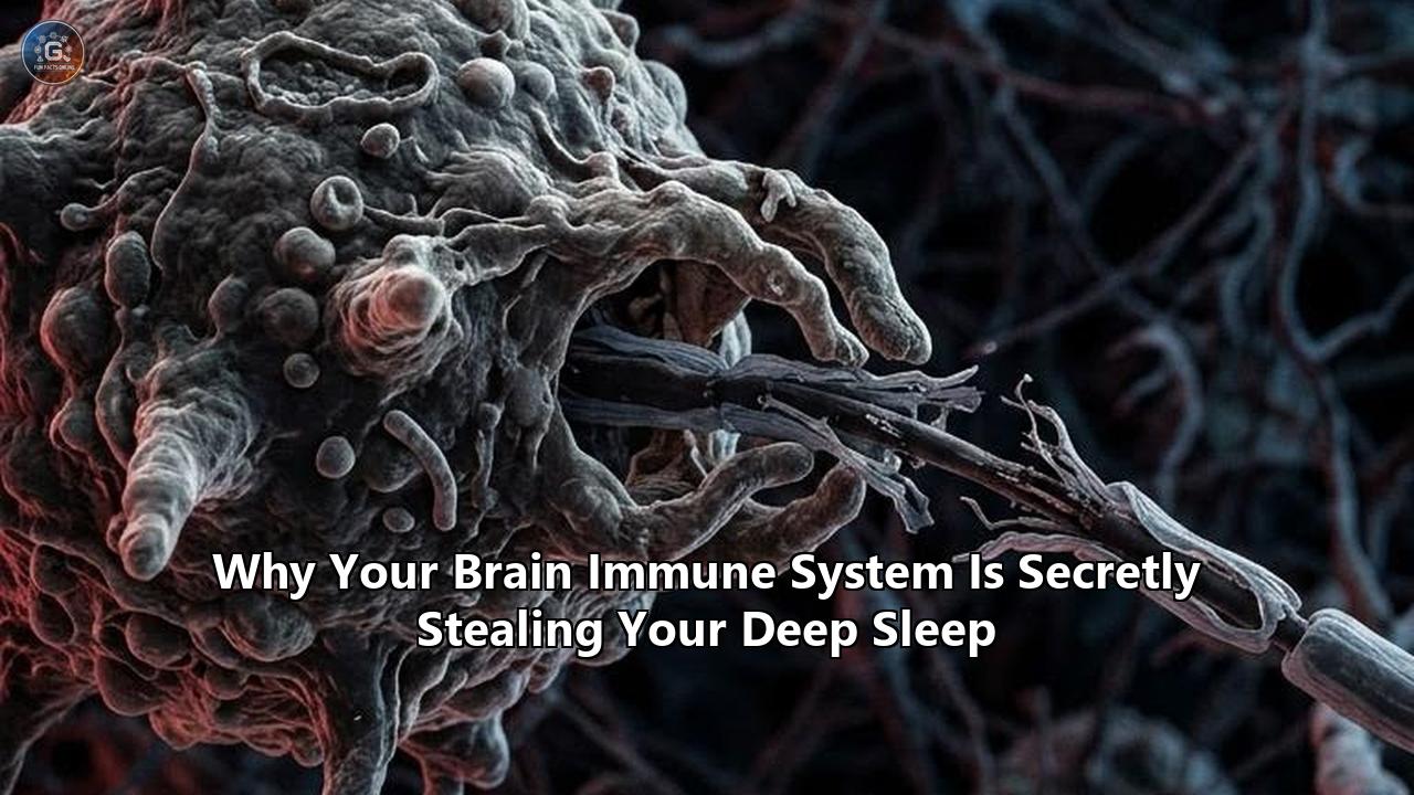
Published in Alzheimer's & Dementia, the study led by Associate Professor Shannon L. Macauley, Ph.D., and first author Nicholas J. Constantino, Ph.D., demonstrates that microglia—the resident immune cells responsible for defending the central nervous system—act as the principal hijackers of slow-wave sleep. When exposed to early pathological triggers, these cells initiate a sustained inflammatory signaling cascade that prevents the brain from entering deep, restorative sleep stages.
Using a pharmacological intervention to temporarily deplete roughly 87% of microglial populations in animal models, the Kentucky team successfully restored over two hours of deep sleep per day. Crucially, this dramatic recovery in sleep architecture occurred without reducing the physical burden of amyloid-beta plaques.
"Basically, we showed that it is not the plaques themselves, or solely dysfunctional neurons, that cause sleep loss but actually microglia," said Macauley. "Microglia are immune cells that, when they respond to plaques, kick off this elaborate cascade of inflammation, as if the microglia are partying all night, and keeping the brain awake."
                       NEUROINFLAMMATORY SLEEP DISRUPTION LOOP

    +-------------------------------------------------------------------+
    |                                                                   |
    v                                                                   |
+-----------------------+      +-----------------------+      +---------+-------------+
| Early Protein Debris  | ---> | Microglial Activation | ---> | Pro-Inflammatory      |
| & Circadian Stress    |      | (Morphological Shift) |      | Cytokines Released    |
+-----------------------+      +-----------------------+      +---------+-------------+
                                                                        |
                                                                        v
+-----------------------+      +-----------------------+      +-----------------------+
| Glymphatic System     | <--- | Loss of Slow-Wave     | <--- | Thalamocortical Loop  |
| Stalled (Waste Builds)|      | (N3 NREM) Delta Sleep |      | Neurons Depolarized   |
+-----------------------+      +-----------------------+      +-----------------------+
The discovery challenges long-standing assumptions about how neurodegenerative diseases impair circadian rhythms and slow-wave sleep, forcing a fundamental split in how biotechnology labs and clinical neuroscientists approach sleep preservation.
Comparing Pathological Drivers: Protein Aggregates vs. Neuroimmune Ignition
For decades, the dominant medical hypothesis held that sleep disruption in conditions like Alzheimer's, Parkinson's, and vascular dementia was a direct mechanical outcome of structural neuronal loss. Under this classical framework, physical accumulations of amyloid-beta plaques and hyperphosphorylated tau tangles were thought to strangle the sleep-promoting centers of the brain—specifically the galanin-producing neurons of the ventrolateral preoptic nucleus (VLPO) and the cholinergic networks of the basal forebrain.
The University of Kentucky findings, alongside parallel research from Texas A&M University, establish a competing paradigm: sleep destruction is an active immunologic suppression rather than a passive structural decline.
+---------------------------------------------------------------------------------------------------+
| STRUCTURAL DAMAGE HYPOTHESIS                                                                      |
| Protein Aggregates (Plaques/Tangles) --> Neuronal Cell Death --> Degraded Sleep Centers          |
+---------------------------------------------------------------------------------------------------+
| NEUROIMMUNE IGNITION HYPOTHESIS (NEW PARADIGM)                                                   |
| Early Pathological Triggers --> Activated Microglia --> Cytokine Cascade --> Sleep Architecture   |
|                                                                              Suppression          |
+---------------------------------------------------------------------------------------------------+
Direct Structural Damage Framework
- Primary Target: Amyloid-beta oligomers, extracellular plaques, intracellular tau tangles.
- Mechanism: Protein toxicity induces apoptosis in wake-sleep regulatory nuclei; damaged axons disrupt thalamocortical signaling pathways physically.
- Therapeutic Goal: Remove sticky protein aggregates via targeted monoclonal antibodies (e.g., lecanemab, donanemab).
- Observed Sleep Impact: Removing physical plaques fails to immediately restore delta power or fix N3 sleep fragmentation, as the underlying neuroimmune storm continues unabated.
Neuroimmune Ignition Framework
- Primary Target: Hyper-reactive microglia, astrocytic inflammatory states, pro-inflammatory cytokine signaling (TNF-$\alpha$, IL-1$\beta$, IL-6).
- Mechanism: Microglia adopt a "stress-primed" or amoeboid morphology, secreting neuro-active signaling molecules that destabilize neuronal resting membrane potentials within sleep circuits.
- Therapeutic Goal: Calm or temporarily suppress central immune cell activity, modulating cellular bioenergetics or signaling receptors.
- Observed Sleep Impact: Halting microglial inflammatory cascades restores up to two hours of daily slow-wave sleep without requiring the physical clearance of protein plaques.
The contrast between these two models is striking. Traditional amyloid-targeting monoclonal antibodies clear protein deposits effectively, yet clinical trials routinely show minimal immediate improvement in patient sleep architecture. In some instances, antibody therapies trigger transient localized neuroinflammation—known as amyloid-related imaging abnormalities (ARIA)—which can temporarily exacerbate sleep fragmentation.
By contrast, targeting the brain immune system sleep axis directly bypasses the slow process of protein clearance, offering an immediate electrophysiological reset to damaged slow-wave circuits.
Electrophysiological Dynamics: How Microglia Shatter the Thalamocortical Loop
Understanding why immune cells suppress deep sleep requires examining the biophysics of slow-wave sleep (SWS), also categorized as stage N3 non-rapid eye movement (NREM) sleep.
Deep sleep is characterized on an electroencephalogram (EEG) by high-amplitude, low-frequency delta oscillations (0.5 to 4 Hz) and slow oscillations (< 1 Hz). These rhythmic waves originate in the neocortex and are synchronized through reciprocal feedback loops with the thalamus. For a brain to enter N3 sleep, thalamocortical neurons must hyperpolarize, allowing them to shift from a single-spike "bursting" transmission mode during wakefulness to a rhythmic, synchronized oscillatory state during deep sleep.
Activated microglia disrupt this fragile electrophysiological transition through localized chemical signaling.
                          THALAMOCORTICAL DISRUPTION MECHANISM

 +------------------------------------------------------------------------------------+
 | WAKEFULNESS / INFLAMED STATE                                                       |
 | Microglia release IL-1beta, TNF-alpha, ROS                                          |
 |    |                                                                               |
 |    v                                                                               |
 | Thalamocortical Neurons remain DEPOLARIZED (resting potential ~ -55mV to -60mV)     |
 |    |                                                                               |
 |    v                                                                               |
 | Fast tonic firing continues; brain CANNOT synchronize into delta waves             |
 +------------------------------------------------------------------------------------+
                                           VS
 +------------------------------------------------------------------------------------+
 | HEALTHY DEEP SLEEP (N3 NREM)                                                      |
 | Minimal inflammatory cytokine pressure                                            |
 |    |                                                                               |
 |    v                                                                               |
 | Thalamocortical Neurons HYPERPOLARIZE (resting potential shifts to -65mV to -70mV) |
 |    |                                                                               |
 |    v                                                                               |
 | Low-frequency delta oscillations (0.5-4 Hz) synchronize across neocortex          |
 +------------------------------------------------------------------------------------+
Cytokine Biphasic Dynamics
In healthy brain physiology, low homeostatic concentrations of interleukin-1 beta (IL-1$\beta$) and tumor necrosis factor-alpha (TNF-$\alpha$) are somnogenic—they actively promote healthy sleep drive. However, when microglia detect pathologically altered proteins or suffer chronic circadian strain, they shift into a pro-inflammatory state.
In this state, cytokine production spikes by orders of magnitude. Chronically elevated TNF-$\alpha$ alters AMPA receptor trafficking at synaptic sites, increasing baseline excitatory postsynaptic currents. Instead of allowing thalamic neurons to hyperpolarize, the cytokine flood maintains resting membrane potentials at an elevated, depolarized state (typically between -55 mV and -60 mV).
Destruction of Delta Power
Because thalamic relay neurons remain partially depolarized by this local immune activity, they cannot enter the hyperpolarized firing mode (-65 mV to -70 mV) required to generate delta waves.
The electrophysiological result is catastrophic for sleep architecture:
- Slow-wave amplitude drops: The synchronized "up-state" and "down-state" transitions across neocortical columns collapse.
- Micro-arousals spike: Neuronal networks remain hypersensitive to minor sensory inputs, fragmenting sleep into short, unstable bouts.
- Spindle-Slow Wave Uncoupling: Sleep spindles (11–16 Hz bursts crucial for memory consolidation) fail to lock onto the crest of slow oscillations, crippling long-term memory formation.
This electrophysiological lockout explains why patients with neurodegenerative conditions or chronic neuroinflammation report feeling unrefreshed even after spending eight hours in bed. Their brains are held in a state of persistent electrophysiological vigilance by overactive microglial signaling.
Comparing Therapeutic Strategies: Cellular Depletion vs. Metabolic Reprogramming vs. Nanoparticle Delivery
The discovery that microglial activation actively steals deep sleep has ignited an intense debate over how to target these cells clinically. Researchers are pursuing four distinct technological strategies, each presenting radically different risk-reward profiles and biological trade-offs.
+----------------------------------------------------------------------------------------------------+
|                                EMERGING THERAPEUTIC APPROACHES                                     |
+--------------------------+-----------------------+------------------------+------------------------+
| METHOD                   | MECHANISM             | ADVANTAGES             | RISKS & TRADEOFFS      |
+--------------------------+-----------------------+------------------------+------------------------+
| 1. Pharmacological       | CSFR1 Blockade        | Rapid sleep recovery   | Vulnerability to CNS   |
|    Ablation              | (e.g., Pexidartinib)  | (+2 hrs SWS in models) | infection; toxicity    |
+--------------------------+-----------------------+------------------------+------------------------+
| 2. Metabolic             | Bioenergetic Shift    | Well-studied drugs     | Off-target metabolic   |
|    Reprogramming         | (Metformin/Stiripentol)| (low toxicity profile) | impact; variable BBB   |
+--------------------------+-----------------------+------------------------+------------------------+
| 3. Extracellular Vesicle | Anti-inflammatory     | High cell specificity; | Complex manufacturing; |
|    Nanoparticle Delivery | miRNA Cargo           | Preserves cell counts  | BBB transit dynamics   |
+--------------------------+-----------------------+------------------------+------------------------+
| 4. Sensory Non-Invasive  | Acoustic/Visual       | Non-pharmacological;   | Inconsistent efficacy; |
|    Entrainment           | Gamma Modulation      | Zero chemical toxicity | Misses cellular origin |
+--------------------------+-----------------------+------------------------+------------------------+
Approach 1: Pharmacological Ablation (CSF1R Inhibitors)
The method deployed by the University of Kentucky team relies on small-molecule inhibitors targeting the Colony-Stimulating Factor 1 Receptor (CSF1R), such as Pexidartinib (PLX3397). Microglia depend entirely on CSF1R signaling for survival; blocking this receptor forces activated microglia into rapid apoptosis, eliminating up to 90% of the population within days.
[CSF1 Receptor] + [Pexidartinib / PLX3397] ---> Microglial Apoptosis (~87% Cleared)
                                             ---> Inflammatory Signaling Ceases
---> +2 Hours SWS Restored- Pros: Unmatched potency. By clearing the cellular source of neuroinflammation, the brain's baseline neurochemical landscape resets quickly, restoring normal electrophysiological sleep cycles.
- Cons: Total microglial ablation is a radical intervention. Microglia are essential for synaptic pruning, clearing metabolic waste, and defending against viral and bacterial CNS pathogens. Prolonged ablation leaves the brain vulnerable to opportunistic infections and long-term tissue damage. Furthermore, systematic CSF1R inhibitors carry significant systemic risks, including severe hepatotoxicity.
Approach 2: Metabolic Reprogramming (Metformin and Stiripentol)
Rather than destroying microglia, researchers like Dr. Macauley are investigating ways to alter their cellular metabolism. When microglia become chronically activated, they undergo a bioenergetic shift reminiscent of the Warburg effect in cancer cells: they abandon oxidative phosphorylation in favor of rapid aerobic glycolysis. This glycolytic state fuels their production of inflammatory cytokines.
[Inflamed Glycolytic Microglia] + [Metformin / Stiripentol] ---> Bioenergetic Reset to OxPhos
                                                             ---> Cytokine Secretion Drops
---> Sleep Architecture Preserved- Pros: Leverages existing, FDA-approved compounds with well-established safety records. Metformin (an AMP-activated protein kinase activator) and Stiripentol (an anti-seizure agent) can force microglia back into an oxidative phosphorylation state, suppressing inflammatory output without killing the cells.
- Cons: These small molecules act globally across multiple tissue types. Achieving therapeutic concentrations in the central nervous system without causing peripheral metabolic disturbances remains a difficult dosing challenge.
Approach 3: Targeted Extracellular Vesicles (EV Nanoparticles)
Pioneered by Dr. Karienn Souza and Dr. Ashok Shetty at Texas A&M University, this approach uses stem-cell-derived extracellular vesicles (EVs) to re-educate "stress-primed" microglia. Stress-primed microglia develop abnormal, hyper-branched morphotypes under chronic sleep deprivation or circadian disruption.
[Stem Cell Derived EVs] ---> Transmit Anti-Inflammatory miRNAs
                        ---> Intercept Stress-Primed Microglia
---> Restore Ramified Surveillance State- Pros: Highly targeted and non-destructive. The EVs carry specialized bio-molecules and microRNAs that dock directly with activated microglia, signaling them to downregulate the NLRP3 inflammasome and return to a quiescent, homeostatic surveillance state. This quenches the inflammatory storm while preserving the brain's cellular defense network.
- Cons: EV production faces massive scaling and manufacturing quality-control hurdles. Ensuring consistent delivery across the blood-brain barrier via intravenous or intranasal administration requires advanced nanomedicine infrastructure.
Approach 4: Non-Invasive Sensory Entrainment (40 Hz Audio-Visual Stimulation)
Non-pharmacological approaches attempt to quiet microglial activation using sensory inputs—such as 40 Hz flickering lights and auditory clicks—to drive specific neural oscillations.
[40 Hz Light/Sound Stimuli] ---> Thalamic Sensory Processing
                             ---> Synchronized Gamma Entrainment
---> Transient Microglial Clearance- Pros: Completely non-invasive with an exceptional safety profile. It requires no pharmaceuticals and can be administered at home via specialized headsets.
- Cons: While sensory entrainment shows promise in altering microglial morphology in early preclinical models, its ability to reverse deep cellular neuroinflammation in human clinical trials has yielded mixed results. Once microglia shift into a fully activated, self-sustaining inflammatory loop, non-invasive physical oscillations are often insufficient to override their chemical signaling.
The Glymphatic Feedback Loop: How Missing Sleep Fuels the Fire
The relationship between the brain immune system sleep axis and neurological health forms a tightly coupled, closed-loop feedback system. Disruption to any part of this circuit rapidly accelerates pathology across the entire network.
During healthy, unperturbed N3 slow-wave sleep, the brain undergoes a structural transformation. Driven by astrocytic aquaporin-4 (AQP4) water channels, the interstitial space between neurons expands by approximately 60%. This expansion allows cerebrospinal fluid (CSF) to surge through brain parenchyma, sweeping away metabolic waste products—including soluble amyloid-beta, hyperphosphorylated tau, alpha-synuclein, and lactic acid—and flushing them into the venous lymphatic system.
                             THE GLYMPHATIC DISRUPTION CYCLE

                   +-------------------------------------------------+
                   | Activated Microglia Secretes Pro-Inflammatory   |
                   | Cytokines (TNF-a, IL-1b)                        |
                   +-------------------------------------------------+
                                            |
                                            v
                   +-------------------------------------------------+
                   | Suppresses Thalamocortical Delta Power &         |
                   | Destroys Slow-Wave Sleep (N3 NREM)             |
                   +-------------------------------------------------+
                                            |
                                            v
                   +-------------------------------------------------+
                   | Interstitial Space FAILS to Expand by 60%;      |
                   | CSF Flushing Stalls                             |
                   +-------------------------------------------------+
                                            |
                                            v
                   +-------------------------------------------------+
                   | Toxic Waste & Metabolic Byproducts Accumulate   |
                   | in Neural Parenchyma                            |
                   +-------------------------------------------------+
                                            |
                                            v
                   +-------------------------------------------------+
                   | Pattern Recognition Receptors (TLR4/NLRP3)      |
                   | Bind Waste --> FURTHER Microglial Activation    |
                   +-------------------------------------------------+
                                            |
                                            +--- (Loop Repeats continuously)
When activated microglia suppress slow-wave sleep, this glymphatic clearance mechanism stalls. The physiological consequences compound rapidly:
- Failure of Convective Fluid Exchange: Without continuous delta wave oscillations, the arterial pulsations that drive CSF through the tissue lose their rhythmic synchronization.
- Debris Retention: Metabolic waste products remain trapped within the neural parenchyma instead of being cleared.
- Receptor Hyper-Activation: Trapped protein oligomers bind to Toll-like receptor 4 (TLR4) and the NLRP3 inflammasome complex on microglial cell membranes.
- Secondary Inflammatory Waves: This binding signals the microglia that damage is accumulating, causing them to release additional pro-inflammatory cytokines.
This feedback loop turns temporary sleep deprivation into an ongoing inflammatory cycle. Once microglia begin suppressing slow-wave sleep, they effectively destroy the brain's ability to clean itself, ensuring that inflammatory debris continues to accumulate and perpetuate microglial activation.
Technical Comparison of Interventions
| Diagnostic/Therapeutic Metric | CSF1R Ablation (Pexidartinib) | Metabolic Reprogramming (Metformin/Stiripentol) | Extracellular Vesicles (EV Nanoparticles) | Monoclonal Antibodies (Lecanemab/Donanemab) |
|---|---|---|---|---|
| Primary Biological Target | Microglial CSF1 Receptor | Glycolytic Enzymes / AMPK Axis | "Stress-Primed" Microglial Morphotypes | Extracellular Amyloid-Beta Plaques |
| Impact on Deep Sleep (SWS) | Immediate High Restoration (+2 hours/day) | Moderate Restoration (via metabolic stabilization) | High Restoration (via inflammatory suppression) | Minimal to No Direct Impact |
| Impact on Amyloid Burden | Unchanged (Plaques remain) | Unchanged | Slowed accumulation | Significant Reduction (Plaques cleared) |
| Cellular Survival Rate | ~13% (87% cellular death) | 100% (Cells preserved) | 100% (Cells preserved) | 100% (Microglia activated during clearance) |
| Risk of Neuro-Infection | Severe / Critical | Negligible | Very Low | Negligible |
| Primary Delivery Route | Oral Small Molecule | Oral Small Molecule | Intranasal / Intravenous | Intravenous Infusion |
| Clinical Translation Stage | Preclinical Animal Models | Off-Label / Phase 2 Trials | Preclinical / Early Seedling Grants | FDA Approved (Clinical Use) |
Diagnostic Paradigms: EEG Sleep Architecture vs. Neuroimmune Fluid Biomarkers
As researchers pivot toward targeting neuroinflammation to salvage sleep, clinicians face a diagnostic dilemma: how do you identify patients whose deep sleep is actively being compromised by microglial overactivation before irreversible neurodegeneration sets in?
Two competing diagnostic frameworks are emerging, each offering distinct vantage points on the brain immune system sleep dynamic.
+----------------------------------------------------------------------------------------------------+
|                                    DIAGNOSTIC FRAMEWORKS                                           |
+---------------------------------------------------+------------------------------------------------+
| PARADIGM A: ELECTROPHYSIOLOGICAL ARCHITECTURE     | PARADIGM B: MOLECULAR & NEUROIMMUNE BIOMARKERS |
+---------------------------------------------------+------------------------------------------------+
| Focus: Functional real-time brain dynamics        | Focus: Direct measurement of immune signaling  |
| Tool: Portable High-Density Home EEG              | Tool: Lumbar Puncture, Plasma & TSPO-PET       |
| Key Metrics: Delta power density, SWS duration,   | Key Metrics: sTREM2, GFAP, YKL-40,             |
|              spindle-slow wave coupling           |              microglial binding density        |
+---------------------------------------------------+------------------------------------------------+
Paradigm A: Electrophysiological Architecture (High-Density Portable EEG)
With recent advancements in dry-sensor technology and home-use high-density EEG, clinicians can now track a patient's sleep architecture longitudinally across weeks or months rather than relying on a single night in a sleep lab.
[Home EEG Device] ---> Longitudinal Delta Power Analysis
                   ---> Identifies Micro-Arousals & SWS Degradation
---> Early Functional Sleep Theft Signal- Key Biomarkers Tracked:
Delta Power Spectral Density: Measuring the total energy within the 0.5–4 Hz band during non-REM sleep. A continuous, unexplained drop in delta power serves as an early functional warning sign of neuroimmune suppression.
Slow-Oscillation Amplitude: Tracking the physical height of slow waves. Decreasing amplitude directly mirrors depolarizing cytokine pressure on thalamocortical loops.
Sleep Fragmentation Index: Quantifying micro-arousals lasting under 15 seconds that break up deep N3 stages.
- Advantages: Non-invasive, relatively inexpensive, and capable of detecting functional changes in brain operation years before memory loss or cognitive impairment manifests on clinical tests.
- Disadvantages: Functional EEG changes are non-specific. A drop in delta power can stem from sleep apnea, severe psychological stress, alcohol consumption, or environmental disturbances, requiring careful differential diagnosis to isolate immunologic causes.
Paradigm B: Neuroimmune Biomarkers & Molecular Imaging
This approach relies on quantifying central nervous system immune activation through direct fluid analysis and targeted neuroimaging.
[Blood/CSF Sampling & TSPO-PET] ---> Quantifies sTREM2, GFAP, & Microglial Ligand Binding
---> Confirms Active Cellular Neuroinflammation- Key Biomarkers Tracked:
Soluble TREM2 (sTREM2): A cleavage product of the Triggering Receptor Expressed on Myeloid Cells 2, shed into CSF and blood plasma when microglia undergo activation.
Glial Fibrillary Acidic Protein (GFAP): A marker of reactive astrogliosis that rises rapidly in response to central inflammatory signaling.
18K kDa Translocator Protein (TSPO) PET Imaging: Radiotracer ligands that bind specifically to outer mitochondrial membrane proteins upregulated in activated microglia, producing a 3D heat map of neuroinflammation across the brain.
- Advantages: Provides definitive, molecular proof of central immune activation and pinpoints the precise anatomical brain regions undergoing neuroinflammatory stress.
- Disadvantages: Highly invasive (requiring spinal taps for CSF access), expensive (TSPO-PET scans cost thousands of dollars per session), and logistically unfeasible for broad population screening.
Rethinking Early Interventions
The realization that the brain's immune system actively suppresses deep sleep alters our understanding of cognitive aging and neurodegenerative prevention. Sleep loss is no longer seen as merely a passive byproduct of brain aging, but as an active immune-mediated suppression that accelerates pathology.
                             THE SHIFT IN CLINICAL STRATEGY

 +----------------------------------------------------------------------------------+
 | OLD APPROACH: SYMPTOMATIC & LATE DEBRIS CLEARANCE                                |
 | Wait for cognitive decline --> Detect protein plaques --> Clear plaques          |
 | (Result: Sleep architecture remains ruined; underlying inflammation persists)     |
 +----------------------------------------------------------------------------------+
                                          VS
 +----------------------------------------------------------------------------------+
 | NEW APPROACH: EARLY NEUROIMMUNE SLEEP PRESERVATION                               |
 | Monitor longitudinal EEG --> Detect delta power drop --> Calm overactive         |
 | microglia via metabolic/EV therapy --> Save deep sleep & preserve brain function |
 +----------------------------------------------------------------------------------+
This paradigm shift opens up several practical avenues for clinical research and therapeutic development:
1. Dual-Action Therapeutic Cocktails
Future treatments for neurodegenerative diseases will likely move away from single-agent mono-therapies. Instead, combination regimens will couple low-dose protein-clearing agents with microglial immunomodulators (such as Metformin, Stiripentol, or EV therapies). The goal is to clear structural aggregates while keeping the brain's sentinel cells calm enough to permit restorative slow-wave sleep.
2. Microglia-Aware Sleep Pharmacology
Traditional hypnotic sleep drugs, such as benzodiazepines and non-benzodiazepine "Z-drugs" (e.g., zolpidem), sedate the brain by enhancing global GABAergic inhibition. However, these drugs do not reduce microglial cytokine output or increase natural delta wave power; in fact, they often reduce time spent in true N3 deep sleep.
Future sleep pharmacology must focus on developing targeted neuro-immunomodulators that induce sleep by quieting inflammatory signaling within thalamocortical networks rather than forcing global neural sedation.
3. Early Longitudinal Screening
Combining portable, home-based EEG monitoring with blood-based neuroimmune biomarkers (such as plasma GFAP and sTREM2) could enable early screening for neurodegenerative risk. By catching microglial sleep theft at its inception—potentially decades before memory loss begins—clinicians can deploy anti-inflammatory and metabolic interventions while neural architecture remains intact.
Future Outlook: Key Milestones in Neuroimmune Sleep Science
The discovery that microglial activation actively steals deep sleep has initiated a new phase of translational neuroscience. Over the next several years, research will focus on advancing these basic science discoveries into actionable human clinical interventions.
                             TIMELINE OF EXPECTED MILESTONES

       2026                        2027--2028                     2029 AND BEYOND
  +-------------------------+  +-------------------------+  +-------------------------+
  | Preclinical Discovery   |  | Phase 1/2 Clinical      |  | Integrated Diagnostic   |
  | University of Kentucky  |  | Human Trials            |  | & Therapeutic Platforms |
  | & Texas A&M Studies     |  | Metformin, Stiripentol, |  | Home EEG + Targeted     |
  | (Microglial sleep loss) |  | & EV Sleep Restoration  |  | Immunomodulation        |
  +-------------------------+  +-------------------------+  +-------------------------+
Key Developments to Watch:
- Human Clinical Translation (2026–2028): Researchers are shifting from preclinical rodent models to early-phase human trials. Priority is being given to repurposing safe, existing metabolic regulators—such as Metformin and Stiripentol—to evaluate their ability to preserve delta power and SWS duration in individuals with early-stage cognitive decline.
- Refining Nanoparticle Delivery (2027–2029): Biotechs are working to standardize the manufacturing and intranasal delivery of stem-cell-derived extracellular vesicles. These trials will test whether EV therapies can quiet stress-primed microglia and restore sleep without compromising the brain's primary immune defenses.
- Validation of EEG Biomarkers (2026–2030): Large-scale clinical studies are evaluating whether continuous home EEG monitoring can reliably differentiate immune-mediated sleep loss from non-inflammatory sleep disturbances. Validating these non-invasive metrics could provide a low-cost, widely accessible tool for early detection.
The discovery that the brain's immune system actively steals deep sleep reframes our understanding of neurodegenerative disease. Sleep destruction is no longer seen as an inevitable consequence of dying brain tissue, but as a reversible outcome of cellular neuroinflammation.
By focusing on calming overactive immune cells rather than solely clearing physical structural damage, medicine is moving closer to preserving deep sleep, maintaining brain waste clearance, and protecting cognitive health long before symptoms appear.
References
- Constantino, N. J., & Macauley, S. L. et al. (2026). Microglia drive sleep deficits in preclinical models of Alzheimer's disease independently of amyloid-beta plaque burden. Alzheimer's & Dementia, University of Kentucky College of Medicine.
- Souza, K., Shetty, A., & Earnest, D. (2026). Circadian dysregulation primes microglial activation: Implications for extracellular vesicle protection against neuroinflammation. Texas A&M Health Dementia & Alzheimer's Research Initiative (DARI).
- Sasidharan, V., & Tucci, V. et al. (2026). Epigenetic and microglial modulation of slow-wave sleep architecture under neuroinflammatory stress. Sleep Medicine Reviews, Fondazione Istituto Italiano di Tecnologia.
- International Journal of Pharmaceutical Sciences Review (2026). Microglial activation, pro-inflammatory cytokine signaling, and thalamocortical disruption in chronic sleep disturbance. IJPS, 18(2), 112–129.
Reference:
- https://www.sciencedaily.com/releases/2026/07/260719035931.htm
- https://lexingtonky.news/2026/07/15/uk-researchers-discover-brain-immune-cells-drive-alzheimer-s-sleep-loss/
- https://www.eurekalert.org/news-releases/1136483
- https://neurosciencenews.com/circadian-rhythm-microglia-dementia-30670/
- https://www.ijpsjournal.com/article/neuroinflammatory-signaling-in-chronic-insomnia-from-microglial-activation-to-sleep-wake-dysregulation
- https://pubmed.ncbi.nlm.nih.gov/41921278/