At the Rambam Health Care Campus in Haifa, Israel, a 70-year-old woman legally blind in one eye recently regained her sight following the successful transplantation of PB-001, a fully synthetic, lab-grown tissue. Manufactured by the biotechnology firm Precise Bio, this operation marked the first successful human transplant of a 3D-bioprinted human cornea produced entirely from cultured human cells.
The procedure, conducted as part of an ongoing Phase 1 clinical trial, was led by Professor Michael Mimouni, Director of the Cornea Unit at the Rambam Eye Institute. The surgical team utilized standard endothelial keratoplasty techniques, placing the lab-generated tissue into the patient's eye exactly as they would a naturally harvested donor graft. The tissue integrated, the cells sustained themselves, and the patient’s visual acuity was restored.
What makes this specific medical event highly significant is not just the restoration of one patient's vision, but the underlying manufacturing process that produced the implant. The tissue was created using a proprietary 4D biofabrication platform that takes a single deceased donor cornea and expands its cellular material to print up to 300 distinct, transplant-ready implants. Aryeh Batt, CEO and Co-Founder of Precise Bio, confirmed that the platform allows researchers to expand human cells in the laboratory and print them with exact structural fidelity, fundamentally unlinking corneal transplantation from the strict one-to-one ratio of human donor availability.
This milestone forces a total reevaluation of regenerative ophthalmology. For decades, the field has struggled to move bioprinted ocular tissues out of the laboratory and into the operating room. This recent trial provides the clinical evidence that engineered tissues can survive, function, and refract light within a living human eye.
The Arithmetic of Corneal Blindness
To understand why this specific bioengineering achievement matters, one must examine the severe structural deficits in the global tissue supply chain. The cornea is the eye’s outermost transparent layer, responsible for protecting the inner structures and providing roughly 65% to 75% of the eye's refractive power. When it becomes cloudy or scarred—due to conditions like Fuchs' endothelial dystrophy, keratoconus, trachoma, or physical trauma—vision degrades rapidly, often leading to total blindness.
Because the human cornea cannot regenerate severe structural damage natively, the only definitive treatment is a keratoplasty—a physical tissue transplant.
The global mathematics surrounding this procedure are grim. Approximately 12.7 million people worldwide suffer from corneal blindness, waiting for a transplant. Yet, according to global eye bank data, there is only one donor cornea available for every 70 patients who need one. In developing nations, the ratio is exponentially worse.
Furthermore, traditional corneal transplantation is bottlenecked by stringent logistical requirements. Once harvested from a deceased donor, a cornea has an extremely narrow window of viability. It must be processed, evaluated for endothelial cell density, tested for infectious diseases, stored in specific media (such as Optisol-GS), and kept within a strict cold chain. The tissue typically expires within 14 days. If a surgical complication occurs, or if the recipient develops a sudden contraindication, the tissue is often wasted.
The rise in elective laser eye surgeries (like LASIK and PRK) has further compounded this shortage, as corneas altered by laser ablation are frequently disqualified from the donor pool. The reliance on a one-to-one, human-to-human transfer model is a mathematically unsolvable crisis. The only viable path forward is to manufacture the tissue.
The Bioink Hack: Solving the Transparency-Strength Paradox
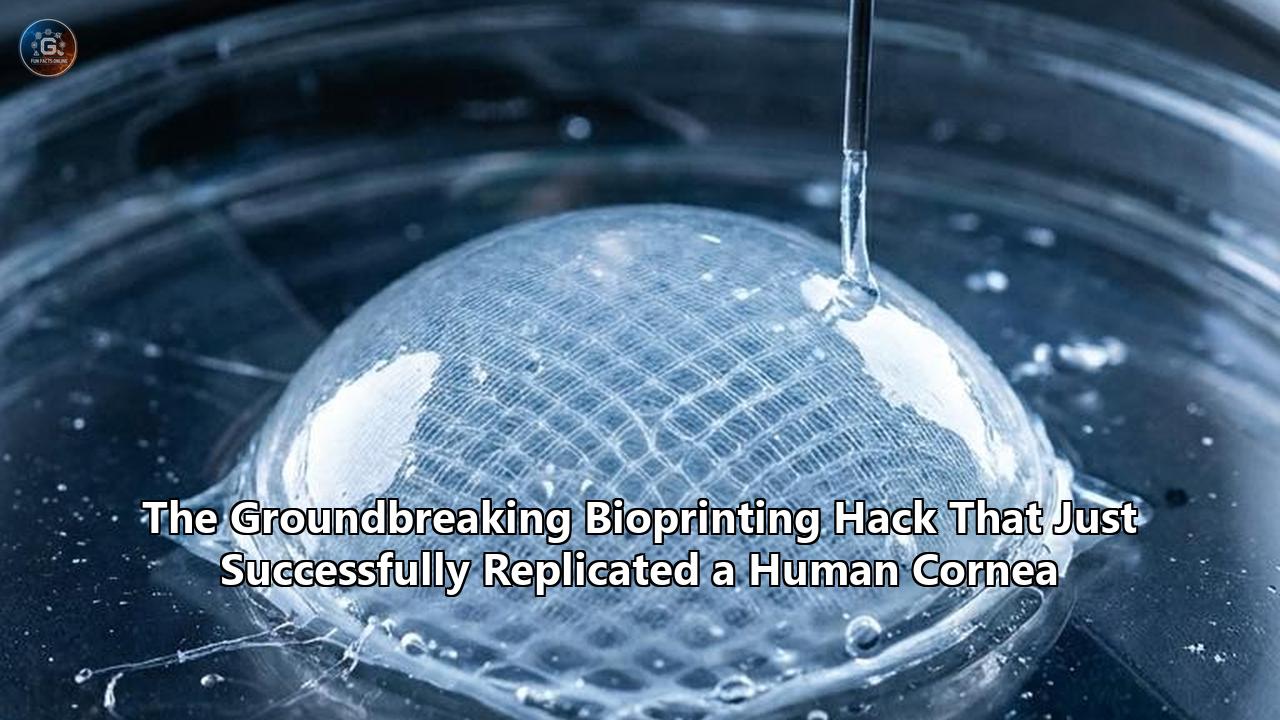
Manufacturing an artificial cornea sounds straightforward in a world where we routinely 3D print titanium joint replacements. However, biological printing requires "bioinks"—substances composed of living cells, structural biopolymers, and growth factors. For years, tissue engineers faced an infuriating biochemical paradox when attempting to print a bioprinted human cornea: the inverse relationship between optical transparency and mechanical strength.
The native human cornea is highly transparent due to the precise, microscopic lattice arrangement of Collagen Type I fibrils. To replicate this, early researchers attempted to use low-concentration collagen bioinks. While these low-density inks achieved the necessary optical clarity, they were physically weak. When extruded through a printer nozzle, they collapsed into gelatinous puddles, utterly failing to maintain the rigid dome-like curvature required to refract light properly.
Conversely, when researchers increased the concentration of collagen to give the tissue enough tectonic strength to hold its shape and withstand surgical suturing, the dense collagen fibrils self-assembled chaotically at neutral pH levels. The resulting tissue was strong but completely opaque, rendering it useless for vision.
The recent clinical breakthroughs were unlocked by a series of biochemical "hacks" that stabilized high-concentration bioinks without sacrificing clarity.
One major method involves altering the photo-initiation process. Researchers discovered that by utilizing high concentrations of highly purified Collagen I mixed with specific photoinitiators like riboflavin (Vitamin B2), they could prevent premature self-assembly. Once the bioink is extruded into the exact curved geometry of a cornea, it is exposed to targeted Ultraviolet (UV) light. The UV irradiation induces rapid crosslinking of the collagen fibers, instantly locking the structural lattice in place while it remains optically clear.
Other entities have bypassed pure collagen entirely. Researchers in South Korea developed a cornea-derived decellularized extracellular matrix (Co-dECM). By taking animal corneas, stripping away all the immunogenic cellular material, and reducing the remaining structural matrix into a hydrogel, they created an ink that is already biochemically optimized for the eye. When subjected to shear stress through a printer nozzle (shear-thinning), the Co-dECM bioink flows smoothly, but its viscosity spikes dramatically once deposited, allowing it to hold its shape.
Additionally, teams at the University of Tampere in Finland and the Swiss Federal Laboratories for Materials Science and Technology (Empa) have successfully experimented with hybrid hydrogels combining hyaluronic acid and customized bio-polymers. These unique formulations keep delicate stem cells alive during the high-pressure printing process while resulting in a tissue stiff enough to handle yet soft enough to integrate seamlessly into the host eye.
Hardware Engineering: Extrusion vs. Digital Light Processing
The hardware utilized to construct these tissues is just as vital as the chemistry. The bioprinting industry is currently divided into two primary fabrication methodologies: Extrusion-based printing and Digital Light Processing (DLP).
Extrusion bioprinting functions similarly to standard desktop 3D printers. A pneumatic or mechanical syringe pushes the bioink through a micro-nozzle, depositing the material layer by layer in concentric circles. This method is highly effective for high-viscosity materials and allows for dense cell encapsulation. Precise Bio’s platform heavily utilizes advanced extrusion techniques, depositing distinct layers that mimic the human cornea’s five anatomical tiers (the epithelium, Bowman's layer, the stroma, Descemet's membrane, and the endothelium).
However, extrusion introduces physical shear stress on the cells passing through the nozzle, which can lower cell viability if not carefully calibrated. To counter this, companies like Pandorum Technologies in India have begun integrating Digital Light Processing (DLP) systems into their workflow, utilizing hardware like the CELLINK LUMEN X.
DLP bioprinting does not push material through a nozzle. Instead, a pool of liquid bioink is exposed to a precisely controlled projector screen that flashes specific patterns of light. The light selectively cures and solidifies the bioink layer by layer in a matter of seconds. Because there is no physical extrusion, the delicate human corneal stromal cells face far less mechanical stress, resulting in higher viability rates and incredibly high-resolution tissue structures.
Immunological Advantages of Synthetic Corneas
One of the most persistent challenges in any transplant surgery is the host’s immune response. While the human cornea is uniquely "immune-privileged"—meaning it lacks direct blood vessels and thus has a lower rejection rate than organs like the liver or heart—graft rejection still occurs in roughly 10% of standard keratoplasty cases. When a patient rejects a donor cornea, the host's immune cells attack the foreign endothelial cells, causing the graft to turn opaque.
A bioprinted human cornea sidesteps much of this risk through careful cellular programming. The implants used by platforms like Precise Bio and KeratOPrinter are engineered to be hypoimmunogenic. By starting with undifferentiated stem cells or carefully vetted allogeneic cell lines, researchers strip away the highly specific human leukocyte antigens (HLAs) that typically trigger an immune response.
Furthermore, because the tissue is fabricated in a tightly controlled cleanroom environment rather than harvested from a deceased human body, the risk of transmitting latent viral infections, prion diseases, or fungal keratitis is reduced to absolute zero. Every printed cornea is highly standardized, traceable, and backed by comprehensive data regarding its cellular origin, exact thickness, and optical properties. When a surgeon requests a synthetic cornea, they receive a clinically perfect product every single time, devoid of the biological variability that plagues natural tissue harvesting.
The Global Competitors: Precise Bio, KeratOPrinter, and Pandorum
While Precise Bio currently holds the clinical milestone for the first human transplant, they are not operating in a vacuum. A fierce, highly capitalized global race is underway to scale this technology for commercial distribution.
In Europe, the KeratOPrinter initiative recently launched with an €8 million funding grant from the European Union. Led by Professor Heli Skottman at Tampere University, the KeratOPrinter consortium is attempting to fully automate the bioprinting process to align with strict Good Manufacturing Practice (GMP) standards.
The KeratOPrinter project is distinct because of its deep integration of artificial intelligence. Bioprinting organic tissue is inherently unpredictable; minor fluctuations in ambient temperature or bioink batch quality can cause catastrophic failures in the microstructural lattice. The KeratOPrinter system utilizes machine learning algorithms combined with computer vision to monitor the extrusion process in real-time. The AI continuously assesses the microstructural patterns of the collagen being deposited, predicting the final optical performance of the cornea before it is even finished printing. If the AI detects a defect, it can recalibrate the print parameters on the fly or flag the batch for disposal, ensuring that only flawless tissues reach the operating room.
In India, a hub for both high rates of corneal blindness and aggressive biotech innovation, Pandorum Technologies has entered human trials for their proprietary "Corneal Lenticule". Pandorum takes a highly personalized approach: they acquire detailed medical scans of a specific patient's damaged eye, generate a custom 3D model, and print a cornea tailored to match the precise curvature and dimensions required by that specific individual. Supported by multi-hospital trials spanning both India and the United States, Pandorum’s photo-crosslinkable cornea-mimetic bioink is positioning the company as a major force in the Asian and North American markets.
Surgical Integration: The Operating Room Experience
A critical factor determining the success of any medical device is how easily physicians can adopt it. If a bioprinted human cornea requires ophthalmic surgeons to learn an entirely new, highly complex operative technique, adoption will stall.
Developers have specifically engineered these bioprinted tissues to mimic the physical handling characteristics of natural donor tissues. Precise Bio’s CEO Aryeh Batt explicitly noted that their PB-001 implant is handled exactly like traditional tissue during Endothelial Keratoplasty (EK) procedures.
During a standard EK (such as a DMEK or DSAEK), the surgeon makes a microscopic incision in the periphery of the eye, removes the patient's diseased endothelial layer, and carefully unfolds the donor tissue into place, using a temporary air or gas bubble to press the new tissue against the inner dome of the eye until it adheres. The synthetic implants are designed to possess the exact tensile strength and elasticity required to survive being folded, inserted through a 3-millimeter micro-incision, and manipulated within the anterior chamber of the eye without tearing.
By designing the product to fit seamlessly into existing surgical workflows, the barrier to entry for global deployment is effectively reduced to simple supply logistics and regulatory clearance.
Regulatory Headwinds and the Economics of Synthetic Tissue
The successful phase 1 trial in Israel has dramatically accelerated the conversation surrounding regulatory frameworks. Tissues produced via bioprinting occupy a unique and complex space in global health regulation. They are neither purely mechanical medical devices (like a pacemaker) nor strictly traditional biologics (like a vaccine or a natural organ transplant).
Regulatory bodies like the U.S. Food and Drug Administration (FDA) and the European Medicines Agency (EMA) are currently establishing clear pathways for 3D-bioprinted organs. Because these corneas contain living, metabolically active human cells suspended in a manufactured matrix, they must undergo the rigorous, multi-phased clinical trial process required of new pharmaceutical drugs, assessing long-term cell viability, toxicity, and rejection rates over years.
Simultaneously, the manufacturing hardware and the bioinks themselves must meet the exacting quality systems regulations of medical devices. This dual-track regulatory burden is exceptionally expensive and time-consuming.
However, the economic upside is staggering. The traditional process of harvesting, processing, testing, storing, and shipping donor corneas involves high operational costs, with eye banks passing these expenses onto the healthcare system. The KeratOPrinter initiative and Precise Bio’s mass-production model demonstrate that once the initial capital expenditure of the printing hardware is covered, the marginal cost of printing an individual cornea drops significantly. A single robotic 3D biofabrication system can potentially supply enough implants for an entire national health system, operating on-demand.
Instead of maintaining vast networks of cold-chain logistics to move fragile human tissue across continents within a 14-day window, hospitals could theoretically order specific corneal grafts directly from regional bio-fabrication hubs, receiving them cryopreserved and ready for use. This shifts corneal transplantation from a scarcity-driven model to a scalable manufacturing model.
What Happens Next?
The ophthalmology sector is closely monitoring the progression of Precise Bio’s Phase 1 clinical trial through the latter half of 2026. The initial success in the 70-year-old patient must now be replicated across a broader cohort to prove long-term durability. Researchers are specifically watching for any delayed immune responses and ensuring that the implanted cells maintain their refractive transparency over a period of 12 to 24 months.
If Phase 2 and Phase 3 trials continue on their current trajectory, EU and FDA fast-track designations for medical devices could see commercial availability of synthetic corneas within the next three to five years.
Furthermore, the technology validated in the eye is already bleeding into other medical disciplines. The eye serves as the perfect proving ground for 3D bioprinting; it is relatively small, immune-privileged, and easily observable without invasive surgery. The techniques perfected in solving the corneal bioink challenge—specifically balancing cell viability with structural integrity—are directly applicable to larger, more complex systems. Precise Bio has publicly stated that following ophthalmic tissues, their 4D printing platform will target cardiology, orthopedics, and nephrology.
The success at the Rambam Eye Institute provides concrete proof that the era of relying solely on human donors for vision restoration is ending. The focus now shifts from proving the science works to aggressively scaling the manufacturing, optimizing the bioinks, and pushing the technology through international regulatory pipelines. The engineering of human sight has officially moved off the laboratory bench and into the clinic.
Reference:
- https://cyberguy.com/future-tech/3d-printed-cornea-restores-sight-world-first/
- https://timesofindia.indiatimes.com/life-style/health-fitness/health-news/how-worlds-first-3d-printed-cornea-restored-a-70-yr-olds-vision/articleshow/125875541.cms
- https://www.3dnatives.com/en/precise-bio-3d-printed-cornea-transplant-12012026/
- https://www.ntoaeye.com/blog/a-breakthrough-in-vision-care-first-ever-3d-printed-cornea-restores-sight.html
- https://www.healio.com/news/ophthalmology/20260227/qa-3d-bioprinted-corneal-implants-may-ease-donor-tissue-shortage
- https://www.voxelmatters.com/keratoprinter-aims-to-restore-vision-for-millions-with-bioprinting-technology/
- https://pmc.ncbi.nlm.nih.gov/articles/PMC6348552/
- https://createdigital.org.au/3d-printed-corneas-people-see/
- https://www.researchgate.net/publication/362559800_Corneal_bioprinting_using_a_high_concentration_pure_collagen_I_transparent_bioink
- https://ro.uow.edu.au/articles/journal_contribution/Corneal_bioprinting_using_a_high_concentration_pure_collagen_I_transparent_bioink/27821445
- https://www.researchgate.net/publication/366243200_Hyaluronic_acid_based_next_generation_bioink_for_3D_bioprinting_of_human_stem_cell_derived_corneal_stromal_model_with_innervation
- https://pmc.ncbi.nlm.nih.gov/articles/PMC9852906/
- https://www.cellink.com/customer-spotlight/on-the-horizon-using-3d-printed-corneas-to-cure-corneal-blindness/