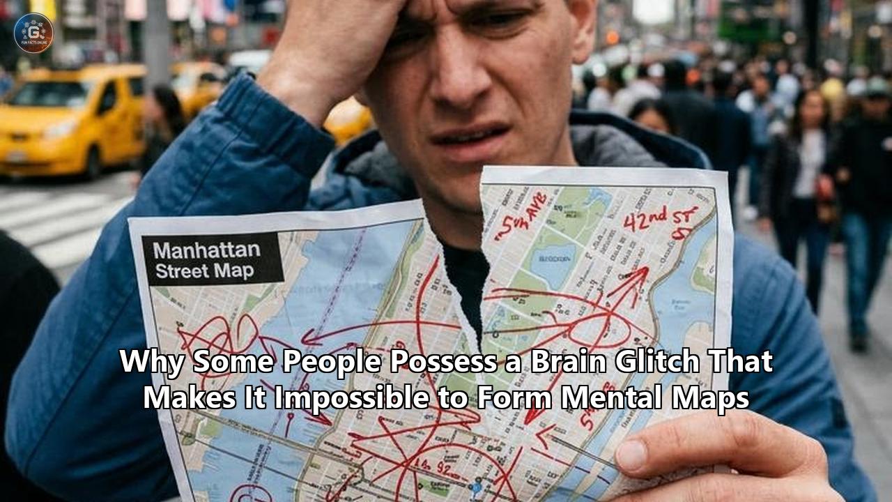
When an otherwise healthy 42-year-old architectural designer arrived at the Neuroimaging Laboratory at the University of Calgary, she brought an unusual dilemma. Despite passing standardized intelligence tests in the 99th percentile and successfully drafting complex two-dimensional technical blueprints, she confessed that if she stepped outside the front entrance of her own home without an active turn-by-turn satellite GPS application running on her phone, she could not locate the grocery store three blocks away. Inside her mind, the physical environment did not exist as a continuous, navigable terrain. When she closed her eyes, there was no internal blueprint, no visual trace of landmarks, and no intuitive sense of how different streets connected behind her back.
Neuroscientists led by Dr. Giuseppe Iaria and collaborating teams at the Paris Brain Institute and Exeter University have documented this phenomenon across international cohorts, providing structural and functional insights into a condition that leaves individuals completely incapable of forming or consulting cognitive maps. Known clinically at the intersection of Developmental Topographical Disorientation (DTD) and congenital visual aphantasia, this neural anomaly alters how researchers conceptualize spatial awareness, memory consolidation, and internal sensory simulation.
For decades, cognitive science operated on the core assumption that spatial navigation and episodic memory share an indissoluble bond forged by visual imagery. If a person needed to travel from point A to point B, the classical model argued, the brain’s inner eye simulated the path ahead, accessing an internal spatial map projected by the hippocampal-entorhinal system and decorated with landmark imagery by the retrosplenial and occipitotemporal cortices.
Recent neuroimaging investigations published in Cortex, Brain, and eLife are dismantling this monolithic view. By examining clinical case studies of individuals living with congenital spatial and imagery deficits, researchers have uncovered an unexpected architecture of human cognition: a functional decoupling between the systems that construct spatial coordinate networks and the neural machinery responsible for conscious sensory visualization.
+-----------------------------------------------------------------------------+
|                     CLASSICAL VS. DECOUPLED SPATIAL COGNITION               |
+-----------------------------------------------------------------------------+
| 1. Classical Model:                                                         |
|    Sensory Input -> Occipitotemporal -> Retrosplenial -> Hippocampus       |
|    (Perception)     (Visual Imagery)    (Translation)     (Cognitive Map)   |
|                                                                             |
| 2. Decoupled Architecture (Aphantasia / DTD Profile):                      |
|    Sensory Input -> Occipital (Hyper-active Sensory Isolation)               |
|                          |--X-- (Severed Top-Down/Translation Loops)        |
|                     Retrosplenial / Prefrontal Network                      |
|                          |--X-- (Functional Disconnect)                     |
|                     Hippocampal-Entorhinal Grid Engine                      |
|    (Result: Propositional/Algorithmic Navigation without Spatial Simulation)|
+-----------------------------------------------------------------------------+
The Anatomy of a Mapless Mind: Patient S.D. and the Clinical Baseline
To understand how the brain's internal cartographic software fails, researchers have looked closely at single-subject and small-cohort case studies that isolate specific cognitive variables. Among the most thoroughly documented recent cases is patient S.D., an adult participant evaluated across a battery of virtual-reality spatial navigation tasks, remote environment recall tests, and high-resolution neuroimaging paradigms.
S.D. presents with a lifelong absence of voluntary visual imagery—a condition categorized as total visual aphantasia. When instructed to close her eyes and visualize the facade of her childhood home, her mother’s face, or the geometry of a local intersection, S.D. reports an utter absence of visual impressions. She experiences what cognitive scientists term "blind mind" phenomenology. Yet, unlike patients with acquired brain lesions from stroke or localized trauma, S.D. possesses no focal tissue damage, no neurodegenerative markers, and normal intellectual functioning.
+-----------------------------------------------------------------------------+
|                 NEUROPSYCHOLOGICAL TESTING PROFILE (PATIENT S.D.)           |
+-----------------------------------------------------------------------------+
| Test Paradigm               | Patient Performance | Control Group Baseline  |
+-----------------------------+---------------------+-------------------------+
| Full-Scale IQ (WAIS-IV)     | 128 (Superior)      | 100 ± 15 (Standard)     |
| Mental Rotation (Shepard)   | Intact (Slower RT)  | Intact (Standard RT)    |
| Egocentric Route Following  | Preserved (Stepwise)| Highly Flexible         |
| Allocentric Vector Survey   | Severely Impaired   | Accurate                |
| Pointing to Unseen Targets  | Chance Level        | High Precision          |
| Autobiographical Memory     | Severely Deficient  | Episodically Rich       |
+-----------------------------+---------------------+-------------------------+
In standard life routines, S.D. functions through systematic behavioral workarounds. She memorized step-by-step verbal rules for her daily commute: drive precisely 1.2 miles, make a right turn at the pharmacy with the green neon sign, proceed past three sets of traffic signals, and turn left immediately after the railroad tracks.
When researchers placed S.D. within controlled experimental paradigms—such as the digital spatial environment of the Sea Hero Quest navigation testing engine—her compensatory systems broke down. When the virtual environment forced her to calculate an immediate shortcut across an open, landmark-free expanse, or when the starting position was rotated 180 degrees to test allocentric (world-centered) spatial knowledge, her performance plummeted to chance levels.
"These individuals are not experiencing a failure of basic perception or intellect," explains Dr. Giuseppe Iaria, who established the first diagnostic registry for Developmental Topographical Disorientation at the University of Calgary. "They perceive the tree, the building, and the road in front of them with perfect clarity. What their brain fails to execute is the automatic, subconscious conversion of those perceptual fragments into a unified coordinate system. They exist in a perpetual present, navigating by static checklists rather than dynamic internal landscapes".
The case of S.D. exposed a critical distinction that neuropsychology long obscured: the profound difference between route-based navigation (procedural stimulus-response associations handled predominantly by the basal ganglia and caudate nucleus) and survey-based navigation (allocentric cognitive mapping anchored in the hippocampal-entorhinal complex). S.D. could execute routes if the chain of sequential visual cues remained unbroken; the moment a road closure interrupted her sequence, the entire spatial model evaporated instantly.
Translating the World: How the Brain Builds Spatial Cartography
To pinpoint why this internal compass breaks down, neuroscientists must examine how typical brains generate a spatial layout. The biological foundation of the cognitive map relies on a complex network of specialized cellular networks operating across the medial temporal lobe, parietal cortex, and midline subcortical structures.
+-----------------------------------------------------------------------------+
|                      THE SPATIAL CODING HIERARCHY                           |
+-----------------------------------------------------------------------------+
|                                                                             |
|  [ PARIETAL & OCCIPITAL CORTICES ]                                          |
|  Processes egocentric (viewer-dependent) visual inputs & scene landmarks.   |
|                                |                                            |
|                                v                                            |
|  [ RETROSPLENIAL CORTEX (RSC) ]                                             |
|  The Translation Engine: Converts egocentric views into allocentric maps.   |
|                                |                                            |
|                                v                                            |
|  [ ENTORHINAL CORTEX ] <--------------------> [ HIPPOCAMPUS ]               |
|  Grid Cells: Hexagonal metric coordinate       Place Cells: Discrete        |
|  system; distance & scale computation.        location tagging & context.   |
|                                |                                            |
|                                v                                            |
|  [ PREFRONTAL CORTEX (PFC) ]                                                |
|  Goal-directed trajectory planning, detour calculation, route selection.    |
|                                                                             |
+-----------------------------------------------------------------------------+
1. The Cellular Coordinate System
At the base of the spatial hierarchy sit the place cells, discovered in the rodent hippocampus by John O’Keefe, and grid cells, identified in the medial entorhinal cortex by Edvard and May-Britt Moser. Place cells fire selectively when an organism occupies a specific physical location in an environment (the cell's "place field"). Grid cells fire in a periodic, tessellated triangular array that blankets the spatial environment, establishing an internal metric coordinate grid akin to longitude and latitude lines.
Working alongside these are:
- Head direction cells (located across the presubiculum, thalamic nuclei, and retrosplenial cortex) that act as an internal biological compass, firing when the head points in a specific azimuth regardless of body orientation.
- Border and boundary vector cells that encode proximity to geometric perimeters, walls, or cliffs.
- Speed cells whose firing rates adjust linearly to movement velocity, enabling continuous path integration.
2. The Egocentric-to-Allocentric Translation Hub
Raw sensory experience is inherently egocentric—it arrives through the retina from a first-person, viewer-dependent perspective ("the doorway is to my left, the desk is to my right"). However, an effective cognitive map must be allocentric—a viewpoint-independent representation where the relationships between landmarks remain constant regardless of where the observer stands ("the desk sits between the doorway and the eastern wall").
The critical translation hub between these two modes of representation is the Retrosplenial Cortex (RSC), designated by Brodmann areas 29 and 30, positioned strategically along the medial parietal surface. The RSC receives visual-spatial feeds from the posterior parietal cortex and occipital scene-selective areas (the Parahippocampal Place Area and Occipital Place Area), translates those viewer-centered snapshots into global coordinates, and routes the transformed vectors directly into the hippocampal-entorhinal grid engine.
When this translational loop operates seamlessly, human beings move through space with an implicit, multi-layered mental model. They can close their eyes and point toward an unseen building behind them, visualize alternate paths through a neighborhood, and rotate three-dimensional layouts within their mind's eye.
The Connectivity Glitch: What High-Resolution 7T fMRI Reveals
When individuals lack these spatial abilities, where does the neural chain break? For years, the inability to find lesions or overt structural pathology on standard 1.5-Tesla and 3-Tesla clinical MRI scans confounded neurologists. Patients with DTD and severe congenital imagery deficits consistently presented with completely normal brain volume, healthy tissue morphology, and intact gray-white matter boundaries.
The resolution to this mystery emerged with the deployment of ultra-high-field 7-Tesla functional Magnetic Resonance Imaging (7T fMRI) and advanced resting-state functional connectivity analyses.
+-----------------------------------------------------------------------------+
|                     7-TESLA fMRI CONNECTIVITY DISSOCIATIONS                 |
+-----------------------------------------------------------------------------+
|                                                                             |
|   HEALTHY CONTROL PHENOTYPE:                                                |
|   [Occipital V1/V2] <========== Synchronous ==========> [Hippocampus/RSC]   |
|   Balanced functional connectivity; dynamic top-down visual simulation.     |
|                                                                             |
|   APHANTASIC / DTD PHENOTYPE:                                               |
|   [Occipital V1/V2]                                    [Hippocampus/RSC]    |
|   (Hyper-active baseline) <--- Decoupled/Desync ---> (Hypo-active network)  |
|   Sensory noise trap; no feedback-evoked imagery; isolated coordinate engine|
|                                                                             |
+-----------------------------------------------------------------------------+
A research team at the Paris Brain Institute, led by cognitive neurologist Dr. Paolo Bartolomeo, utilized 7T fMRI to examine the precise functional micro-circuitry of participants with aphantasia during mental imagery tasks and spatial recollection. Their findings, alongside multi-cohort investigations from the University of Bonn and the University of Calgary, identified three critical anomalies in how the brain processes these tasks:
1. Hippocampal-Prefrontal Functional Desynchronization
Resting-state fMRI reveals that in individuals with developmental topographical disorientation, the intrinsic functional coupling between the right hippocampus and the medial/dorsolateral prefrontal cortex (PFC) is significantly attenuated.
In typical brains, this hippocampal-prefrontal highway allows spatial coordinate data generated by place and grid cells to be held in working memory, evaluated by executive systems, and transformed into forward route planning. In DTD patients, while the hippocampus may still register local positional transitions, its real-time dialogue with the prefrontal cortex is fractured. The brain cannot monitor spatial trajectory shifts on the fly.
2. The Retrosplenial Translation Bottleneck
Task-based fMRI studies have uncovered localized hypo-activation within the retrosplenial cortex and the parahippocampal place area when mapless individuals attempt to translate visual landmarks into allocentric space.
The physical hardware of the retrosplenial cortex is anatomically intact, but functional connectivity between the RSC and the posterior parietal networks is functionally suppressed. The translation engine stalls: first-person visual observations cannot be translated into allocentric vector formats.
3. The Occipital Hyper-Activation Paradox and the Fusiform Imagery Node
Investigating the neural mechanics of aphantasia mental mapping deficits, researchers at the University of Bonn and the Paris Brain Institute uncovered an unexpected paradox. When individuals with complete aphantasia attempt to retrieve visual memories or project spatial scenes, their primary visual cortices (V1, V2, and V4) display heightened, hyper-active baseline activity compared to neurotypical controls.
Simultaneously, top-down feedback connectivity originating from the frontoparietal control network and the Fusiform Imagery Node (FIN)—a specialized locus in the left ventral temporal cortex—fails to modulate early sensory areas.
"Visual mental imagery relies entirely on top-down signals traveling backward from higher-order associative regions into sensory cortices," notes Dr. Bartolomeo. "In aphantasia, this top-down signaling pathway is functionally interrupted. The primary visual cortex is essentially running at maximum baseline gain, acting as a sensory noise trap that prevents subtle internal feedback projections from crystallizing into conscious mental imagery".
+-----------------------------------------------------------------------------+
|                      THE VISUAL IMAGERY RETRIEVAL CIRCUIT                   |
+-----------------------------------------------------------------------------+
|                                                                             |
|  [ Prefrontal Executive Control / FIN ]                                     |
|             |                                                               |
|             | (Top-Down Feedback Drive: FRACTURED IN APHANTASIA)            |
|             v                                                               |
|  [ Early Visual Cortices (V1 / V2 / V4) ]                                   |
|             | (Isolated Hyper-Activity: Trapped in sensory noise)           |
|             X                                                               |
|             v                                                               |
|  [ Conscious Mental Scene Construction (Fails to Emerge) ]                  |
|                                                                             |
+-----------------------------------------------------------------------------+
Because conscious visual mental imagery serves as the primary canvas through which most humans preview routes and inspect landmarks, this neural interruption compromises spatial planning.
Aphantasia vs. DTD: Untangling the Spectrum of Spatial Deficits
A central breakthrough emerging from recent clinical trials is that spatial cognitive disorders are not uniform. Cognitive scientists have traditionally conflated visual imagery, spatial manipulation, and cognitive map formation under the umbrella of "visuospatial cognition." However, large-scale behavioral screening and psychometric clustering demonstrate that the intersection of aphantasia mental mapping disruptions reveals distinct neuropsychological phenotypes.
+-----------------------------------------------------------------------------+
|                THE VISUOSPATIAL NEUROPSYCHOLOGICAL TAXONOMY                 |
+-----------------------------------------------------------------------------+
| Phenotype                 | Visual Imagery | Coordinate Map | Spatial Logic |
+---------------------------+----------------+----------------+---------------+
| 1. Pure Aphantasic        | Absent         | Intact         | High/Intact   |
| 2. Pure DTD Subject       | Vivid/Intact   | Absent         | Variable      |
| 3. Comorbid Aphantasia+DTD| Absent         | Absent         | Deficient     |
| 4. Typical Neurotype      | Intact         | Intact         | Intact        |
+---------------------------+----------------+----------------+---------------+
VISUAL IMAGERY AXIS
▲
│ (Pure DTD)
│ Can visualize landmarks vividly;
│ Cannot map their geometric positions.
│
──────────────────┼──────────────────► COGNITIVE MAPPING
│ AXIS
(Comorbid) │ (Pure Aphantasia)
Cannot see maps;│ No visual imagery;
Cannot build or │ Calculates vector grids abstractly.
access vectors. │Phenotype 1: The Pure Aphantasic (Spatial Vector Preserved)
These individuals have no voluntary visual imagery. If asked to imagine a square, a horse, or their bedroom, their mind remains visually dark. However, when tested on mental rotation tasks (e.g., Shepard-Metzler block rotation) or blindfolded metric positioning, they perform with exceptional accuracy, albeit taking slightly longer response times.
Their hippocampal-entorhinal grid system operates correctly, and their retrosplenial cortex successfully builds allocentric coordinate arrays. They track locations using abstract, non-visual spatial vectors, proving that the brain's internal cartography does not strictly require conscious visual rendering to function.
Phenotype 2: The Pure DTD Patient (Visual Imagery Intact, Coordinate Engine Offline)
These individuals report exceptionally vivid, cinematically detailed visual mental imagery. They can picture every brick of their schoolhouse, every detail of a church spire, and the distinct colors of storefronts. Yet, they have an absolute inability to position these landmarks in relation to one another.
If they walk out of a store, they have no intrinsic understanding of whether the church is to their left, right, or directly behind them. Their visual recollection is functionally decoupled from the metric grid of the hippocampus. They become hopelessly lost inside hotels, office complexes, or even single-story suburban houses they have inhabited for years.
Phenotype 3: The Comorbid Aphantasia-DTD Profile (The Total Mapless State)
Representing the most profound expression of spatial blindness, individuals in this category possess neither visual mental imagery nor an intact allocentric coordinate engine.
They cannot imagine routes visually, nor can they calculate geometric vectors without external reference points. For these individuals, navigation is entirely dependent on rigid, propositional language rules and ubiquitous digital technology.
In a 2026 investigation published in Cortex by Takamura, Bartolomeo, and colleagues, researchers tracked the eye movements of aphantasics and control visualizers exploring mental maps of familiar environments. Intriguingly, pure aphantasics made rapid saccadic eye movements that mirrored the physical geometry of the map they were thinking about, despite reporting that their conscious visual experience was completely black.
This critical finding proved that their brains were accessing spatial structural coordinates below the threshold of subjective awareness. In individuals with true DTD, however, these spatial saccades were chaotic and disorganized, confirming an absence of underlying metric representation.
The Compensatory Architecture: How Mapless Minds Survive Daily Life
Living without an internal spatial coordinate network forces the human brain to construct alternative cognitive scaffolding. Individuals with severe spatial deficits develop intricate, non-cartographic strategies to navigate their environments.
+-----------------------------------------------------------------------------+
|                   COMPENSATORY NAVIGATION SCENT-TRAILS                      |
+-----------------------------------------------------------------------------+
|                                                                             |
|  1. Propositional / Semantic Scripting:                                     |
|     "Origin -> Mile 0.8 -> Red Signpost (Turn 90° Right) -> Target"         |
|     (Pure linguistic/numerical logic; non-spatial sequence)                 |
|                                                                             |
|  2. Proprioceptive / Somatosensory Kinematics:                              |
|     Muscle memory of limb extension, stair counting, physical cadence.      |
|                                                                             |
|  3. Digital Prosthetics:                                                    |
|     Continuous active turn-by-turn GPS, digital compass tracking.           |
|                                                                             |
+-----------------------------------------------------------------------------+
1. Semantic Rule Scripting
Instead of consulting an allocentric map, mapless individuals navigate via semantic scripts. They convert space into linear linguistic code:
- "Exit building through primary glass door."
- "Walk forward until sidewalk intersects asphalt."
- "Count exactly five street lamps, then turn right at the concrete planter."
- "Target door is the second entrance on the left."
This propositional system functions reliably under static conditions. However, unlike a true cognitive map, it possesses zero topological flexibility. If a construction barrier blocks the third street lamp, the algorithm fails completely. Because the individual lacks a geometric model of the surrounding street grid, they cannot construct a detour around the block. Stepping 50 feet off the known line plunges them into total disorientation.
2. Muscle Memory and Kinesthetic Dead Reckoning
Some mapless individuals rely heavily on motor cortex routines and proprioception—the somatosensory awareness of their own body moving through space. They do not calculate distances; they remember physical sequences of effort: the sensation of climbing two flights of stairs, taking thirty strides down a corridor, and extending a hand to open a door.
This kinesthetic navigation is mediated by the basal ganglia and supplementary motor area (SMA), bypassing the medial temporal lobe entirely.
3. The Digital Prosthesis: Total Dependency on Satellite Systems
The widespread adoption of mobile GPS technology over the past two decades has served as a cognitive equalizer for those with topographical disorientation. Prior to the smartphone era, individuals with congenital DTD often restricted their lives to extremely narrow physical radii, frequently turning down career promotions or educational opportunities that required commuting through unfamiliar territory.
Today, real-time satellite mapping apps act as a synthetic hippocampus. Yet, this reliance introduces vulnerability. A dead battery, a disrupted cellular signal inside an underground parking garage, or a software glitch can immediately strand a mapless individual in what feels like an unrecognizable wilderness, even within their own home district.
+-----------------------------------------------------------------------------+
|                      THE COST OF COGNITIVE COMPENSATION                     |
+-----------------------------------------------------------------------------+
| Navigation Strategy    | Flexibility  | Cognitive Load | Error Vulnerability |
+------------------------+--------------+----------------+--------------------+
| Allocentric Visual Map | Maximum      | Low (Automatic)| Minimal            |
| Semantic Scripting     | Extremely Low| High (Manual)  | Catastrophic       |
| Kinesthetic Memory     | Rigid        | Medium         | High               |
| Active Satellite GPS   | High         | Minimal        | High (Tech-Failure)|
+------------------------+--------------+----------------+--------------------+
Cascading Cognitive Effects: Memory, Identity, and Face Processing
The inability to generate mental maps is rarely an isolated neurological trait. Because the neural structures responsible for spatial navigation—the hippocampus, entorhinal cortex, parahippocampal gyrus, and retrosplenial cortex—also form the core engine of episodic memory and identity consolidation, spatial blindness often triggers broad cognitive ripples across related domains.
+-----------------------------------------------------------------------------+
|                   THE SHARED NEURAL INFRASTRUCTURE ENGINE                   |
+-----------------------------------------------------------------------------+
|                                                                             |
|                    [ HIPPOCAMPAL-RETROSPLENIAL CORE ]                       |
|                                    │                                        |
|         ┌──────────────────────────┼──────────────────────────┐             |
|         ▼                          ▼                          ▼             |
|  SPATIAL MAPPING           EPISODIC MEMORY            PROSOPAGNOSIA         |
|  Allocentric cartography,  (SDAM): Inability to       Ventral stream        |
|  grid networks,            relive past events in      overlap: Face and     |
|  topographical vectors.    first-person imagery.      place recognition.    |
|                                                                             |
+-----------------------------------------------------------------------------+
1. Severely Deficient Autobiographical Memory (SDAM)
A striking majority of individuals presenting with congenital visual aphantasia and DTD also meet clinical criteria for Severely Deficient Autobiographical Memory (SDAM). While their semantic memory remains sharp—they know facts about their lives, their birthdates, where they went to school, and their professional credentials—they cannot mentally re-experience past events.
When a neurotypical individual recalls their wedding day or a college graduation, the hippocampus re-activates the original spatial and perceptual scene, replaying an internal visual sequence. For an aphantasic individual with spatial mapping deficits, the past is stored strictly as conceptual knowledge.
Under fMRI, these patients demonstrate reduced hippocampal engagement during autobiographical retrieval tasks, paired with an inability to recreate scene context. Spatial navigation and mental time travel rely on the exact same neural mechanism: the ability to construct a mental arena within which objects, people, and trajectories can be manipulated.
+-----------------------------------------------------------------------------+
|                   AUTOBIOGRAPHICAL RETRIEVAL DISSOCIATION                   |
+-----------------------------------------------------------------------------+
| Dimension                | Neurotypical Memory    | SDAM / Mapless Memory   |
+--------------------------+------------------------+-------------------------+
| Retrieval Mode           | Experiential Replay    | Conceptual / Fact-Based |
| Visual Perspective       | 1st / 3rd Person Scene | Non-Visual Knowledge    |
| Emotional Resonance      | Viscerally Re-evoked   | Detached Semantic Value |
| Neural Driver            | Hippocampal-Occipital  | Prefrontal-Semantic Hub |
+--------------------------+------------------------+-------------------------+
2. The Constructive Episodic Simulation Deficit
According to the Constructive Episodic Simulation Hypothesis, proposed by Harvard neuroscientist Dr. Daniel Schacter, our ability to imagine novel future events depends on recombining elements from our past spatial and episodic stores.
When researchers instruct mapless aphantasics to imagine a hypothetical event occurring next year—such as walking along an unfamiliar beach—they describe the scenario in abstract, list-like properties ("there will be sand, water, and warm temperatures") rather than an integrated sensory scene. Their internal stage cannot be constructed without an active spatial scaffold.
3. Prosopagnosia and Landmark Agnosia Overlap
Clinical data reveal an overlap between developmental topographical disorientation, aphantasia, and developmental prosopagnosia (face-blindness).
The fusiform gyrus—home to the Fusiform Face Area (FFA) and the Fusiform Imagery Node (FIN)—sits parallel to the parahippocampal place area along the ventral temporal processing stream. Neurodevelopmental variations that alter axonal guidance, synaptic pruning, or functional connectivity along this ventral highway frequently impact both the recognition of human faces and the identification of environmental landmarks.
A patient struggling with aphantasia mental mapping decoupling will often fail to recognize that two distinct photographs depict the same street corner from different angles, just as a prosopagnosic struggles to recognize the same face under varying lighting.
+-----------------------------------------------------------------------------+
|                     VENTRAL TEMPORAL PROCESSING PATHWAY                     |
+-----------------------------------------------------------------------------+
|                                                                             |
|                       [ VENTRAL OCCIPITOTEMPORAL STREAM ]                   |
|                                       │                                     |
|               ┌───────────────────────┴───────────────────────┐             |
|               ▼                                               ▼             |
|   [ FUSIFORM GYRUS (FFA / FIN) ]                [ PARAHIPPOCAMPAL GYRUS ]   |
|   • Facial Identity Mapping                     • Landmark Recognition      |
|   • Visual Imagery Generation                   • Scene Context Analysis    |
|   • Structural Face Coding                      • Parahippocampal Place Area|
|               │                                               │             |
|               └───────────────────────┬───────────────────────┘             |
|                                       ▼                                     |
|                  [ CONGENITAL VENTRAL VARIATIONS ]                          |
|             (Produces Comorbid: Prosopagnosia + DTD + Aphantasia)           |
|                                                                             |
+-----------------------------------------------------------------------------+
The Diagnostic Dilemma and Evolutionary Questions
Why did clinical medicine take so long to formally identify Developmental Topographical Disorientation and congenital aphantasia? The term "aphantasia" was only coined in 2015 by neurologist Dr. Adam Zeman at the University of Exeter, while DTD received its first comprehensive clinical description from Dr. Giuseppe Iaria in 2008.
1. The Masking Imperative
Because spatial navigation occurs entirely inside subjective mental space, individuals who lack an internal compass rarely realize their experience is atypical throughout childhood. They assume that everyone else relies on verbal street signs, meticulous landmark counting, or parental guidance.
By adulthood, many have developed subconscious masking strategies. They consistently volunteer to drive only along fixed, memorized routes, make excuses to avoid traveling to unfamiliar locations alone, or quietly follow companions in public spaces. Because their general intelligence, language, and executive functions are intact, routine clinical examinations fail to uncover any abnormalities.
+-----------------------------------------------------------------------------+
|                      THE 20-YEAR DIAGNOSTIC GAP CYCLE                       |
+-----------------------------------------------------------------------------+
|  Childhood: Assumes everyone navigates via memorized text/numbers.          |
|      │                                                                      |
|      v                                                                      |
|  Adolescence: Realizes peers navigate new towns intuitively; hides deficit. |
|      │                                                                      |
|      v                                                                      |
|  Adulthood: Develops strict behavioral masking, GPS dependency, route lock. |
|      │                                                                      |
|      v                                                                      |
|  Clinical Encounter: Standard neurological scans (1.5T MRI) return normal.  |
|      │                                                                      |
|      v                                                                      |
|  Resolution: Diagnosis achieved via specialized functional neuro-testing.   |
+-----------------------------------------------------------------------------+
2. The Evolutionary Question
If spatial navigation and episodic memory are essential to mammalian survival, why do genetic variations that disrupt these systems persist in approximately 1% to 4% of the human population?
Evolutionary neurobiologists suggest that cognitive diversity provides compensatory computational advantages at the group level. Recent studies demonstrate that individuals with aphantasia show:
- Reduced Vulnerability to Visual Trauma: They exhibit near-total immunity to intrusive visual flashbacks following psychological trauma, making them remarkably resilient to classical post-traumatic stress symptomatology.
- Enhanced Abstract and Mathematical Reasoning: Many individuals with aphantasia excel in high-level coding, linguistics, and mathematical logic, domains where reliance on concrete visual-spatial imagery can introduce cognitive clutter.
- Resistance to False Memory Inductions: Without a malleable internal visual sketchpad, these individuals are significantly less prone to false memory distortion during forensic eyewitness paradigms.
The human brain possesses redundant, polygenic pathways for solving real-world problems. What appears as a spatial deficit from one perspective functions as an alternate cognitive profile optimized for abstract, non-pictorial information processing.
+-----------------------------------------------------------------------------+
|                   EVOLUTIONARY TRADE-OFF ARCHITECTURE                       |
+-----------------------------------------------------------------------------+
| Cognitive Trait           | Typical Visualizer       | Aphantasic / DTD     |
+---------------------------+--------------------------+----------------------+
| Real-Time Spatial Mapping | High (Vector-based)      | Low (Script-based)   |
| Trauma Susceptibility     | High (Visual Flashbacks) | Low (Somatic/Logic)  |
| Abstract Logic Execution  | Concrete/Visual Scaffold | Direct Propositional |
| Eyewitness Accuracy       | Prone to Imagery Bias    | High Factual Fidelity|
+---------------------------+--------------------------+----------------------+
Emerging Interventions and Research Directions
As research moves past initial characterizations of spatial blindness, laboratories worldwide are shifting toward targeted diagnostics, neuromodulation, and assistive interface design.
+-----------------------------------------------------------------------------+
|                     FUTURE THERAPEUTIC & RESEARCH HORIZONS                  |
+-----------------------------------------------------------------------------+
|                                                                             |
|  1. NON-INVASIVE NEUROMODULATION:                                           |
|     HD-tDCS / rTMS targeting the Retrosplenial Hub & Prefrontal Axes.       |
|                                                                             |
|  2. AUGMENTED REALITY (AR) ADAPTIVE SENSORY INTERFACES:                     |
|     Converts raw spatial metrics into real-time egocentric breadcrumb cues. |
|                                                                             |
|  3. 7-TESLA RESTING-STATE CONNECTIVITY BIOMARKERS:                          |
|     Pediatric screening tools for early detection of DTD / aphantasia.      |
|                                                                             |
+-----------------------------------------------------------------------------+
1. Targeted Non-Invasive Brain Stimulation
Researchers are investigating whether targeted neurostimulation can enhance connectivity between the medial temporal lobe and neocortical hubs.
Using high-definition transcranial direct current stimulation (HD-tDCS) and repetitive transcranial magnetic stimulation (rTMS), experimental protocols target the retrosplenial cortex and dorsolateral prefrontal cortex during active virtual maze training. Early exploratory trials suggest that synchronized rhythmic theta-band stimulation across the temporo-parietal network can transiently improve allocentric boundary processing, even in subjects with lifelong spatial disorientation.
2. Augmented Reality (AR) Sensory Substitution
Traditional satellite GPS presents visual maps that still require cognitive translation. Next-generation navigation aids utilize smart glasses equipped with real-time AR engines that project continuous, egocentric breadcrumb trails directly onto the user's physical field of view.
By eliminating the need to consult a 2D map and translate it into a 3D environment, AR systems bypass the damaged or non-functional retrosplenial translation engine entirely, feeding forward-path trajectories straight into the user's preserved egocentric perceptual pathways.
+-----------------------------------------------------------------------------+
|                     AR COGNITIVE BYPASS PROTOCOL                            |
+-----------------------------------------------------------------------------+
|                                                                             |
|   TRADITIONAL MAP NAVIGATION (Requires Intact Retrosplenial Hub):           |
|   2D Screen Map ──> [Egocentric/Allocentric Translation (FAILS)] ──> Route  |
|                                                                             |
|   AR HEAD-UP OVERLAY (Bypasses Retrosplenial Hub):                          |
|   Physical Retinal Field ──> Direct In-Sight Virtual Path ──> Execution     |
|                                                                             |
+-----------------------------------------------------------------------------+
3. Pediatric Biomarker Identification and Universal Screening
The early identification of spatial processing deficits in school-aged children remains a central clinical priority. Because young children with DTD cannot articulate their inability to form cognitive maps, they are often misdiagnosed with attention-deficit disorders, generalized learning disabilities, or emotional anxiety when they display panic in unfamiliar school environments.
Standardized digital navigation assessments (such as virtual maze batteries) and resting-state fMRI connectivity metrics are being tested across pediatric clinics to identify spatial variations before children face diagnostic hurdles.
Advancing therapies and educational strategies tailored for aphantasia mental mapping variations will require abandoning the historical assumption that all human minds construct reality through visual imagery.
By understanding the distinct pathways that construct the human mind's cartography, neuroscience is mapping not only the physical routes we travel, but the complex, variable internal landscapes that guide our experience of the world.
Reference:
- https://www.geographyrealm.com/how-developmental-topographical-disorientation-affects-the-ability-of-some-people-to-make-mental-maps/
- https://parisbraininstitute.org/news/aphantasia-might-be-linked-alterations-brain-connectivity
- https://pmc.ncbi.nlm.nih.gov/articles/PMC11437436/
- https://pubmed.ncbi.nlm.nih.gov/24976168/
- https://pmc.ncbi.nlm.nih.gov/articles/PMC6028313/
- https://aphantasia.com/research
- https://elifesciences.org/for-the-press/7ead8959/study-finds-link-between-functional-brain-connectivity-and-aphantasia
- https://yorkspace.library.yorku.ca/items/7fc017f8-663f-48fe-bd18-9eb12154f3af
- https://neurocarto.geography.wisc.edu/2024/04/04/aphantasia/
- https://academic.oup.com/cercor/article/34/4/bhae104/7639054
- https://aphantasia.com/topic/spatial-imagery
- https://osf.io/3gtnf/overview
- https://elifesciences.org/reviewed-preprints/107265
- https://www.researchgate.net/publication/263512184_Developmental_topographical_disorientation_and_decreased_hippocampal_functional_connectivity
- https://www.medrxiv.org/content/10.1101/2025.05.23.25328072v1
- https://pmc.ncbi.nlm.nih.gov/articles/PMC11755474/
- https://www.youtube.com/watch?v=rIGgfzBwKMM
- https://www.semanticscholar.org/paper/Developmental-Topographical-Disorientation.-Iaria-Burles/333d405951864d7de8ebeee76ce441df0659ae58