A research consortium of biophysicists and vascular engineers has just published unprecedented high-speed birefringence imaging of human circulatory flow, capturing the exact, fragile physical threshold that prevents the fluid inside our veins from spontaneously solidifying. The findings, emerging this week from coordinated studies in biomechanics and applied physics, map the non-local spatial boundaries of blood’s shear-thinning properties with cellular precision.
Researchers have essentially photographed the microscopic forces that keep our circulation in a liquid state. By utilizing dynamic solid-liquid birefringence—a technique originally designed to visualize stress patterns in industrial polymers—scientists can now see the invisible structural transitions happening inside moving blood. The results confirm a long-suspected but rarely visualized reality: human blood operates perpetually on the absolute brink of a phase transition.
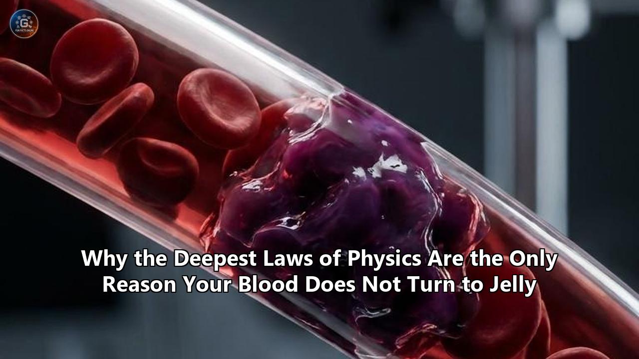
Without the continuous application of highly specific physical laws governing non-Newtonian fluids, your blood would immediately cease to flow, collapsing into a gelatinous matrix. The heart is not merely a pump pushing a liquid; it is a mechanical agitator violently disrupting a solid.
This new visualization data provides the most detailed look yet at the yield stress tipping point of human plasma and red blood cells. It forces a fundamental reevaluation of how we treat cardiovascular disease, aneurysms, and catastrophic trauma. Doctors are no longer viewing circulation merely as a biological plumbing issue. Instead, they are approaching the vascular network through the lens of thermodynamics, fluid mechanics, and quantum-level electrostatic repulsion.
The Edge of Solidification: Mapping the Yield Stress Tipping Point
To understand the magnitude of this week’s imaging breakthrough, one must discard the assumption that blood acts like water. Water is a Newtonian fluid. Its viscosity remains constant regardless of the forces applied to it. Whether it is sitting still in a glass or rushing through a fire hose, water fundamentally remains water.
Blood actively defies this behavior. It is a multiphase suspension consisting of erythrocytes (red blood cells), leukocytes (white blood cells), and thrombocytes (platelets) suspended in a complex protein-rich plasma. Because of this dense particulate composition, blood behaves as a pseudoplastic, non-Newtonian fluid.
At rest, or under extremely low shear stress, blood behaves more like a solid than a liquid. The red blood cells, which are flexible biconcave discs, naturally tend to stick together. They stack flat against one another like coins tightly rolled in a paper wrapper, forming microscopic structures called rouleaux. As these rouleaux structures interlock, they form a three-dimensional lattice throughout the plasma. If blood flow stops, this lattice rapidly achieves a yield stress threshold—the minimum amount of force required to make a material flow. Below this threshold, blood is physically a gel.
The heart’s primary job is to generate enough mechanical shear stress to shatter these rouleaux structures before they become permanent. With every systolic contraction, a shockwave of kinetic energy races through the arterial tree. This energy forces the stacked red blood cells to peel apart, deform, and slide past one another. The harder the fluid is pushed, the thinner and more slippery it becomes. This property is known as shear-thinning.
The new birefringence imaging data visually confirms how these stress interactions operate at the sub-millimeter level. The imaging technique passes polarized light through a vascular phantom—a synthetic blood vessel—and a specialized nanocellulose dispersion mimicking blood. As the artificial heart pulses, the light refracts differently based on the localized stress fields. For the first time, researchers watched the precise millisecond where the non-Newtonian gel matrix shattered under shear stress, transitioning into a highly fluid state to squeeze through capillaries smaller than the cells themselves.
When Ketchup Mechanics Keep You Alive
The mechanical principles keeping humans alive share a direct mathematical lineage with common household fluids like ketchup, paint, and toothpaste. All of these are Bingham plastics or Casson fluids.
Getting ketchup out of a glass bottle requires a sharp tap. The fluid refuses to move until a specific force threshold is crossed, at which point its viscosity suddenly drops and it flows rapidly. Blood operates on the exact same Casson fluid model, a mathematical equation originally developed to characterize pigment-oil suspensions in printing ink varnishes.
The Casson model dictates that a fluid possesses a yield stress that must be overcome before flow initiates. In the human body, this yield stress is a literal life-or-death metric. If the yield stress of blood becomes too high—due to dehydration, disease, or abnormal protein levels—the heart cannot generate enough shear force to break the rouleaux structures in the body's furthest extremities. The blood effectively turns to gel inside the capillaries, leading to tissue death, necrosis, and ischemia.
Biophysicists utilize the Casson model, alongside the more complex Carreau-Yasuda model, to calculate the infinite-shear viscosity and zero-shear viscosity of blood. Recent computational fluid dynamics studies have demonstrated that relying on simple Newtonian models to simulate blood flow vastly underestimates the danger of low-shear regions in the body. When blood enters a wide section of a diseased artery, the flow slows down. The shear rate plummets. In that exact localized zone, the blood begins to rapidly thicken, reverting back toward its gel state.
This behavior is entirely dictated by the physics of human biology. Biological survival requires a material that can exist simultaneously as a liquid in high-velocity arteries and as a near-solid in the sluggish venous return of the lower legs. This dual nature is what allows a complex organism to regulate internal pressure, halt bleeding when punctured, and direct oxygen exactly where it is needed most.
Red Blood Cell Rouleaux and the Electrostatic Dance
Why do red blood cells want to stack together in the first place, and what prevents them from permanently fusing into a solid mass? The answer lies in the delicate balance of intermolecular forces, specifically van der Waals forces and electrostatic repulsion.
Fibrinogen, a massive, elongated protein found in blood plasma, is the primary driver of rouleaux formation. At low velocities, fibrinogen molecules act like molecular bridges, binding to the surfaces of adjacent red blood cells. The attractive van der Waals forces between these cellular structures encourage them to aggregate, minimizing their thermodynamic free energy. The cells want to rest. They want to stick together.
However, each red blood cell is coated in a layer of sialic acid, giving the cell membrane a net negative electrical charge. This creates an electrostatic boundary known as the Zeta potential. Because all red blood cells carry this negative charge, they inherently repel each other.
This creates a spectacular physical standoff inside your veins. The attractive forces of the fibrinogen proteins are constantly fighting the repulsive electrostatic forces of the Zeta potential.
When the heart pumps, the kinetic shear force tilts the balance, allowing the electrostatic repulsion to push the cells apart so they can flow freely. When the heart rests between beats, or when blood pools in the deep veins of the legs during a long flight, the shear force drops to zero. The fibrinogen bridges overcome the electrostatic repulsion, and the blood begins to gel.
If the Zeta potential is compromised—often due to systemic inflammation, heavy metal toxicity, or severe infection—the negative charge on the red blood cells drops. The cells clump together aggressively, raising the yield stress of the blood so high that even the heart’s maximum output cannot liquefy it. This condition, known as hyperviscosity syndrome, forces the cardiovascular system into catastrophic failure.
The Physics of Human Biology: Bridging Thermodynamics and Hemodynamics
Understanding the physics of human biology requires looking past the biological descriptions of cells and tissues to see the raw mechanical forces governing their behavior. Hemodynamics—the study of blood flow—is essentially applied thermodynamics and fluid mechanics.
Every time a red blood cell deforms to pass through a capillary, it absorbs and dissipates mechanical energy. The cellular membrane is viscoelastic; it stores energy elastically like a rubber band and dissipates it viscously like honey. This energy transfer generates minute amounts of heat, contributing to the thermal regulation of the human body.
Recent research into spatial fractional derivative models has revealed that the physical behavior of blood is non-local. This means that the viscosity and flow characteristics of blood at a specific point in an artery are not just determined by the conditions at that exact point. They are influenced by the “memory” of the shear stress the blood experienced just milliseconds earlier upstream.
Because red blood cells take a fraction of a second to relax back to their resting biconcave shape after being stretched, the fluid retains a structural memory of its previous deformation. When mapping blood flow through narrow, diseased arteries, physicists now apply fractional calculus to account for this non-local memory effect. The new mathematical frameworks prove that biological fluids are deeply entangled with time-dependent physical laws. You cannot understand how blood will act in the brain without calculating the physical trauma it just experienced passing through the carotid artery.
Computational Fluid Dynamics: Predicting the Aneurysm's Breaking Point
The integration of advanced physics into biological research is rapidly altering how surgeons evaluate vascular threats, particularly cerebral aneurysms. An aneurysm is a balloon-like bulge in the wall of a blood vessel, most commonly found in the brain or the abdominal aorta.
Historically, physicians assessed the rupture risk of an aneurysm based on its size. Large aneurysms were operated on; small ones were watched. But clinical reality proved this approach inadequate. Many small aneurysms ruptured unexpectedly, while large ones remained stable for decades. The missing variable was not the size of the biology, but the mechanics of the fluid.
By utilizing computational fluid dynamics (CFD), biophysicists now create 3D patient-specific simulations of blood flow inside the brain. These models run on the same supercomputing architecture used to test the aerodynamics of hypersonic jets. By inputting the Carreau-Yasuda non-Newtonian blood parameters and mapping the specific geometry of a patient's arterial network, algorithms can predict exactly where the physical forces are eroding the vessel wall.
When blood rushes into the sac of an aneurysm, the flow pattern fractures. The fluid spins into chaotic vortices. In the center of the aneurysm, the flow velocity drops near zero. Because blood is shear-thinning, this low-velocity zone causes the blood viscosity to spike exponentially. The fluid thickens, forming a sluggish, gel-like core.
Simultaneously, at the neck of the aneurysm, the blood whips around the curve at high speed, exerting massive Wall Shear Stress (WSS) against the fragile tissue. The non-Newtonian nature of blood exacerbates the danger: the highly viscous gel core forces the fast-moving liquid outward, grinding it against the arterial wall.
Recent computational studies evaluating ischemic stroke and aneurysm hemodynamics emphasize that assuming blood behaves like a Newtonian fluid creates dangerous blind spots in medical models. Newtonian models systematically overestimate the safety of complex vascular geometries because they fail to account for how the blood thickens and redirects shear stress when it slows down. By integrating the exact physics of phase-transition into medical software, surgeons can pinpoint the specific square millimeter of an artery that will fail under the mechanical load.
New Imaging Techniques: Dynamic Birefringence and the Invisible Forces of Flow
Until this month's breakthroughs, visualizing these complex fluid mechanics in real time was mathematically possible but experimentally impossible. Blood is opaque. You cannot shine a laser through it to map its internal velocity with standard optical tools.
To solve this, experimental physicists turned to dynamic solid-liquid birefringence. Birefringence is an optical property where a material has a refractive index that depends on the polarization and propagation direction of light. It is commonly seen in clear plastics; when you bend a plastic ruler between polarized lenses, rainbow-colored stress bands appear.
Researchers built transparent, artificially elastic blood vessels out of specialized polymer gels. Instead of using actual opaque blood, they developed a birefringent nanocellulose dispersion—a clear liquid that perfectly mimics the exact shear-thinning and yield stress properties of human blood.
By pumping this fluid through the synthetic vessels using a mechanical device that replicates the human cardiac cycle, and illuminating the entire system with polarized light, the team successfully visualized the invisible stress interactions. The resulting imagery is striking. With every pulse, vivid shockwaves of mechanical stress illuminate the fluid and the vessel walls simultaneously.
This technique provides an unprecedented look at how the solid boundary of a blood vessel and the liquid boundary of the blood interact during a heartbeat. When a vessel narrows due to plaque, the birefringence data shows massive spikes in localized stress, not just on the vessel wall, but propagating deep into the fluid itself. This proves that the cardiovascular system operates as a single, unified mechanical entity. The physics of the container and the fluid cannot be separated.
The Diabetic Disconnect: When the Gel Threshold Fails
The precise balance between flowing liquid and stagnant gel is uniquely vulnerable to metabolic changes, specifically high blood glucose levels. Diabetes is often viewed primarily as an endocrine disorder, a failure of insulin regulation. However, the most devastating long-term consequences of diabetes—blindness, kidney failure, and lower-limb amputations—are fundamentally rheological and biophysical failures.
When blood glucose levels remain chronically elevated, excess sugar molecules bind to proteins in the blood and on the surface of red blood cells through a process called glycation. This fundamentally alters the physical properties of the fluid.
Glycated red blood cells become stiffer. They lose their viscoelastic elasticity. When they arrive at the capillary beds, they can no longer deform smoothly to slide through the microscopic tunnels. Furthermore, the elevated glucose levels alter the plasma viscosity and disrupt the Zeta potential, causing the cells to aggregate more aggressively. The yield stress of diabetic blood is significantly higher than that of healthy blood.
In the microvasculature of the toes or the retinas, the blood pressure drops significantly. The shear rate falls. Because diabetic blood has a higher yield stress, it reaches its gel-transition point prematurely. The blood literally stops flowing before it reaches the end of the capillary bed. It thickens into a solid matrix, starving the surrounding tissue of oxygen.
This biophysical reality is driving a new generation of therapeutic interventions. Recently, bioengineers introduced a breakthrough “smart” hydrogel designed to treat diabetic wounds that refuse to close. The hydrogel utilizes tiny cellular messengers called vesicles, combined with microRNAs, to locally manipulate the microenvironment of the wound.
Instead of just covering the injury, this smart gel actively interfaces with the damaged endothelial cells, overriding the signals caused by high glucose. It suppresses the expression of certain proteins that inhibit blood vessel growth, drastically accelerating angiogenesis—the formation of new blood vessels. By physically restructuring the local tissue architecture, the gel lowers the local resistance, allowing the thickened diabetic blood to finally push through and deliver the oxygen necessary for tissue regeneration.
Gluing Blood on Purpose: The Strange Future of Embolic Agents
While doctors spend immense effort ensuring blood does not solidify in the heart or brain, there are critical moments in medicine when rapidly inducing this gel phase is exactly what is needed. When a patient suffers a severe hemorrhage, or when a surgeon needs to starve a cancerous tumor of its blood supply, the goal is to force the blood to cross the phase boundary and solidify instantly.
Traditionally, surgeons used mechanical coils or toxic chemical solvents to block blood vessels. These methods are imprecise and carry severe risks of damaging surrounding healthy tissue. However, a deep understanding of the physics of human biology has led to the development of highly advanced liquid embolic agents that manipulate the electrical charge of blood to freeze it in place.
Recent bioengineering studies highlight the development of water-soluble polymers designed to glue blood into a gel utilizing pure electrostatic interaction. These polymers are composed of specific cationic (positively charged) and aromatic sequences.
When this polymer is injected into a bleeding artery via a microcatheter, it does not trigger the biological coagulation cascade. It bypasses biology entirely and acts on the physics. The positively charged polymer immediately seeks out the negatively charged Zeta potential of the red blood cells and the plasma proteins. Through intense electrostatic adhesion and cation-π interactions, the polymer physically binds the flowing cells together in a fraction of a second.
The blood instantly transitions from a shear-thinning fluid into a solid-like gel. It plugs the artery perfectly, conforming to the exact microscopic geometry of the vessel. Because the process relies on physical electrostatic binding rather than biological clotting factors, it works instantaneously, even in patients who are on heavy blood thinners. This technique represents a massive paradigm shift in trauma surgery, proving that manipulating the fundamental physical state of blood offers capabilities that standard pharmacology cannot match.
The Architectural Ice Age: 3D-Printing the Vascular Network
The exact mechanical properties that make blood so resilient inside the body make it notoriously difficult to replicate or support outside the body. For decades, the holy grail of regenerative medicine has been creating lab-grown organs. Scientists can successfully grow heart muscle tissue and liver cells in Petri dishes, but keeping a full-sized 3D organ alive requires a vascular network. You have to pump blood through it, or the center of the organ dies of hypoxia.
Creating synthetic blood vessels that can withstand the complex fluid dynamics and non-Newtonian shear forces of human blood has proven exceptionally difficult. The vessels must be microscopically thin, yet strong enough to handle pulsatile pressure, and smooth enough that they do not trigger the blood's innate tendency to gel upon contacting a foreign surface.
Researchers have turned to extreme phase transitions to solve this engineering bottleneck. By utilizing the physics of freezing water, engineers have developed a method to 3D-print synthetic blood vessel networks using ice as a temporary template.
Drawing inspiration from the way water expands and solidifies into branching crystalline structures, researchers use advanced 3D printers to sculpt incredibly intricate, branching networks out of ice. They then pour a viscous, tissue-compatible gel around the ice structure. Once the surrounding gel cures into a solid, the team melts the ice and drains the water.
What remains is a perfect, hollow network of microscopic channels embedded within an artificial tissue matrix. Endothelial cells—the cells that line natural blood vessels—are then flushed into these hollow channels. Because the channels were formed by the physical phase transition of water, they possess the precise geometry and smoothness required to allow non-Newtonian blood analogues to flow without triggering the yield-stress gelation response.
This icy bio-printing technique bypasses the limitations of trying to print hollow tubes directly. By manipulating phase boundaries—freezing water to create space, then melting it to allow fluid dynamics to take over—engineers are building the literal foundations for artificial organ transplants.
The Evolutionary Advantage of Metastability
Why would evolution select for a fluid that is constantly on the verge of catastrophic solidification? Why not pump a simple, low-viscosity Newtonian fluid that is immune to gelation?
The answer is survival under trauma. A Newtonian fluid cannot seal its own container. If you puncture a pipe carrying water, the water will continue to leak until the pressure drops to zero. If the human body utilized a Newtonian fluid, a simple paper cut would be fatal. The blood would flow out with zero resistance.
The deep physics of human biology require metastability—a state of precarious balance. By existing perfectly on the borderline between liquid and solid, blood is primed to instantly change its physical state the moment the environment demands it.
When a blood vessel is severed, the internal pressure drops, and the shear rate plummets. The blood escaping the wound instantly slows down. Because it is a non-Newtonian fluid, this drop in velocity causes an immediate, massive spike in viscosity. The blood thickens right at the exit point. Simultaneously, the biological coagulation cascade triggers, polymerizing fibrinogen into a solid fibrin mesh that catches the already-thickening red blood cells.
The physical shear-thinning property of blood buys the biological clotting mechanism the crucial seconds it needs to build a permanent seal. Without the underlying fluid mechanics of yield stress and rouleaux aggregation, the biological clotting factors would just be washed out of the wound before they could take hold. The physical laws of the universe are the scaffolding upon which the biological survival mechanisms are built.
Redefining the Parameters of Circulation
The synthesis of dynamic birefringence imaging, computational fluid dynamics, and fractional calculus is permanently altering the medical perspective on human circulation. The cardiovascular system is no longer viewed purely through the lens of cardiology or hematology. It is an active laboratory of high-level physics.
The realization that blood flow incorporates non-local spatial memory—that the fluid remembers the mechanical stress it experienced heartbeats ago—opens entirely new avenues for diagnostics. By scanning the geometry of a patient's carotid artery, supercomputers can now theoretically calculate the exact physical state of the blood as it reaches the cerebral microvasculature. Doctors can identify areas where the shear rate is dangerously close to zero, predicting capillary death long before a patient experiences cognitive decline.
Furthermore, as researchers continue to map the exact electrostatic parameters of the Zeta potential, we are nearing the ability to chemically fine-tune the yield stress of a patient's blood in real time. Instead of using blunt biological blood thinners like heparin, which block clotting factors and increase the risk of severe bleeding, future therapeutics may subtly increase the electrostatic repulsion between red blood cells. By slightly boosting the negative charge, doctors could lower the yield stress of the blood, allowing it to flow easily through diseased, narrowed arteries without entirely disabling the body's ability to seal a wound.
Unresolved Boundaries and the Next Generation of Vascular Medicine
Despite the massive leap forward provided by this week's birefringence imaging data, profound questions remain regarding the quantum and thermal interactions at the phase boundary.
Current mathematical models, even those utilizing fractional derivatives, struggle to accurately simulate blood flow under extreme temperature variations. When treating severe frostbite, or when surgeons intentionally cool a patient's body during complex cardiac surgery to slow cellular metabolism, the blood behaves unpredictably. The drop in temperature alters the kinetic energy of the water molecules in the plasma, radically shifting the van der Waals forces between the fibrinogen proteins. Mapping the exact thermal threshold where low-temperature blood irreversibly transitions into a gel remains a critical objective for cryogenic medicine.
Additionally, the role of hybrid nanoparticles in regulating blood flow is emerging as a major frontier. Recent numerical analyses have demonstrated that injecting specific magnetic nanoparticles into the bloodstream, and then applying an external magnetic field, can artificially manipulate the velocity and temperature of non-Newtonian blood flow deep within the body. This magneto-hydrodynamic approach could eventually allow doctors to use targeted magnetic fields to physically push stagnant blood through a blocked artery or rapidly heat up a localized gelled mass to restore circulation.
The mapping of the yield stress tipping point is not an ending, but a foundational baseline. We now possess the visual and mathematical proof that our blood is not merely flowing; it is constantly fighting an intricate, high-stakes battle against the fundamental forces of physics. The next decade of vascular medicine will rely on our ability to not just observe these physical laws, but to master and manipulate the precise thresholds that keep the human machine in motion.
Reference:
- https://www.researchgate.net/publication/325488821_A_REVIEW_ON_RHEOLOGY_OF_NON-NEWTONIAN_PROPERTIES_OF_BLOOD
- https://pubs.aip.org/aip/pof/article-abstract/37/8/081907/3358007
- https://pubs.aip.org/aip/pof/article/37/6/063113/3349334/Numerical-analysis-of-blood-flow-and-heat-transfer
- https://www.aimspress.com/article/doi/10.3934/biophy.2026004?viewType=HTML
- https://pmc.ncbi.nlm.nih.gov/articles/PMC12644918/
- https://www.sciencedaily.com/releases/2025/08/250807233035.htm
- https://www.researchgate.net/publication/364379764_Gluing_blood_into_gel_by_electrostatic_interaction_using_a_water-soluble_polymer_as_an_embolic_agent
- https://www.snexplores.org/article/innovation-2024-frozen-movie-inspired-ice-3d-printing-blood-vessels