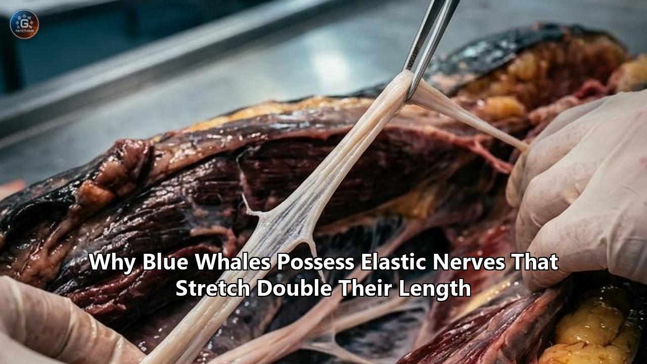
Researchers investigating the biomechanics of giant marine mammals have unraveled the multi-tiered structural geometry that allows rorqual whales—most notably the blue whale (Balaenoptera musculus)—to stretch their peripheral nerves to more than double their resting length. The findings, resolved through high-resolution micro-computed tomography (micro-CT) and mechanical strain testing, explain a biological paradox that has long puzzled neurobiologists: how the largest animals on Earth repeatedly deform their nervous systems during feeding without inducing catastrophic neurological failure.
When a 100-ton blue whale opens its jaws to engulf a swarm of krill, its ventral groove blubber expands like an elastic accordion, taking in a volume of seawater that can exceed the whale's own body mass. This sudden engulfment stretches the tissues in the floor of the mouth and tongue by up to 160%. In any standard vertebrate, such an extreme displacement would shred peripheral nerve fibers, snap blood vessels, and inflict permanent paralysis. Yet, the nerves supplying the blue whale's tongue and throat extend rapidly under load and recoil instantly as the water is expelled, maintaining uninterrupted electrical conduction throughout the dive.
The discovery reveals an evolutionary solution fundamentally distinct from any other vertebrate nervous system. By examining the micro-architecture of these tissues, biophysicists have shown that blue whale nerves do not stretch at the cellular level in the way an elastic band does. Instead, they rely on a nested, two-tier structural hierarchy—combining macro-scale sine-wave folding with micro-scale fascicular undulations—encased inside an elastomeric outer sleeve reinforced with a collagenous "check ligament".
Understanding this mechanism does more than solve a mystery of marine megafaunal evolution; it provides a direct blueprint for bio-inspired materials, soft robotics, and flexible neural implants designed to withstand severe mechanical strain.
The Biophysical Limit: Why Stretch Kills Normal Nerves
To understand why the blue whale's neural adaptation is so extraordinary, one must examine how peripheral nerves behave across the rest of the animal kingdom.
In terrestrial mammals, including humans, peripheral nerves are taut, relatively inelastic structures. They are composed of individual nerve fibers (axons) bundled into fascicles, wrapped in protective connective tissue sheaths: the endoneurium surrounding individual axons, the perineurium bounding fascicles, and the epineurium enclosing the entire nerve trunk. While mammalian nerves possess a slight degree of natural slack—visible under a microscope as tiny, wavy folds called the Spiral Bands of Fontana—this structural buffer only accommodates minor physiological movements.
MAMMALIAN NERVE STRAIN SPECTRUM
[ 0% Resting ] --------> [ 6-10% Elastic Strain Limit ] --------> [ 12-20% Conduction Failure ] --------> [ >29% Physical Rupture ]
When a human peripheral nerve is subjected to mechanical tension, the response follows a rigid stress-strain curve:
- 0% to 6% Strain: The resting microscopic waviness (Spiral Bands of Fontana) straightens out. The nerve experiences negligible mechanical stress.
- 6% to 10% Strain: The connective tissue sheaths begin to bear load. Ischemia (blood flow restriction) begins as intraneural blood vessels are compressed, reducing oxygen delivery to axons.
- 10% to 20% Strain: Nerve action potentials degrade rapidly. The myelin sheaths insulating the axons begin to shear, and the Nodes of Ranvier—the gaps responsible for saltatory signal conduction—are physically distorted.
- Beyond 20% to 29% Strain: The nerve suffers irreversible mechanical failure. Axons snap, the perineurium ruptures, and permanent neurological deficit or paralysis ensues.
For a terrestrial mammal, a 30% stretch is a traumatic injury. For a blue whale, a 100% stretch is an ordinary Tuesday lunchtime.
If blue whale nerves were built like standard mammalian nerves, a single lunge-feeding event would sever the vagus and hypoglossal nerve branches supplying the floor of the mouth, paralyzing the animal's feeding apparatus and causing immediate starvation.
The Physics of the Lunge: Engulfment Mechanics in Giant Rorquals
The requirement for hyper-extensible neural tissue is driven directly by the extreme mechanics of rorqual lunge feeding. Rorquals—a family of baleen whales that includes blue, fin, humpback, and minke whales—are the only vertebrates that feed by taking in a volume of water larger than their own body dimensions.
                  LUNGE-FEEDING CYCLE IN BLUE WHALES

    [ High-Speed Approach ]  --->  Accelerates to 3-5 m/s toward krill patch
              |
              v
    [ Jaw Opening & Engulfment ] -> Mandible opens ~80-90°; water rushes in
              |                    Ventral groove blubber balloons 160%
              v                    Nerves untether and uncoil to >200% length
    [ Expulsion & Filtration ] ---> Jaws close; tongue/VGB contract
              |                    Seawater forced out through baleen
              v                    Nerves passively recoil into folded core
During a feeding lunge, a 30-meter blue whale accelerates to speeds of 3 to 5 meters per second toward a dense swarm of krill. As it nears the prey, it opens its lower jaw to an angle approaching 80 to 90 degrees. The forward momentum forces seawater into the oral cavity, creating an immense hydrodynamic drag force that expands the ventral groove blubber (VGB)—a specialized layer of pleated, expandable tissue extending from the chin down to the umbilicus.
The physical parameters of this event are staggering:
- Volume Intake: A mature blue whale can engulf up to 90 metric tons (90,000 liters) of water and krill in a single gulp lasting less than 10 seconds.
- Tissue Deformation: The ventral floor of the mouth expands by roughly 160% in circumference and nearly 40% in length.
- Tongue Inversion: The massive tongue, which is largely non-muscular and highly extensible, is inverted and flattened into a thin pouch that coats the expanded floor of the mouth.
This dynamic transformation turns the lower jaw and throat into a giant, ballooning pocket. The major nerve trunks traversing this area—principally the ventral branches of the trigeminal, facial, hypoglossal, and vagus nerves—must bridge the distance between the fixed cranium and the distended body wall.
Because these nerves are embedded directly within the expanding blubber matrix, they are forced to elongate from a resting length of roughly 1.5 meters to an extended length exceeding 3 meters during every single lunge.
Comparative Analysis: Four Evolutionary Solutions to Nerve Elongation
Nature has confronted the challenge of nervous system deformation in several distinct animal lineages. Comparing these strategies highlights why the mechanism found in blue whale nerves represents a unique evolutionary architecture.
===================================================================================================
COMPLEXITY & EXTENSIBILITY MATRIX OF NERVOUS SYSTEM ADAPTATIONS
===================================================================================================
Organism / Group      Max Strain Limit  Primary Mechanism                     Key Structural Feature
---------------------------------------------------------------------------------------------------
Standard Mammal       6% - 12%          Microscopic Unfolding                 Spiral Bands of Fontana
(e.g., Human, Dog)                      (Minimal slack)                       Stiff Collagen Epineurium

Ballistic Reptile/    20% - 40%         Anatomical Slacking & Loop Tethering  Pre-formed Macroscopic
Amphibian                               (Tissue displacement)                 Slack Loops in Cavities
(e.g., Chameleon)

Hyperextensible       150% - 275%       Viscoelastic Material Strain          Unmyelinated Axons;
Invertebrate                            (Seasonal hormonal softening)         Low-pressure Axoplasm
(e.g., Locust Female)

Rorqual Cetacean      100% - 115%       Two-Tier Hierarchical Unfolding +     Sine-generated Wave Core;
(e.g., Blue Whale)                      Elastomeric Recoil Sheath             Elastin-Collagen Sleeve
===================================================================================================
1. The Standard Mammalian Model: Taut Strain Resistance
In terrestrial mammals, the peripheral nervous system prioritizes structural stability and rapid signal transmission over flexibility. The outer epineurium is dominated by dense, cross-linked collagen fibers that resist mechanical deformation.
To handle minor joint movements (such as flexing an elbow or knee), the nerve slides within a loose connective tissue bed (the mesoneurium) and straightens its microscopic axonal undulations. The primary tradeoff of this approach is an extreme vulnerability to traction injuries: if the strain exceeds 10%, conduction drops, and if it exceeds 20%, axonal loss occurs.
2. The Ballistic Tongue Model (Chameleons and Frogs)
Chameleons and specialized amphibians project their tongues rapidly over long distances to capture prey. To accommodate this projection, the hypoglossal nerves supplying the tongue must span significant distances.
However, micro-anatomical studies show that chameleon nerves do not actually stretch elastically under high tension. Instead, the nerve is folded into loose, serpentine loops within large, lubricated anatomical cavities in the throat when at rest. When the tongue is fired, these pre-stored slack loops simply straighten out. The tradeoff is space: storing long, coiled nerve loops requires bulky internal cavities, a spatial luxury that cannot be accommodated within the dense, pressurizing blubber layer of a blue whale.
3. The Unmyelinated Invertebrate Model (Female Locusts)
During egg-laying, the female desert locust (Locusta migratoria) burrows her abdomen deep underground, extending her body and her abdominal nervous system to between 200% and 275% of their original length.
Biomechanical testing has revealed that locust nerves achieve this hyperextensibility through true viscoelastic deformation of the neural tissue itself. However, this solution is limited by two key constraints:
- Insect axons are unmyelinated, meaning they lack the delicate, rigid myelin sheath wraps that insulate mammalian nerves. Unmyelinated axons can endure much higher shearing forces without losing the structural integrity of a myelin wrap.
- The extension is slow and heavily controlled by hormonal changes during specific reproductive phases, rather than being subjected to rapid, cyclic loading hundreds of times a day.
4. The Rorqual Model: Dual-Scale Hierarchical Unfolding
Blue whales cannot adopt the insect strategy because their axons require thick myelin insulation to maintain high conduction velocities across multi-meter body lengths. Nor can they adopt the chameleon strategy, as loose nerve loops would entangle or burst under the crushing hydrodynamic pressures of lunge feeding.
Instead, blue whale nerves utilize a nested hierarchical unfolding system. The nerve fibers themselves remain strain-free, protected inside a thick, elastomeric tube that handles the tensile mechanics.
Architectural Deep-Dive: How Blue Whale Nerves Stretch Double Their Length
The structural secret of blue whale nerves lies in a distinct anatomical separation between the mechanical load-bearing elements and the electrical signal-conducting elements.
In a typical nerve, the electrical axons and the structural sheath are fused into a tight, integrated cable. In a blue whale, the nerve is organized like a flexible electrical wire housed within a thick, rubbery garden hose.
                     CROSS-SECTION OF A BLUE WHALE NERVE

   +-----------------------------------------------------------------+
   | OUTER ELASTOMERIC SHEATH                                        |
   |  - Thick Elastin Core: Provides elastic recoil force            |
   |  - Parallel Collagen Outer Limit: Acts as high-stiffness stop   |
   |                                                                 |
   |    +-------------------------------------------------------+    |
   |    | INNER FOLDED NERVE CORE (Sine-Generated Curve)        |    |
   |    |                                                       |    |
   |    |    ~~~~~ Fascicle Wave Scale 1 (Macro-buckle) ~~~~~  |    |
   |    |    ||||| Fascicle Wave Scale 2 (Micro-undulation) ||| |    |
   |    |                                                       |    |
   |    +-------------------------------------------------------+    |
   +-----------------------------------------------------------------+
The Outer Sheath: Elastin Meets Collagen
The outer wall of the nerve trunk is exceptionally thick, accounting for more than 50% of the nerve's total cross-sectional area. This wall is composed of two distinct extracellular matrix proteins working in tandem:
- Elastin Matrix: The primary structural component of the inner wall is elastin, a highly springy protein that can undergo large, reversible elastic deformation. When the whale lunges, the elastin fibers stretch linearly under low mechanical force, storing elastic potential energy.
- Collagen "Check Ligament": Woven around and through the elastin is a dense network of stiff, non-extensible collagen fibers. In the resting state, these collagen fibers are crimped and loose. As the nerve approaches ~115% elongation (more than double its length), the collagen fibers pull completely taut. At this point, the nerve's stiffness increases exponentially, acting as a mechanical "check ligament" that physically prevents further extension, protecting the internal tissues from over-stretching and tearing.
The Two-Tiered Wavy Core
Inside this elastomeric hose sits the central nerve core containing the axon fascicles. Micro-CT imaging conducted by researchers at the University of British Columbia revealed that this core is not straight; it is heavily folded in a complex three-dimensional pattern.
Crucially, the nerve core employs two distinct length scales of waviness to handle extension and contraction:
   RESTING STATE (Unstretched)
   Macro-Wave:  /\  /\  /\  /\  (Sine-generated curve / river meander)
   Micro-Wave:  vvvvvvvvvvvvvvvv  (Tight fascicle undulations along the curve)

   FULLY EXTENDED STATE (115% Strain)
   Macro-Wave:  ----------------  (Macro-wave completely straightened)
Micro-Wave: ---------------- (Micro-undulations unfurled; axons remain unstrained)- Tier 1: Macro-Scale Core Buckling (The Sine-Generated Curve)
At rest, the entire central nerve core is buckled into a 3D serpentine shape that resembles a meandering river. Mathematicians categorize this specific geometry as a sine-generated curve. This shape minimizes the maximum bending strain along the path of the core. When the outer sheath stretches during a lunge, this macro-wave simply uncoils and straightens out, providing roughly 75% to 80% of the total nerve elongation without putting any direct tensile strain on the internal fibers.
- Tier 2: Micro-Scale Fascicular Undulation
A fundamental law of beam physics dictates that when a rod or tube is bent into a curve, the material on the outer edge of the bend is stretched, while the material on the inner edge is compressed.
If the whale's nerve core relied solely on macro-scale folding, the tight bends of the sine wave would create localized stretch strains exceeding 60% on the outer edges of the curves—easily high enough to damage the delicate nerve fascicles. To prevent this localized bending strain, individual fascicles inside the core possess a second, smaller level of microscopic undulation. On the outer edge of a core bend, these micro-folds unfurl to supply extra slack; on the inner edge, they stay compressed.
By partitioning the mechanical deformation across two structural tiers, the actual nerve axons inside blue whale nerves experience virtually zero direct mechanical tension throughout the entire stretch cycle. When the whale closes its jaw and expels the water, the outer elastin sheath recoils entropic energy, smoothly repacking the nerve core back into its original, folded resting geometry.
Neurophysiological Trade-offs: Electrical Conduction Under Severe Strain
While the two-tier nested geometry solves the mechanical problem of stretch, it introduces a severe neurophysiological challenge: how to maintain fast, efficient electrical signaling along a nerve that continuously changes its length, geometry, and internal diameter.
===================================================================================================
MECHANICAL & ELECTRICAL TRADE-OFF ANALYSIS IN HYPER-EXTENSIBLE NERVES
===================================================================================================
Parameter               Resting State (Unstretched)           Extended State (115% Stretch)
---------------------------------------------------------------------------------------------------
Physical Path Length    Short (~1.5 meters)                   Long (~3.2 meters)
Nerve Core Geometry     Folded 3D Sine Wave                   Straightened Cable
Axonal Cross-Section    Normal Diameter ($d$)                 Thinning Diameter ($d - \Delta d$)
Internal Resistance     Baseline Axial Resistance ($R_a$)     Increased $R_a$ due to thinning
Conduction Delay        Low propagation latency               Slightly increased signal travel time
Myelin Status           Compressed, overlapping sheaths       Extended, sliding lamellae
Nodes of Ranvier        Standard width (~1 $\mu$m)             Widened gap (within safe limit)
===================================================================================================
The Cable Theory Challenge
In neurobiology, nerve impulse propagation velocity ($v$) along a myelinated axon is governed by cable theory:
$$v \propto d_{axon}$$
where $d_{axon}$ is the internal diameter of the axon. The axial electrical resistance ($R_a$) of the axoplasm is inversely proportional to the cross-sectional area:
$$R_a = \frac{4\rho}{\pi d_{axon}^2}$$
In a hypothetical scenario where an axon is physically stretched and thinned out, two negative electrical effects occur simultaneously:
- The physical distance the electrical signal must travel doubles.
- The axonal diameter ($d_{axon}$) shrinks, causing axial resistance ($R_a$) to surge and slowing signal conduction velocity.
The Blue Whale Counter-Strategy
Because blue whale nerves elongate via geometric unfolding rather than true axonal elongation, the individual axons do not undergo significant cross-sectional thinning. However, the physical path length does increase during a lunge, which inherently increases total signal transit time from the brainstem to the throat muscles.
To handle this dynamic path length, the cetacean nervous system employs three specialized micro-cellular adaptations:
- Myelin Lamellar Sliding: Microscopic studies of mammalian nerve strain demonstrate that as a nerve straightens, the concentric layers of lipid-rich myelin surrounding the axon slide past one another—a process known as lamellar shear. In rorquals, the myelin sheaths possess enhanced fluid-membrane properties that allow extensive inter-lamellar sliding without delaminating or unspooling from the axon.
- Dynamic Node Widening: At the Nodes of Ranvier, where voltage-gated sodium channels generate action potentials, the outer membrane sheaths widen slightly during elongation. The ion-channel clusters remain intact within the nodal membrane, preserving saltatory conduction even when the nerve is fully extended.
- High Baseline Axon Diameters: Rorquals possess exceptionally large motor axon diameters relative to terrestrial mammals. By maintaining a large baseline diameter, the nerve ensures that even at maximum elongation, the internal resistance ($R_a$) remains low enough to drive rapid motor signals to the ventral groove blubber muscles, triggering jaw closure and throat contraction.
Engineering Paradigms: Biomimicry vs. Current Stretchable Electronics
The mechanical architecture of blue whale nerves offers a major structural model for bio-inspired engineering. Materials scientists and roboticists have spent decades attempting to build stretchable, conductive cables for soft robotics, wearable health monitors, and bio-electronic prosthetics.
Comparing current artificial engineering approaches against the blue whale model illustrates how far natural evolution has outpaced synthetic design.
===================================================================================================
COMPARATIVE PARADIGMS: ARTIFICIAL STRETCHABLE CONDUCTORS VS. BLUE WHALE ARCHITECTURE
===================================================================================================
Technology Paradigm     Max Strain  Fatigue Resistance    Primary Mechanism of Failure
---------------------------------------------------------------------------------------------------
Serpentine Metal        30% - 50%   Low to Moderate       Interfacial delamination;
Traces on Elastomer                 (10,000 cycles)       micro-cracking from stress concentrations

Liquid Metal Channels   100% - 300% Moderate              Fluidic leakage; micro-pinhole punctures;
(e.g., EGaIn in Silicone)           (50,000 cycles)       high pressure impedance under deformation

Conductive Hydrogels    100% - 500% Poor                  Dehydration over time; ion leaching;
                                    (1,000 cycles)        low mechanical toughness under shear

Blue Whale Nerve        >115%       Exceptional           Biological self-repair; structural failure
Architecture                        (Millions of cycles)  prevented by non-linear collagen stop
===================================================================================================
Synthetic Approach 1: Serpentine Thin-Film Metal Traces
The classical engineering approach to stretchable electronics involves depositing ultra-thin metallic wires (gold, copper, or aluminum) in two-dimensional serpentine patterns onto an elastic silicone substrate. When the substrate stretches, the metal serpentines unbend in-plane.
- The Limitation: These systems suffer from intense stress concentrations at the apex of each curve. Under repeated cyclic stretching, microscopic fatigue cracks develop at these high-stress points, leading to sudden electrical open-circuits. Furthermore, max strain rarely exceeds 30% to 50% without substrate delamination.
Synthetic Approach 2: Liquid Metal Microfluidic Channels
Engineers often inject liquid metals—such as Eutectic Gallium-Indium (EGaIn)—into hollow elastomer micro-channels. As the elastomer tube stretches, the liquid metal flows inside the channel, maintaining continuous electrical contact even at strains exceeding 200%.
- The Limitation: Liquid metal channels are vulnerable to mechanical punctures, chemical degradation, and fluid displacement under localized pinching. When compressed, the internal cross-sectional area drops sharply, causing wide fluctuations in electrical resistance.
Synthetic Approach 3: Conductive Hydrogels
Hydrogels infused with ionic salts or conductive polymer networks can match the low mechanical stiffness of soft biological tissue while enduring high extensions.
- The Limitation: Hydrogels suffer from low mechanical toughness, susceptibility to drying out, and rapid performance degradation under continuous mechanical shear.
The Biomimetic Whale Paradigm: Hierarchical Core-Sheath Composite
The discovery of the rorqual nerve structure has inspired a new biomimetic paradigm in soft bio-electronics: the dual-scale hierarchical core-sheath conductor.
                     BIOMIMETIC WHALE-INSPIRED CONDUIT

    [ Elastomeric Polyurethane Sleeve ] -> Mimics outer elastin wall
    [ Woven Kevlar/Nylon Fibers ]       -> Mimics non-linear collagen check ligament
    [ Floating Liquid Metal Trace ]     -> Mimics 3D sine-generated nerve core
    [ Micro-Corrugated Sheath ]         -> Mimics fascicular micro-undulations
Instead of bonding conductive wires directly to an elastic base material, modern bio-inspired designs isolate the conductor inside an unattached fluidic or gel-lubricated central channel. The central wire is pre-formed into a 3D sine-generated wave, while the outer elastic casing is reinforced with braided non-extensible polymer threads (acting as synthetic collagen).
This design offers three major engineering advantages:
- Zero Strain on the Conductor: The electrical element experiences no tensile strain until the outer sheath reaches its hard mechanical stop.
- Immunity to Fatigue: By distributing bending stresses across a multi-tier wave hierarchy, peak localized stress is reduced by over 80%, extending the fatigue lifespan of the conductor over millions of strain cycles.
- Strain-Independent Resistance: Because the inner conductor uncoils rather than thins out, electrical resistance remains virtually constant during extension—a critical requirement for soft robotic sensors and neural prosthetic interfaces.
Evolutionary Scaling and Bioenergetics: Why Gigantism Demanded Stretchy Nerves
The emergence of hyper-extensible blue whale nerves was not an isolated anatomical quirk; it was an evolutionary absolute requirement for the emergence of marine gigantism.
                 EVOLUTIONARY CASCADE OF RORQUAL GIGANTISM

   Natural Selection for Large Body Size (Prey Abundance in Upwelling Zones)
                                    |
                                    v
            Requirement for Mass Filter Feeding (Lunge Engulfment)
                                    |
                                    v
     Evolution of Expandable Ventral Groove Blubber & Unfused Jaw Symphysis
                                    |
                                    v
             [ HYDRODYNAMIC CRITICAL MECHANICAL BOTTLENECK ]
        Extremely large tissue strain threatens peripheral nerves with rupture
                                    |
                                    v
   Co-Evolution of Two-Tier Hierarchical Elastic Nerves & Sensory Chin Organ
The positive allometry of lunge feeding
Biomechanical studies led by marine biologists have demonstrated that engulfment capacity in rorqual whales scales with positive allometry relative to body size. This means that as a whale grows longer, its mouth volume expands far faster than its overall body mass:
$$V_{engulf} \propto L_{body}^{3.5}$$
A blue whale that is twice as long as a minke whale can swallow nearly ten times the volume of water. This explosive volume scaling makes lunge feeding enormously energetic and efficient: a single lunge allows a adult blue whale to harvest up to 500,000 calories from a krill patch, far exceeding the metabolic cost of the high-speed dive.
However, this scaling law created a severe biomechanical limit:
- Larger engulfment volumes required disproportionately larger expansions of the floor of the mouth.
- Larger tissue expansion meant that the tissue strain in the ventral groove blubber increased from ~50% in small ancestral whales to over 150% in modern blue whales.
- Without a fundamental redesign of peripheral nerve architecture, evolutionary selection for larger body size would have halted at the strain limit of standard mammalian peripheral nerves.
The Sensory Chin Organ Synergy
The evolution of stretchy nerves also made possible another critical anatomical innovation discovered in rorquals: the sensory chin organ.
Embedded between the two unfused jawbones at the tip of the chin, this organ consists of a dynamic, gel-filled matrix packed with mechanoreceptors connected to the brainstem via the mandibular branch of the trigeminal nerve. As the jaw opens and the ventral pouch balloons outward, the sensory organ detects real-time changes in hydrodynamic pressure and tissue tension, sending rapid feedback signals to the brain to coordinate the precise moment of jaw closure.
Because the trigeminal nerve supplying this chin organ must traverse the entire length of the expanding lower jaw, it requires the exact same hyper-extensible two-tier wave structure. Without elastic nerves, this real-time sensory feedback loop would be physically severed at the start of every lunge, leaving the whale blind to the mechanical state of its own mouth.
Future Frontiers: Unresolved Questions in Cetacean Neurobiology
The discovery of two-tier wave dynamics and elastomeric sheath recoil in blue whale nerves has answered how these ocean giants feed, but it has opened up fundamental questions at the intersection of biophysics, cell biology, and clinical medicine.
===================================================================================================
KEY RESEARCH FRONTIERS IN EXTENSIBLE NEURAL SYSTEMS
===================================================================================================
Research Domain         Core Unresolved Question              Potential Application / Impact
---------------------------------------------------------------------------------------------------
Schwann Cell            How do myelinating cells maintain    Treating demyelinating human diseases
Mechanobiology          adhesion to axons during fast,       (e.g., Multiple Sclerosis, Guillain-Barré)
                        cyclic 100% shear deformation?       under mechanical strain

Extracellular Matrix    What chemical cross-links enable     Engineering long-life synthetic elastomeric
Recoil Physics          elastin to recoil rapidly without    grafts and bio-artificial tendons
                        viscoelastic hysteresis or fatigue?

Biomimetic Nerve        Can we synthesize core-sheath        Advanced peripheral nerve repair conduits
Repair Conduits         geometry to bridge massive surgical  capable of spanning large limb gaps
                        gaps in human trauma victims?        without tension necrosis
===================================================================================================
1. Schwann Cell Mechanobiology under Extreme Shear
In standard cell culture, peripheral glial cells (Schwann cells) detach from axons or undergo apoptosis when exposed to continuous mechanical shear. In blue whales, Schwann cells wrap around axons that are repeatedly uncoiled and repacked millions of times over an 80-year lifespan.
Researchers are currently investigating the specific cell-adhesion molecules (such as specialized integrins and dystroglycan complexes) that tether whale Schwann cells to the axonal membrane, hoping to discover cellular stabilization pathways that could protect human nerve cells from traumatic compression or stretch injury.
2. Clinical Applications: Tension-Free Peripheral Nerve Repair
When a human peripheral nerve is severed due to trauma, surgeons face a major challenge: if the gap between the severed ends is too wide, pulling the nerve ends together creates high mechanical tension, leading to ischemia, scar tissue formation, and repair failure.
By applying the blue whale core-sheath model, medical researchers are developing biomimetic nerve guidance conduits. These synthetic sleeves incorporate internal sine-wave micro-channels that allow regenerating human axons to grow in a wavy, pre-folded configuration. This ensures that as the patient moves the surrounding joint, the newly regenerated nerve uncoils freely without experiencing direct tensile stress, vastly improving recovery outcomes for severe limb injuries.
3. Soft Robotic Control Systems
As soft robotics moves away from rigid metal joints toward fully deformable, fluidically actuated systems, engineers face a major hurdle: transmitting power, data, and sensor signals through artificial limbs that stretch like rubber.
By embedding whale-inspired hierarchical conductors into soft biomimetic underwater vehicles and flexible robotic limbs, roboticists are creating a new class of untethered machines capable of navigating complex, high-pressure ocean environments without structural or electrical failure.
A Paradigm-Shifting Evolutionary Achievement
The elastic properties of blue whale nerves demonstrate how physical architecture can overcome biological limits. Where standard vertebrate anatomy encounters a strict ceiling—where stretching a nerve by a fraction of its length causes permanent neurological damage—evolution solved the problem not by changing the underlying chemistry of the nerve cell, but by reimagining its micro-geometry.
By enclosing a two-tier wavy nerve core inside a rubbery elastin tube guarded by a non-linear collagen check ligament, nature decoupled electrical signaling from mechanical stress. This mechanical innovation allowed rorqual whales to unlock the massive energy bounty of ocean upwelling zones, drive the positive allometry of lunge feeding, and grow into the largest living creatures in the history of the planet.
As researchers continue to translate these biological mechanics into materials science, medicine, and robotics, the biomechanics of the blue whale will continue to shape human technology—proving that the secrets of extreme engineering are often already written in the living anatomy of nature.
Reference:
- https://science.ubc.ca/news/gigantic-whales-have-stretchy-%E2%80%98bungee-cord%E2%80%99-nerves
- https://news.stanford.edu/stories/2015/05/stanford-biologists-discover-that-large-whales-have-nerves-that-stretch-like-bungee-cords
- https://www.eurekalert.org/news-releases/715518
- https://baleinesendirect.org/en/copious-meal-for-two-blue-whales/
- https://www.noigroup.com/noijam/the-very-stretchy-nerves-of-the-blue-whale/
- https://www.calacademy.org/explore-science/stretchy-nerves
- https://pubmed.ncbi.nlm.nih.gov/28216319/
- https://engineering.yale.edu/news-and-events/news/flexible-electronics-stretching-possibilities-soft-robots
- https://www.researchgate.net/publication/364261482_The_Biomechanics_of_Ultra-Stretchable_Nerves
- https://oceanbites.org/elastic-nerves-make-rorqual-whales-the-mister-fantastic-of-the-ocean/
- https://cdnsciencepub.com/doi/10.1139/cjz-2014-0311
- https://baleinesendirect.org/en/une-deuxieme-visite-pour-phoenix-et-eline/
- https://www.divephotoguide.com/underwater-photography-scuba-ocean-news/whales-have-bungee-cord-nerves-capture-fish/
- https://www.researchgate.net/publication/385760149_Bio-Inspiration_Unveiled_Dissecting_Nature's_Designs_Through_the_Lens_of_the_Female_Locust's_Oviposition_Mechanism
- https://pmc.ncbi.nlm.nih.gov/articles/PMC3019093/
- https://pubs.acs.org/doi/10.1021/acsbiomaterials.6b00469
- https://imechanica.egr.uh.edu/node/23712
- https://ioplus.nl/en/posts/the-future-of-robotics-flexible-soft-natural
- https://biox.stanford.edu/highlight/stanford-biologists-discover-large-whales-have-nerves-stretch-bungee-cords
- https://www.dora.lib4ri.ch/empa/dload/empa%3A43157/PDF/Laschi-2026-Soft_robotics-(published_version).pdf.pdf)
- https://pmc.ncbi.nlm.nih.gov/articles/PMC12942800/