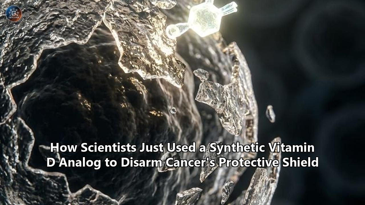
On May 25, 2026, a clinical milestone published in Nature Cancer quietly shook the oncology community. For years, pancreatic ductal adenocarcinoma (PDAC) has stood as one of the most impenetrable fortresses in medicine, largely because the tumor does not exist in isolation. Instead, it wraps itself in a dense, fibrous "shield" of connective tissue that blocks chemotherapies from entering and prevents the immune system from mounting an attack.
Now, a first-in-human clinical trial (NCT03520790) led by researchers at the Dana-Farber Cancer Institute, built on foundational basic science from the Salk Institute, has demonstrated that this protective shield can be pharmacologically disarmed. By introducing a highly specialized, synthetic vitamin D analog called paricalcitol, scientists successfully "reprogrammed" the tumor’s protective microenvironment.
Rather than attempting to violently tear down this protective barrier—a strategy that has repeatedly backfired in previous clinical efforts—the researchers used the synthetic analog to chemically pacify the cells constructing the shield. The early data is striking: in a small cohort of patients with previously untreated metastatic pancreatic cancer, adding paricalcitol to standard-of-care chemotherapy yielded a 42% partial tumor response rate, compared to just 9% in the chemotherapy-and-placebo control group. Furthermore, multiple patients in the paricalcitol cohort remained progression-free at the one-year mark, while none in the placebo group did.
This is not a story about dietary supplements or standard vitamin D pills. It is a narrative of precision molecular engineering, epigenetic rewiring, and a major shift in how oncologists approach therapeutic resistance. To understand how a drug originally developed for chronic kidney disease is now cracking the code of pancreatic cancer, one must look behind the scenes at the cellular mechanics, the historical failures of "stromal depletion," and the spatial genomic technologies that finally proved the concept in human tissue.
The Biological Wall: Understanding the Desmoplastic Stroma
To appreciate why this development is a turning point, it is necessary to understand the unique biology of pancreatic ductal adenocarcinoma. In most epithelial cancers, such as breast or lung cancer, the bulk of the tumor mass is made up of malignant cancer cells. Pancreatic cancer is different.
In a typical PDAC tumor, up to 80% to 90% of the tumor mass consists of non-malignant, supportive tissue known as the stroma. This phenomenon, called desmoplasia, is a highly active, dense, and fibrotic cellular scar that the body builds around the cancer—and that the cancer actively exploits.
[Normal Pancreas] ───(Oncogenic Stress/KRAS)───► [Activated Stellate Cells/CAFs]
                                                           │
                                            ┌──────────────┴──────────────┐
                                            ▼                             ▼
                              [Densely Packed Collagen]       [Immunosuppressive Cytokines]
                              (Collapses Blood Vessels)       (Excludes Cytotoxic CD8+ T-Cells)
                                            │                             │
                                            └──────────────┬──────────────┘
                                                           ▼
                                              [THE DESMOPLASTIC SHIELD]
                                           (Physical & Chemical Barrier)
The principal architects of this stroma are pancreatic stellate cells (PSCs) and cancer-associated fibroblasts (CAFs). In a healthy pancreas, stellate cells are quiescent, fat-storing cells that act as local caretakers, maintaining the structural matrix of the organ. However, when pancreatic cells mutate (almost universally driven by the KRAS oncogene) or suffer chronic inflammation, they release a cascade of distress signals, including Transforming Growth Factor-beta (TGF-β), Platelet-Derived Growth Factor (PDGF), and Tumor Necrosis Factor-alpha (TNF-α).
In response to these signals, quiescent stellate cells undergo a dramatic transdifferentiation. They shed their lipid droplets and morph into highly active, myofibroblastic CAFs. These activated fibroblasts begin churning out massive quantities of extracellular matrix proteins:
- Fibrillar Collagens (Types I and III): Creating a physical mesh of tough, rope-like fibers.
- Fibronectin and Laminin: Providing structural pathways that cancer cells can use to migrate and metastasize.
- Hyaluronic Acid: A highly water-binding glycosaminoglycan that swells within the tumor, creating immense physical pressure.
The Physics of Tumor Resistance
This cellular machinery has devastating consequences for treatment. The accumulation of collagen and hyaluronic acid increases the tumor's interstitial fluid pressure (IFP). In a normal tissue, IFP is close to zero. In a pancreatic tumor, this pressure can skyrocket to over 100 mmHg.
Because the pressure inside the tumor is so high, it physically collapses the delicate capillaries and blood vessels feeding the area. This leads to two major clinical bottlenecks:
- Drug Exclusion: Intravenous chemotherapies like gemcitabine or paclitaxel cannot diffuse down the pressure gradient. The drugs flow through the surrounding healthy tissue but are physically barred from entering the core of the tumor.
- Hypoxia and Acidification: The lack of blood flow starves the tumor of oxygen, creating an incredibly hostile, hypoxic, and acidic microenvironment. While this would kill normal cells, it forces cancer cells to undergo metabolic adaptations that make them highly aggressive and resistant to radiation.
Beyond the physical barrier, CAFs also orchestrate a chemical shield. They secrete chemokines such as CXCL12, which binds to CXCR4 receptors on immune cells. This signaling pathway actively excludes cytotoxic CD8+ T-cells—the primary cells capable of killing cancer—from the tumor microenvironment. Instead, the stroma recruits immunosuppressive myeloid-derived suppressor cells (MDSCs) and regulatory T-cells (Tregs), transforming the tumor into an immune desert.
The CAF Paradox: Why Destroying the Shield Backfired
For years, the obvious therapeutic strategy was simple: destroy the stroma. If the stroma is a physical wall protecting the cancer, then knocking down the wall should allow chemotherapy to flood in and eradicate the tumor.
In the early 2010s, this hypothesis led to high-profile drug development campaigns and clinical trials targeting the stroma. Two main approaches were tested:
1. Sonic Hedgehog (Shh) Pathway Inhibition
The Sonic Hedgehog signaling pathway is a primary driver of stromal recruitment in pancreatic cancer. Researchers developed small-molecule inhibitors, such as IPI-926 (saridegib), designed to block Shh signaling and deplete the CAFs.
The preclinical results in mice were highly promising, showing decreased stromal density and increased drug delivery. However, when moved into Phase II clinical trials in humans, the results were catastrophic.
Patients receiving the Shh inhibitor alongside gemcitabine actually performed worse than those receiving chemotherapy alone. The trials had to be halted early because the treated patients experienced accelerated disease progression, rapid metastasis, and decreased overall survival.
2. Enzymatic Depletion of Hyaluronic Acid
Another major effort targeted the physical matrix directly using PEGylated recombinant human hyaluronidase (PEGPH20) to dissolve hyaluronic acid and lower the interstitial fluid pressure.
While early-phase trials showed some promise in patients with high baseline hyaluronic acid levels, the Phase III HALO-301 trial ultimately failed to show an overall survival benefit. Furthermore, it significantly increased the risk of severe thromboembolic events.
The Epiphany: Reprogramming vs. Ablation
These failures forced a fundamental reassessment of tumor biology. Basic scientists, including the team at the Salk Institute led by Ronald Evans, PhD, realized that the stroma is not merely a passive partner to the tumor; it is also a host-protective response.
[STROMAL ABLATION] (Failed Strategy)
Destroy Fibroblasts ──► Loss of Structural Cage ──► Angiogenesis & Hyper-Vascularity ──► Rapid Metastasis

[STROMAL REPROGRAMMING] (Paricalcitol Strategy)
Apply VDR Agonist ──► Revert CAFs to Quiescence ──► Normal Vascularity & Permeability ──► Chemotherapy Delivery
The body builds the fibrotic stroma in an attempt to wall off, sequester, and contain the developing cancer, much like it uses scar tissue to wall off an infectious abscess or a chronic wound. When researchers completely obliterated the fibroblasts and the extracellular matrix, they removed this physical "cage."
Without the stroma to restrain them, the pancreatic cancer cells became highly vascularized, developed a more aggressive, mesenchymal phenotype, and metastasized throughout the body far more rapidly.
The goal, therefore, could not be destruction. It had to be re-education. Researchers needed a way to pacify the activated, inflammatory, and pro-fibrotic fibroblasts—reverting them back to their quiescent, resting state—without actually killing them or reducing their numbers.
This is where the biology of the vitamin D receptor entered the spotlight.
Unlocking the Stroma: The Rise of Synthetic Vitamin D Cancer Therapeutics
The realization that the vitamin D receptor (VDR) could serve as a master switch to reprogram the pancreatic stroma was born out of a surprising discovery in Ronald Evans’ laboratory at the Salk Institute. While mapping the genomic expression profiles of pancreatic tissue, Evans and his team discovered that the VDR is highly expressed in tissue-resident fibroblasts, specifically pancreatic stellate cells.
In a healthy pancreas, these quiescent stellate cells are packed with lipid droplets containing vitamin A and vitamin D, which keep them in a resting state. But when a tumor begins to form, the local inflammatory signals deplete these lipid stores, shut down VDR expression, and drive the cells into their activated, myofibroblastic CAF state.
Evans hypothesized that if they could chemically reactivate the VDR, they could force these angry, activated fibroblasts to revert back to their peaceful, quiescent, lipid-storing state. This would "re-educate" the stroma, loosening the physical collagen mesh and shutting down the secretion of immunosuppressive chemokines, while keeping the structural cage intact.
However, translating this concept into a viable therapy presented two major pharmacological hurdles that standard, over-the-counter vitamin D supplements simply could not overcome.
1. The Catabolic Sink of Activated Fibroblasts
When a patient takes a standard vitamin D3 supplement (cholecalciferol), the liver converts it to 25-hydroxyvitamin D [25(OH)D], and the kidneys (or local tissues) convert it to the active hormone, 1,25-dihydroxyvitamin D [1,25(OH)₂D], also known as calcitriol.
Calcitriol is a highly potent ligand for the VDR. However, activated stellate cells and CAFs protect themselves from VDR signaling by overexpressing CYP24A1. This enzyme is a highly efficient hydroxylase whose sole purpose is to catabolize active 1,25(OH)₂D into inactive metabolites like calcitroic acid, effectively neutralizing the hormone before it can bind to the receptor.
[Natural Calcitriol] ──► [CYP24A1 Enzyme (Overexpressed in CAFs)] ──► [Inactive Calcitroic Acid] (No VDR Activation)

[Paricalcitol (Analog)] ──► [Resists CYP24A1 Degradation] ──► [Binds VDR] ──► [Epigenetic Reprogramming]
To break through this biochemical defense mechanism, scientists needed a synthetic analog designed to resist enzymatic degradation.
Paricalcitol (commercially known as Zemplar, developed by AbbVie) is a synthetic vitamin D analog originally approved by the FDA in 1998 for the prevention and treatment of secondary hyperparathyroidism in chronic kidney disease. The chemical structure of paricalcitol features two critical modifications compared to natural calcitriol:- The 19-Nor Modification: The methylene group at carbon 19 is removed.
- Side-Chain Modification: The side chain is modified with a double bond between carbon 22 and 23, and a methyl group at carbon 24.
These subtle structural changes make paricalcitol a poor substrate for the CYP24A1 enzyme. It can enter the microenvironment of the tumor, bypass the catabolic sink, and bind directly to the VDR within the fibroblasts with high affinity.
2. The Lethal Risk of Hypercalcemia
The second hurdle is systemic toxicity. The primary physiological role of natural vitamin D is to maintain calcium homeostasis by increasing calcium absorption in the intestines and mobilizing calcium from the bones.
If an oncologist attempts to use natural calcitriol or high-dose vitamin D3 to achieve the concentrations required to reprogram tumor stroma, it inevitably triggers severe, life-threatening hypercalcemia (excess calcium in the blood). This leads to acute renal failure, calcification of soft tissues, cardiac arrhythmias, and death.
Paricalcitol is classified as a low-calcemic VDR agonist. Because of its unique structural modifications, it exhibits highly selective genomic activity. When paricalcitol binds to the VDR, the receptor undergoes a conformational change that favors its interaction with transcriptional co-regulators involved in anti-inflammatory and anti-fibrotic gene pathways, rather than the co-activators that drive intestinal calcium absorption.
This allows paricalcitol to be administered at therapeutic doses capable of remodeling the tumor stroma with a much lower, clinically manageable risk of hypercalcemia. This therapeutic strategy—deploying a synthetic vitamin D cancer approach—safely achieves what high-dose natural supplementation never could.
The Molecular Choreography of VDR-Mediated Reprogramming
When paricalcitol reaches an activated cancer-associated fibroblast, it initiates a precise sequence of genetic events at the nuclear level.
           [Paricalcitol (Ligand)]
                     │
                     ▼
          [Vitamin D Receptor (VDR)]
                     │
                     ▼ (Heterodimerization)
         [Retinoid X Receptor (RXR)]
                     │
                     ▼
       [Vitamin D Response Elements (VDREs)]
                     │
       ┌─────────────┴─────────────┐
       ▼                           ▼
[Gene Repression]          [Epigenetic Silencing]
- ACTA2 (α-SMA)            - Bromodomain (BRD) proteins displaced
- COL1A1/COL3A1            - Chromatin condensation at fibrotic loci
- TGF-β1 & CXCL12          - Histone deacetylation
       │                           │
       └─────────────┬─────────────┘
                     ▼
          [CAF Reprogramming to
            Quiescent State]
1. Heterodimerization and DNA Binding
Upon entering the cell cytoplasm, paricalcitol binds to the ligand-binding domain of the monomeric VDR. This binding induces a conformational shift that triggers the phosphorylation of the receptor and its translocation into the nucleus.
Once inside the nucleus, the paricalcitol-VDR complex recruits the Retinoid X Receptor (RXR) to form a heterodimer. This active VDR-RXR complex then scans the genome, binding specifically to DNA sequences known as Vitamin D Response Elements (VDREs).
2. Epigenetic Reconfiguration
The binding of the VDR-RXR complex to VDREs initiates local chromatin remodeling. In activated CAFs, the chromatin regions containing pro-fibrotic and inflammatory genes are highly open and accessible (euchromatin), allowing for rapid transcription.
The VDR-RXR complex recruits corepressor complexes, including Histone Deacetylases (HDACs) and methyltransferases. These enzymes strip acetyl groups from histone tails, causing the chromatin to wrap tightly around the histones (heterochromatin) and physically blocking transcription factor access.
3. Key Gene Targets of Reprogramming
This epigenetic silencing shuts down the primary pathways responsible for maintaining the CAF activated state:
- Downregulation of ACTA2 (α-SMA): ACTA2 encodes Alpha-Smooth Muscle Actin, the contractile protein that gives CAFs their physical rigidity and allows them to exert mechanical force on the tumor matrix. Inhibiting α-SMA relaxes the physical tension within the stroma.
- Inhibition of Collagen Synthesis (COL1A1 and COL3A1): The production of new, dense collagen fibers is halted, preventing the continuous reinforcement of the physical shield.
- Suppression of Pro-inflammatory and Pro-fibrotic Cytokines: The VDR complex directly represses the transcription of TGFB1 (TGF-beta 1), IL6 (Interleukin-6), and CXCL12. This stops the feedback loop that recruits more stellate cells and shuts down the chemical signals excluding immune cells.
Through this molecular choreography, paricalcitol does not kill the fibroblasts. Instead, it safely and effectively silences their aggressive transcriptional program, transitioning them into a state that closely resembles their healthy, quiescent precursors.
Behind the Scenes of the Clinical Trial: NCT03520790
While the preclinical animal models developed at the Salk Institute in 2014 were compelling, moving this concept from the lab bench to a patient’s bedside was a long, complex journey. The clinical translation was led by Brian Wolpin, MD, MPH, and Kimberly Perez, MD, at the Dana-Farber Cancer Institute, culminating in the Phase 1b trial published in May 2026.
This study had to answer a critical, multi-layered question: Can a synthetic vitamin D analog be safely combined with highly toxic, standard-of-care chemotherapy in metastatic pancreatic cancer patients, and will it actually remodel the human stroma as predicted?
Trial Design and Patient Cohorts
The trial enrolled 36 patients with previously untreated, biopsy-proven metastatic pancreatic ductal adenocarcinoma. This was a highly vulnerable patient population with advanced, rapidly progressing disease.
The patients were randomized into three distinct treatment arms:
| Arm | Chemotherapy Backbone | VDR Agonist Intervention | Cohort Size |
|---|---|---|---|
| Arm A (Control) | Gemcitabine ($1000 \text{ mg/m}^2$) + Nab-Paclitaxel ($125 \text{ mg/m}^2$) | Placebo | 12 patients |
| Arm B (Intravenous) | Gemcitabine ($1000 \text{ mg/m}^2$) + Nab-Paclitaxel ($125 \text{ mg/m}^2$) | IV Paricalcitol ($25 \text{ mcg}$, $3\times/\text{week}$) | 12 patients |
| Arm C (Oral) | Gemcitabine ($1000 \text{ mg/m}^2$) + Nab-Paclitaxel ($125 \text{ mg/m}^2$) | Oral Paricalcitol ($16 \text{ mcg}$, daily) | 12 patients |
The Logistics of Paired Biopsies
To definitively prove that paricalcitol was achieving its biological target, the trial investigators had to perform a logistically complex and invasive procedure: paired tumor biopsies.
- Baseline Biopsy: Collected during the initial screening process, before any therapeutic intervention.
- On-Treatment Biopsy: Collected after four to six weeks of combination therapy.
Performing core needle biopsies of pancreatic tumors, especially in patients with metastatic disease, is highly challenging. The pancreas is situated deep in the retroperitoneum, surrounded by major blood vessels, the duodenum, and the spleen.
These biopsies had to be performed using endoscopic ultrasound (EUS) or CT guidance. The researchers had to obtain enough viable tissue not just for standard pathology, but for advanced genomic and spatial transcriptomic profiling.
The fact that the team successfully gathered paired, high-quality tissue samples from these patients is a major technical achievement that provided the key biological proof of the treatment's mechanism.
The Spatial Revolution: Inside the Treated Biopsies
The most scientifically compelling data from the Nature Cancer paper emerged from what the researchers found inside those paired human biopsies. Rather than relying on bulk tissue sequencing—which grinds up all the cells in a sample into a genomic "smoothie," erasing their spatial relationships—the team used cutting-edge Spatial Transcriptomics and Multiplex Immunofluorescence (mIF).
Traditional Bulk Sequencing:
[CAFs + Cancer Cells + Immune Cells] ──► Ground Up ──► Average RNA Signal (Smoothie)

Spatial Transcriptomics / Multiplex IF:
[Tissue Section] ──► Mapped in Situ ──► Exact Geographic Coordinates of Every Cell
                                       - Shows CAFs losing α-SMA
                                       - Shows CD8+ T-cells migrating to Cancer Core
These techniques allowed the researchers to create high-resolution, color-coded geographical maps of the tumor microenvironment before and after paricalcitol treatment.
1. Verification of CAF Reprogramming
Using antibodies against Alpha-Smooth Muscle Actin ($\alpha\text{-SMA}$), a marker of activated CAFs, and cytokeratin, a marker of pancreatic cancer cells, the team measured the density and activation state of the stroma.
In the placebo arm (Arm A), the on-treatment biopsies showed no change or an increase in the dense, tightly packed $\alpha\text{-SMA}^+$ fibroblast mesh surrounding the cancer nests.
In the paricalcitol-treated arms (Arms B and C), the results were starkly different. The proportion of highly active, $\alpha\text{-SMA}^+$ myofibroblastic fibroblasts was significantly reduced.
Crucially, however, the total number of fibroblasts remained constant. This confirmed that paricalcitol was successfully deactivating the cells without destroying them, maintaining the integrity of the physical cage while turning off its pathological, drug-blocking behavior.
2. Opening the Gates to Immune Infiltration
The most striking finding from the spatial analysis was the sudden movement of the immune system. Pancreatic cancer is typically a "cold" tumor, meaning cytotoxic T-cells are completely excluded from the tumor core and are left idling in the outer stromal margins.
Placebo Core:   [Cancer Cells] <--- [DENSE STROMAL WALL (No T-Cells)] <--- [T-Cells blocked outside]
Treated Core:   [Cancer Cells  <--intermingled with--  Cytotoxic CD8+ T-Cells] (Stromal wall relaxed)
By staining for CD8 (a marker for cytotoxic T-cells) and utilizing spatial distance algorithms, the researchers tracked the exact positions of these immune cells relative to the cancer cells:
- Before Treatment: CD8+ T-cells were located hundreds of micrometers away from the cytokeratin-positive cancer cells, physically blocked by the stromal wall.
- After Paricalcitol Treatment: The on-treatment biopsies revealed a significant increase in the density of CD8+ T-cells deep within the tumor nests.
- Spatial Colocalization: The mathematical distance between T-cells and cancer cells dramatically shrank. The T-cells were no longer trapped outside; they had crossed the relaxed stromal barrier and were physically interacting with, and destroying, the malignant cancer cells.
The Biomarker Breakthrough: High VDR Expression as a Predictor of Success
One of the most important, actionable details uncovered in the clinical trial is the variation in VDR expression among patients.
While all humans express the vitamin D receptor, the density of this receptor on the surface of tumor-associated fibroblasts varies significantly from person to person. When the researchers analyzed the baseline biopsies, they discovered a clear correlation between the levels of stromal VDR expression and how well the patients responded to the paricalcitol-chemotherapy cocktail.
                  [Baseline Patient Biopsy]
                             │
               ┌─────────────┴─────────────┐
               ▼                           ▼
      [High VDR Expression]       [Low VDR Expression]
               │                           │
               ▼                           ▼
    - Robust CAF Reprogramming   - Minimal Stromal Remodeling
    - Deep Tumor Shrinkage       - Standard Chemo Efficacy
    - Longest Overall Survival   - Average Progression Timeline
In patients with high VDR expression who received paricalcitol:
- The transition of fibroblasts from activated ($\alpha\text{-SMA}^+$) to quiescent was highly pronounced.
- They experienced the deepest tumor shrinkage.
- They achieved the longest overall survival times observed in the study.
Conversely, patients whose stroma had low VDR expression showed minimal changes in their tumor microenvironment and progressed at a rate similar to the control group.
This discovery provides a clear roadmap for future clinical development. Rather than administering synthetic vitamin D cancer treatments to every pancreatic cancer patient, oncologists can use immunohistochemical staining of a standard pre-treatment biopsy as a companion diagnostic.
This allows clinicians to select and treat only those patients whose tumors are biologically primed to respond, sparing others the potential toxicities and cost of an ineffective therapy.
Safety, Tolerability, and the Clinical Management of Hypercalcemia
Because paricalcitol is a vitamin D analog, the primary safety concern throughout the trial was hypercalcemia. While paricalcitol is low-calcemic compared to natural calcitriol, it is not completely non-calcemic, especially when administered alongside aggressive chemotherapy regimens.
In the clinical trial, the safety profile was closely monitored:
- General Tolerability: The addition of paricalcitol did not exacerbate the classic toxicities of gemcitabine and nab-paclitaxel, such as myelosuppression (neutropenia, thrombocytopenia), peripheral neuropathy, or severe fatigue.
- Hypercalcemia Events: In the oral paricalcitol cohort (Arm C), 5 out of the 12 patients experienced Grade 2 to Grade 4 hypercalcemia.
The Clinical Protocols for Managing Calcium Spikes
The development of hypercalcemia in these patients was not a reason to abandon the therapy. Instead, the investigators implemented highly effective, standard clinical management protocols:
[Patient Serum Calcium > 10.5 mg/dL] (Hypercalcemia Detection)
                  │
                  ▼
        [Hold Paricalcitol Dose] ──► [Initiate Aggressive Hydration (Normal Saline)]
                  │
                  ▼ (If Persistent)
[Administer Bisphosphonates or Calcitonin] ──► [Step-Down Dose Reduction upon Normalization]- Dose Interruptions and Reductions: If a patient's serum calcium rose above $10.5 \text{ mg/dL}$, the paricalcitol dose was temporarily held. Once calcium levels normalized, the drug was resumed at a reduced dose level (e.g., stepping down from $16 \text{ mcg}$ to $12 \text{ mcg}$ daily for the oral formulation).
- Aggressive Hydration: Patients were treated with intravenous normal saline to promote renal calcium excretion.
- Pharmacological Interventions: In cases of persistent or severe hypercalcemia, clinicians administered low-dose bisphosphonates (such as zoledronic acid) or calcitonin, which rapidly shifted calcium back into the bone matrix.
By utilizing these standard protocols, all hypercalcemic events were successfully managed without any long-term kidney damage, cardiac events, or treatment-related deaths. This proved that the synthetic vitamin D cancer strategy is highly feasible and safe for integration into standard oncological care.
The Broader Landscape: Reprogramming other Impenetrable Cancers
While pancreatic ductal adenocarcinoma is the primary proving ground for this therapy, the success of the paricalcitol trial opens up a highly promising front in the war against other stroma-rich, treatment-resistant malignancies.
Any tumor that relies on a dense fibrotic stroma to shield itself from the immune system and drugs could theoretically be targeted using synthetic vitamin D cancer therapies.
                             [PARICALCITOL]
                                   │
         ┌─────────────────────────┼─────────────────────────┐
         ▼                         ▼                         ▼
   [Cholangiocarcinoma]   [Triple-Negative Breast]   [Hepatic Fibrosis & HCC]
   Relax stroma to allow  Break up rigid collagen    Target stellate cells to
   gem/cis infiltration   mesh for checkpoint drugs  prevent tumor progression
1. Cholangiocarcinoma (Bile Duct Cancer)
Like pancreatic cancer, cholangiocarcinoma is characterized by an extremely dense, desmoplastic stroma. The tumor-associated fibroblasts in bile duct cancers are highly active, secreting massive amounts of extracellular matrix that collapse intratumoral blood vessels and drive therapeutic resistance. Preclinical work has already shown that the VDR is highly expressed in cholangiocarcinoma-associated fibroblasts, suggesting that paricalcitol could be directly pivoted to this disease.
2. Triple-Negative Breast Cancer (TNBC)
TNBC is notoriously aggressive and lacks the estrogen, progesterone, and HER2 receptors that allow for targeted therapies. Many TNBC tumors wrap themselves in a dense collagen matrix that excludes immune cells, rendering modern immune checkpoint inhibitors (such as pembrolizumab) ineffective. Combining paricalcitol with chemotherapy and immunotherapy could break up this rigid matrix, allowing T-cells to flood the breast tumor and clear the cancer.
3. Hepatocellular Carcinoma (HCC) and Liver Fibrosis
Primary liver cancer almost always develops in the context of chronic liver fibrosis or cirrhosis, driven by activated hepatic stellate cells (HSCs). These HSCs are highly homologous to pancreatic stellate cells and express massive levels of the VDR.
By using paricalcitol, researchers have shown in preclinical models that they can block liver fibrosis, prevent the formation of a pro-tumorigenic niche, and increase the efficacy of tyrosine kinase inhibitors like sorafenib or lenvatinib.
The Economics and Politics of Repurposing a Generic Drug
Behind the scientific excitement of this trial lies a complex socioeconomic reality: paricalcitol is an off-patent, generic drug. First approved by the FDA in 1998, a generic supply of paricalcitol is highly abundant and incredibly inexpensive compared to modern, newly patented oncology drugs.
While this is fantastic news for global healthcare systems and patient access, it presents a significant hurdle for clinical development.
New Patented Oncology Drug:
[High Profit Potential] ──► [Massive Pharma Funding] ──► [Rapid Phase III Trials]

Repurposed Generic (Paricalcitol):
[Low Profit Potential]  ──► [Pharma Hesitancy]      ──► [Reliance on Philanthropy & Grants]
                                                       (Stand Up To Cancer, NCI, Salk)
The "Valley of Death" for Off-Patent Drugs
Major pharmaceutical companies are rarely motivated to fund large, multi-million-dollar Phase III clinical trials for generic, off-patent compounds because they cannot secure exclusive patents to recoup their investments. This often leaves promising, scientifically sound ideas stranded in the "Valley of Death"—the gap between preclinical proof-of-concept and the definitive clinical trials required to change the official standard of care.
The translation of paricalcitol from a 2014 Salk Institute paper to a 2026 clinical success story was only made possible through a collaborative funding model:
- Academic and Philanthropic Partnerships: Organizations like Stand Up To Cancer (SU2C), the March of Dimes, and the Lustgarten Foundation for Pancreatic Cancer Research stepped in to provide the critical, non-profit funding required to design and launch the trial.
- Government Grants: The National Cancer Institute (NCI) provided critical grant funding to support the highly complex spatial transcriptomics and correlate molecular studies.
This funding structure highlights a major systemic challenge in modern medicine: we need robust, alternative, non-corporate pathways to fund clinical trials for repurposed drugs. Without these philanthropic and academic coalitions, breakthroughs like paricalcitol might never make it out of academic laboratories.
What to Watch For Next: Upcoming Milestones and Unresolved Questions
As the medical community digests the results of the Nature Cancer paper, the roadmap for this therapy is rapidly expanding. Several key milestones and unresolved scientific questions will define the next five years of research:
1. The Phase II/III Registrational Trials
Because NCT03520790 was a small, safety-focused Phase 1b trial, it was not mathematically powered to prove overall survival efficacy. The immediate next step is the launch of multi-center, randomized Phase II and Phase III clinical trials designed to compare paricalcitol-chemotherapy combinations directly against chemotherapy alone in hundreds of patients. These trials will establish whether this stromal reprogramming truly extends the life of pancreatic cancer patients.
2. Integration with Modern Immunotherapy
The finding that paricalcitol allows CD8+ T-cells to penetrate the deep layers of pancreatic tumors is incredibly exciting for the field of immuno-oncology.
Currently, pancreatic cancer is highly resistant to immune checkpoint inhibitors like anti-PD-1 (pembrolizumab) or anti-CTLA-4 (ipilimumab). A major upcoming trial design will evaluate a "triple-threat" combination:
$$\text{Chemotherapy} + \text{Paricalcitol (Stroma Reprogrammer)} + \text{Pembrolizumab (Immunotherapy)}$$
By relaxing the physical shield with paricalcitol, researchers hope to finally allow these powerful immunotherapies to target and destroy pancreatic tumors.
3. Refining Next-Generation VDR Agonists
While paricalcitol is highly effective, it was not originally optimized for cancer therapy. Medicinal chemists at the Salk Institute and other research centers are actively developing next-generation synthetic vitamin D analogs. These novel compounds are engineered to have:
- Zero Calcemic Activity: Completely eliminating the risk of hypercalcemia, allowing for even higher therapeutic dosing.
- Increased CYP24A1 Resistance: Creating molecules that are entirely impervious to the enzymatic degradation pathways of the tumor, maximizing their functional lifespan in the microenvironment.
Rewriting the Narrative of Cancer Therapy
For decades, the dominant philosophy of cancer treatment has been direct, violent eradication: search for the cancer cells and kill them with chemotherapy, radiation, or targeted molecular inhibitors. While this approach has saved millions of lives, it has met its match in highly complex, fibrotic, and adaptive malignancies like pancreatic cancer.
The breakthrough utilizing a synthetic vitamin D analog to disarm the tumor's protective shield represents a profound shift in perspective. Instead of focusing solely on the seed—the cancer cell—scientists are successfully treating the soil.
By pacifying the host-derived fibroblasts and calming the local inflammatory environment, this therapy converts a hostile, highly resistant tumor into a vulnerable, manageable disease. As this approach moves into larger clinical trials, it offers a real, scientifically validated beacon of hope for patients facing some of the most challenging diagnoses in medicine.
Reference:
- https://www.dana-farber.org/newsroom/news-releases/2026/from-discovery-to-patient-care-vitamin-d-analog-shuts-down-pancreatic-cancers-shield-in-clinical-trial
- https://bioengineer.org/from-salk-institute-breakthrough-to-bedside-vitamin-d-analog-disarms-pancreatic-cancers-defenses-in-clinical-trial/
- https://www.cancer.gov/news-events/cancer-currents-blog/2018/vitamin-d-supplement-cancer-prevention
- https://www.salk.edu/news-release/from-salk-institute-discovery-to-patient-care-vitamin-d-analog-shuts-down-pancreatic-cancers-shield-in-clinical-trial/
- https://www.oncozine.com/from-bench-to-bedside-vitamin-d-analog-paricalcitol-remodels-pancreatic-tumor-microenvironment-in-early-clinical-trial/
- https://www.salk.edu/news-release/modified-vitamin-d-shows-promise-as-treatment-for-pancreatic-cancer/
- https://www.dana-farber.org/newsroom/news-releases/2026/from-discovery-to-patient-care-vitamin-d-analog-shuts-down-pancreatic-cancers-shield-in-clinical-trial
- https://oncodaily.com/oncolibrary/vitamin-d-and-cancer
- https://pmc.ncbi.nlm.nih.gov/articles/PMC13116322/
- https://pmc.ncbi.nlm.nih.gov/articles/PMC8914466/
- https://pmc.ncbi.nlm.nih.gov/articles/PMC6201256/
- https://www.researchgate.net/publication/405236365_Gemcitabine_and_nab-paclitaxel_with_or_without_the_VDR_agonist_paricalcitol_for_metastatic_pancreatic_cancer_a_randomized_multiarm_run-in_phase_trial?_tp=eyJjb250ZXh0Ijp7InBhZ2UiOiJzY2llbnRpZmljQ29udHJpYnV0aW9ucyIsInByZXZpb3VzUGFnZSI6bnVsbCwic3ViUGFnZSI6bnVsbH19
- https://bioquicknews.com/from-salk-institute-discovery-to-patient-care-vitamin-d-analog-shuts-down-pancreatic-cancers-shield-in-clinical-trial/
- https://www.broadinstitute.org/publications/broad1377581
- https://bioengineer.org/from-salk-institute-breakthrough-to-bedside-vitamin-d-analog-disarms-pancreatic-cancers-defenses-in-clinical-trial/