A quiet catastrophe has been unfolding undetected inside the brains of millions of people living with dementia. For over a century, neuroscientists have known that Alzheimer’s disease systematically destroys the brain's neural networks, eroding memory, personality, and cognitive function. Yet, the exact biological executioner—the precise sequence of chemical events that ultimately forces a living neuron to die—has remained one of the most stubborn black boxes in modern medicine.
A study published in Nature Communications by researchers at King’s College London, in collaboration with the UK Dementia Research Institute, has finally illuminated this dark corner of neuropathology. The international team has discovered a previously overlooked form of programmed cell death that targets the very core of the neuron: its nucleus.
This newly identified cell death pathway is called karyoptosis.
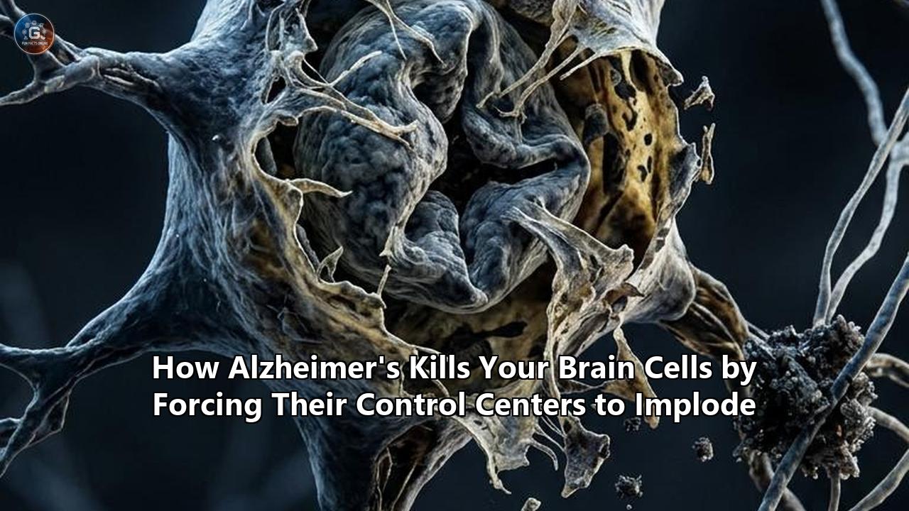
Derived from the Greek words karyon (kernel or nucleus) and ptosis (falling or drooping), karyoptosis describes a devastating molecular chain reaction. Under the stress of toxic protein accumulation, the structural integrity of the nucleus—the cell’s command center containing its genetic instructions—begins to warp, shrivel, and physically disintegrate. Once the nucleus implodes, the neuron loses its ability to regulate its own functions, leading to irreversible cell death.
This discovery marks a profound shift in our understanding of how alzheimers affects brain cells. For decades, therapeutic efforts have focused on clearing the external toxic waste of the disease—namely, amyloid-beta plaques and tau tangles. Karyoptosis, however, represents an internal structural collapse that occurs inside the neuron. It is a process that can potentially be decoupled from the protein buildup itself. By understanding the molecular switches that trigger this nuclear implosion, scientists believe they can develop a entirely new class of drugs designed to halt neuron loss even when toxic proteins are present.
The Mystery of the Vanishing Neurons
To understand why the discovery of karyoptosis is so significant, one must first appreciate the historical and clinical limitations of dementia research.
Since Alois Alzheimer first documented the disease in 1906, researchers have been guided by the presence of two hallmark pathologies: extracellular amyloid-beta plaques and intracellular neurofibrillary tangles made of hyperphosphorylated tau protein. The prevailing theory for decades, known as the "amyloid cascade hypothesis," posited that the accumulation of amyloid-beta in the brain was the primary driver of the disease. According to this model, amyloid plaques initiate a toxic cascade that leads to tau tangles, synaptic dysfunction, chronic neuroinflammation, and eventually, widespread neuronal death.
Yet, this hypothesis has consistently run into a frustrating clinical reality. Modern monoclonal antibody therapies, such as lecanemab and donanemab, are highly effective at clearing amyloid-beta plaques from the brain. Despite this successful clearance, these drugs only offer modest benefits, slowing cognitive decline by roughly 25 to 35 percent. They do not stop the progression of the disease, nor do they reverse existing damage.
Once the toxic cascade is set in motion, the loss of brain cells continues apace. This clinical disconnect suggested that clearing the external trigger is not enough if the internal execution mechanisms of the cell remain active.
"The death and loss of cells in the brain drives many symptoms experienced by people living with dementia," explains Dr. Rebecca Casterton, a neuroscientist at King’s College London and the study's first author. "Our study uncovers a new series of chemical events which can coordinate cell death in brain cells. We have started to lay out the road map of how karyoptosis works".
Before this discovery, the scientific community lacked a complete explanation of how toxic proteins translate into cell death. Neurons are evolutionary anomalies: unlike skin or liver cells, which constantly divide and replace themselves, neurons are "postmitotic." They are designed to last an entire lifetime. Because of this longevity, neurons have evolved highly robust survival mechanisms that make them exceptionally resistant to classical forms of cell death, such as apoptosis.
For years, this left a massive paradox at the heart of neurology: if neurons are so structurally resilient and actively resist standard self-destruction programs, how does Alzheimer's kill them so ruthlessly? The answer, it turns out, lies in karyoptosis—a pathway that bypasses the neuron's standard defenses by attacking the structural scaffolding of its control center.
Karyoptosis: Step-by-Step Anatomy of an Implosion
The nucleus of a eukaryotic cell is not merely a passive bag of DNA. It is a highly dynamic, pressurized organelle. It must maintain a perfect spherical or oval shape to orchestrate the complex transport of molecules, protect the genome from physical stress, and regulate which genes are turned on or off.
To maintain this shape, the nucleus relies on a specialized structural support system located just beneath its double-membrane envelope. This support system is the nuclear lamina, a dense, fibrous meshwork of proteins called lamins. Among these, LaminB1 is the primary structural anchor. You can think of LaminB1 as the load-bearing pillars of a dome. If those pillars are structurally compromised, the entire dome buckles and collapses under its own weight.
[Healthy Neuron]
       ↓
[Toxic Protein Accumulation (Amyloid/Tau)]
       ↓
[Activation of p38 MAP Kinase (Molecular Switch)]
       ↓
[Phosphorylation & Degradation of LaminB1 (Pillars Crumble)]
       ↓
[Nuclear Envelope Loss of Tension & Buckling]
       ↓
[Nucleus Shrivels, Warps, and Disintegrates (Karyoptosis)]
       ↓
[Active Ejection of Nuclear Debris in Vesicles]
       ↓
[Neuronal Death & Cognitive Decline]
When karyoptosis is triggered inside a neuron, the physical transformation is dramatic. Using advanced high-resolution imaging and computational algorithms, the King’s College London team documented the exact chronological sequence of this nuclear demolition:
- Toxic Accumulation: Harmful, misfolded proteins (such as amyloid-beta or tau) accumulate inside the neuron's cytoplasm faster than the cell's waste-clearance systems (autophagy and the proteasome) can degrade them.
- Kinase Activation: The sheer physical and chemical stress of this protein buildup activates a specific stress-response enzyme called p38 MAP kinase (p38 MAPK).
- Lamina Targeting: Activated p38 MAPK acts as a molecular switch, targeting the structural support protein LaminB1. It chemically modifies LaminB1, marking it for rapid degradation.
- Structural Failure: As LaminB1 is destroyed, the nuclear lamina loses its tension. The nuclear envelope, once a tight and protective barrier, begins to warp, wrinkle, and buckle inward.
- Nuclear Shriveling: The nucleus progressively shrinks, losing its rounded shape. Crucially, this physical collapse of the nucleus occurs before any detectable damage to the DNA itself, showing that structural failure is the driver, not a consequence, of cell death.
- Disintegration and Ejection: The shriveled nucleus eventually breaks apart entirely. Rather than bursting violently or quietly dissolving, the neuron actively packages the fragmented pieces of its own nucleus into tiny, membrane-bound bubbles (vesicles) and ejects them outward through its cell membrane before finally dying.
This active ejection of nuclear debris is one of the most distinctive features of karyoptosis. It represents a highly coordinated, albeit self-destructive, attempt by the dying cell to manage its terminal crisis.
How Alzheimer's Affects Brain Architecture: The Human Evidence
To determine whether karyoptosis was merely an artificial phenomenon observed in laboratory dishes or a genuine driver of human disease, the researchers conducted a rigorous analysis of donated post-mortem human brain tissue.
Using sophisticated computational algorithms, the team examined approximately 3,000 individual brain cells from 28 patients who had died with either advanced Alzheimer's disease or frontotemporal dementia (FTD). They focused their analysis on the frontal cortex, the brain region located directly behind the forehead that is responsible for executive functions, planning, decision-making, and personality.
The results were stark.
In the brains of healthy, age-matched older adults, only about 15 percent of the analyzed frontal cortex cells showed any physical signs of nuclear wrinkling or degradation. This baseline rate is attributed to normal age-related wear and tear on the nuclear pore complexes and lamina over many decades.
However, in the frontal cortex of patients who died with Alzheimer’s, 35 percent of the neurons exhibited advanced signs of karyoptosis.
+--------------------------------------------------------------+
|     Frontal Cortex Neurons Showing Signs of Karyoptosis      |
+--------------------------------------------------------------+
| Healthy Aged Controls:  [██████░░░░░░░░░░░░░░░░░░░░] 15%     |
| Alzheimer's Patients:   [██████████████░░░░░░░░░░░░] 35%     |
+--------------------------------------------------------------+
This represents a more than doubling of nuclear structural collapse in the diseased brain. The data provides direct, physical evidence of how alzheimers affects brain cells by targeting their genomic control centers.
When we talk about how alzheimers affects brain function, we are ultimately talking about the loss of physical connections (synapses) and the death of these vital postmitotic neurons. The fact that over a third of the neurons in a critically damaged brain region are actively undergoing nuclear implosion explains the profound cognitive deficits associated with late-stage dementia. When the nuclear command center is destroyed, the neuron can no longer synthesize the proteins required to maintain its synapses, communicate with its neighbors, or repair its daily metabolic damage. It is a slow, structural death that leaves empty space where memories once resided.
Decoupling Toxic Waste from Cell Death: The Therapeutic Breakthrough
Perhaps the most exciting implication of the King’s College London study is that karyoptosis can be biochemically blocked.
In a series of laboratory experiments using primary rat cortical neurons, the researchers induced toxic protein buildup by blocking the cells' trash-removal mechanisms. As expected, this block led to a rapid accumulation of misfolded proteins, triggering the p38 MAP kinase pathway, the destruction of LaminB1, and the rapid onset of karyoptosis.
The researchers then introduced a compound designed to block the activity of the p38 MAP kinase enzyme.
The results were remarkable. Even though the cells were still heavily congested with toxic, clumping proteins, blocking the p38 MAPK pathway significantly delayed nuclear disintegration and prevented the neurons from dying. The nuclei of the treated cells maintained their round, healthy shapes, and their structural integrity remained intact.
[Normal p38 MAPK Activation]      --->   LaminB1 Destroyed   --->   Karyoptosis (Cell Death)
[p38 MAPK Blocked by Inhibitors]  --->   LaminB1 Preserved   --->   Neuron Survives (Despite Toxic Build-up)
This represents a fundamental paradigm shift in neurodegenerative drug discovery.
Historically, the scientific consensus has been that the only way to save neurons is to prevent or clear the accumulation of toxic proteins like amyloid-beta and tau. However, karyoptosis research suggests that we do not necessarily have to clear the toxic waste to save the cell. If we can interrupt the chemical cascade that connects protein accumulation to nuclear collapse, we can shield the nucleus from damage.
"By specifically targeting the interaction between p38 MAP kinase and LaminB1 we may slow down the process of cell death, buying time for more pinpointed therapies against specific neurodegenerative diseases," says Dr. Manolis Fanto, a Reader in Functional Genomics at the Institute of Psychiatry, Psychology and Neuroscience at King’s College London.
This "buying time" strategy is critical. If clinical trials can validate a therapy that prevents karyoptosis, it could transform Alzheimer’s from a terminal, rapidly progressive dementia into a manageable, slow-moving chronic condition. A patient’s brain might still accumulate some protein plaques over time, but if their neurons' command centers remain structurally sound, those neurons can continue to function, preserve synapses, and maintain cognitive function.
Comparing the Pillars of Cell Death: Apoptosis, Necroptosis, and Karyoptosis
To put karyoptosis in perspective, it is helpful to compare it to other known forms of cellular demise.
Over several decades, biology has identified several distinct programs that a cell can use to die. Each program leaves behind a unique set of chemical fingerprints and physical changes. For years, scientists tried to squeeze the neuronal loss seen in Alzheimer's into these pre-existing boxes, but the pieces of the puzzle never quite fit.
| Feature | Apoptosis | Necroptosis | Karyoptosis |
|---|---|---|---|
| Primary Trigger | Development, DNA damage, physiological clearance | Severe infection, tissue trauma, acute inflammation | Toxic protein accumulation, structural nuclear stress |
| Initial Site of Collapse | Cytoplasm and cell membrane | Outer cell membrane | Cell Nucleus (Command Center) |
| Key Molecular Actors | Caspases (e.g., Caspase-3), Bcl-2 family | RIPK1, RIPK3, MLKL | p38 MAP kinase, LaminB1 |
| Fate of the Nucleus | Late-stage chromatin condensation and fragmentation | Random degradation as cell lyses | Early-stage shriveling and physical disintegration |
| Cellular Outcome | Clean engulfment by macrophages without inflammation | Cell bursts violently, spilling toxic contents, causing inflammation | Quiet death; cell packages nuclear fragments into vesicles and expels them |
| Role in Neurons | Actively resisted by mature, adult brain cells | Occurs under extreme inflammatory or ischemic stress | Primary driver of progressive, long-term dementia death |
Why Apoptosis Fails to Explain Alzheimer's
Apoptosis is the textbook form of programmed cell death. It is a highly ordered, energy-dependent process that the body uses during development to sculpt organs (such as removing the webbing between fingers in a developing embryo) or to safely clear out damaged or cancerous cells. When a cell undergoes apoptosis, it activates specialized enzymes called caspases that systematically chop up its proteins and DNA, causing the cell to shrink and be neatly consumed by neighboring immune cells.
However, adult neurons are highly resistant to apoptosis. Because neurons cannot easily divide and replace themselves, they have evolved strict molecular brakes that prevent them from accidentally triggering this self-destruct sequence. If our brain cells died of apoptosis at the slightest sign of stress, our cognitive reserve would vanish in youth. Thus, while some markers of apoptosis have been detected in Alzheimer's brains, they are not abundant enough to explain the massive, widespread neuron loss that characterizes the disease.
Why Necroptosis is Only Part of the Story
Necroptosis is a form of programmed necrosis. Unlike the quiet, orderly process of apoptosis, necroptosis is a inflammatory event. It is often triggered by immune signals like Tumor Necrosis Factor-alpha (TNF-α) under conditions of severe infection or injury. During necroptosis, a complex of proteins (the necrosome) punches physical holes in the outer cell membrane, causing the cell to swell and violently burst, spilling its toxic internal contents into the surrounding tissue and igniting a massive local firestorm of neuroinflammation.
While necroptosis does occur in the Alzheimer’s brain, particularly in areas experiencing chronic, late-stage inflammation, it is a highly destructive process that typically happens in response to severe, acute external stress. It does not fully explain the slow, progressive, and often quiet "attrition" of individual neurons that occurs over twenty or thirty years of disease development.
Karyoptosis: The Missing Evolutionary Link
This is where karyoptosis fills a crucial gap.
Karyoptosis is uniquely adapted to explain the slow-motion collapse of mature, postmitotic cells. It begins not at the outer cell membrane, nor with the activation of apoptotic caspases, but deep inside the nuclear command center. Because it targets LaminB1—a structural protein that mature neurons cannot easily replace—it exploits a fundamental physical vulnerability of aging cells.
Furthermore, the physical packaging and quiet ejection of the fragmented nucleus in tiny, membrane-bound bubbles explain why the dying process can occur with minimal immediate tissue disruption, quietly eroding the brain’s cognitive architecture over decades.
The Broader Landscape: A War on Nuclear Order
To fully comprehend the discovery of karyoptosis, it must be viewed alongside other major developments in neurobiology.
Over the last few years, a consensus has emerged that the nucleus is the ultimate, decisive battleground in the war against Alzheimer’s disease. The physical collapse of the nucleus (karyoptosis) is the structural endpoint of a broader, systemic loss of nuclear stability.
For example, a study published in Cell by researchers at the Massachusetts Institute of Technology (MIT) presented a massive, multimodal atlas of gene expression and regulation spanning 3.5 million individual cells from the brains of over 100 human donors.
Led by Dr. Li-Huei Tsai and Dr. Manolis Kellis, the MIT team profiled both the "transcriptome" (which genes are active) and the "epigenome" (the physical structure of the chromosomes that determines which DNA regions are accessible).
They made a startling discovery: the progression of Alzheimer's is characterized by a profound breakdown of nuclear compartmentalization.
[Healthy Nucleus: Epigenomic Stability]
  - Chromatin neatly organized into compartments.
  - Harmful/inappropriate genes securely locked down.
  - Cell identity (neuron or microglia) preserved.

                       VS.

[Alzheimer's Nucleus: Epigenomic Erosion]
  - Compartmentalization breaks down.
  - Locked-down compartments open up, releasing harmful genes.
  - Open compartments repress, locking down survival genes.
  - Cell loses its molecular memory and identity.
In a healthy brain cell, the nucleus maintains rigorous compartmentalization. It keeps certain regions of the genome tightly wound up and locked away (heterochromatin) so that inappropriate or harmful genes are never expressed. Meanwhile, other regions containing essential survival and functional genes are kept open and accessible (euchromatin).
The MIT researchers found that as Alzheimer's advances, this epigenomic stability is lost. Normally closed compartments break open, exposing genes that should remain silent, while normally open, functional compartments are shut down.
"The message is clear: Alzheimer's is not only about plaques and tangles, but about the erosion of nuclear order itself," noted Dr. Kellis. "Cognitive decline emerges when chromatin guardians lose ground to the forces of erosion, switching from resilience to vulnerability at the most fundamental level of genome regulation. And when our brain cells lose their epigenomic memory marks and epigenomic information at the lowest level deep inside our neurons and microglia, it seems that Alzheimer's patients also lose their memory and cognition at the highest level".
The Structural and Epigenetic Intersection
When we connect the dots between the KCL study on karyoptosis and the MIT study on epigenomic erosion, a complete, unified theory of nuclear destruction emerges:
- The Epigenetic Phase: Early in the disease, toxic stress causes "chromatin guardians" to fail, leading to a loss of epigenomic memory and compartmentalization within the nucleus. The cell physically "forgets" how to operate, leading to early cognitive symptoms and synaptic failure.
- The Structural Phase: As the stress intensifies, the physical boundaries of the nucleus are targeted. The activation of p38 MAP kinase marks LaminB1 for destruction.
- The Implosion (Karyoptosis): With the loss of both epigenomic stability on the inside and structural lamina support on the outside, the nuclear envelope buckles, shrivels, and collapses, ending the neuron's life.
This unified view reinforces the idea that we must think about how alzheimers affects brain cells not as a simple, outside-in disease where plaques crush neurons, but as an inside-out crisis of nuclear decay and structural failure.
From the Lab to the Clinic: The Long Road to Targeted Therapies
The discovery of karyoptosis has opened up an entirely new therapeutic frontier. However, translating these laboratory insights into a safe, effective, and widely available clinical treatment for human patients is a complex, multi-year journey fraught with significant biological and pharmacological challenges.
The most immediate drug target presented by the King’s College London research is the p38 MAP kinase pathway.
Because p38 MAPK acts as the critical molecular switch that marks LaminB1 for destruction, blocking this enzyme represents the most direct way to halt karyoptosis.
The Challenge of Kinase Inhibition
Drugging kinases is notoriously difficult.
Kinases are a massive family of enzymes that act as "on/off" switches throughout the human body, regulating everything from immune responses and cell growth to wound healing and metabolic function. Because they are so ubiquitous, historically, general kinase inhibitors have struggled in clinical trials due to severe, systemic toxicity.
For instance, early generation p38 MAPK inhibitors were developed in the early 2000s to treat chronic inflammatory diseases like rheumatoid arthritis and Crohn's disease. While they were highly effective at blocking inflammation in laboratory models, they failed in human clinical trials because they caused severe liver toxicity, skin rashes, and central nervous system side effects. The human body simply could not tolerate having such a fundamental, systemic molecular switch shut down entirely.
The Next-Generation Solution: Precision Targeting
To overcome these historical hurdles, modern drug development is shifting away from broad, blunt-force inhibitors toward highly precise, next-generation molecular targeting.
Instead of shutting down the entire p38 MAP kinase enzyme throughout the body, the goal is to develop therapies that specifically block the physical interaction interface between p38 MAPK and LaminB1.
[Traditional Approach]
  - Broad p38 MAPK Inhibitor ---> Shuts down all p38 MAPK activity ---> Severe systemic toxicity.

[Modern Precision Approach]
  - Specific Interaction Blocker ---> Blocks only p38 MAPK binding to LaminB1 ---> Saves the nuclear envelope, spares other vital functions.
By selectively blocking this single, specific docking site, researchers can prevent p38 MAPK from tagging LaminB1 for destruction, while leaving all other crucial, life-sustaining functions of the kinase completely untouched. This targeted approach would theoretically minimize side effects and make the therapy highly tolerable for elderly patients who must take the drug over many years.
"Our next goal is to develop ways to selectively target the interaction between p38 MAP kinase and LaminB1 in humans," states Dr. Fanto. "By specifically targeting this interaction, we may slow down the process of cell death, buying time for more pinpointed therapies against specific neurodegenerative diseases".
Other Neurological Diseases in the Crosshairs
While the immediate focus of this research is on Alzheimer’s disease, the discovery of karyoptosis has profound implications for a wide range of other neurodegenerative disorders.
Many of the most devastating brain diseases are characterized by the toxic buildup of misfolded, clumping proteins. For instance:
- Frontotemporal Dementia (FTD): Characterized by the progressive loss of neurons in the frontal and temporal lobes, FTD is often driven by the accumulation of tau or TDP-43 proteins. The KCL team's analysis of FTD patient tissue confirmed that karyoptosis is heavily active in this condition as well, with similar rates of nuclear shriveling in the frontal cortex.
- Amyotrophic Lateral Sclerosis (ALS): Often referred to as Lou Gehrig's disease, ALS is a progressive motor neuron disease linked to the toxic accumulation of TDP-43 or SOD1 proteins. Scientists have long noted that motor neurons in ALS patients exhibit strange, warped nuclear structures, suggesting that karyoptosis may be a major execution pathway for motor neuron loss.
- Parkinson’s Disease: Characterized by the loss of dopamine-producing neurons in the substantia nigra, Parkinson's is driven by the buildup of alpha-synuclein protein aggregates known as Lewy bodies.
Because karyoptosis appears to be a common, fundamental response of postmitotic cells to toxic protein overload, a therapeutic intervention that protects the nuclear envelope could theoretically be used as a "broad-spectrum" neuroprotective treatment across multiple diseases.
Instead of developing separate, highly specific drugs for Alzheimer's, FTD, ALS, and Parkinson's, researchers could potentially deploy a single, master class of nuclear-envelope stabilizers to protect neurons throughout the aging nervous system.
What to Watch for Next: Upcoming Milestones
The identification of karyoptosis marks the end of a ten-year research journey at King's College London, but it is also the beginning of an entirely new era of dementia drug discovery.
As this research transition from the laboratory bench to the clinic, there are several key milestones and unresolved questions that the scientific community will be watching closely over the next few years:
1. High-Throughput Screening for Small Molecules
The immediate next step for researchers is to conduct high-throughput drug screens to identify small molecules that can physically disrupt the binding of p38 MAPK to LaminB1. These screens will utilize automated robotic systems to test hundreds of thousands of chemical compounds against purified proteins, looking for "hits" that can shield LaminB1 from kinase phosphorylation.
2. Validation in Advanced Human Models
Before moving into human clinical trials, candidate compounds must be validated in advanced preclinical models. This will involve testing drugs on human induced pluripotent stem cell (iPSC)-derived neurons. By reprogramming skin cells from Alzheimer's patients into living human neurons, researchers can test whether blocking the karyoptosis pathway preserves human nuclear architecture and prevents cell death in a highly realistic, patient-specific environment.
3. Blood-Brain Barrier Penetration
One of the most significant hurdles in neuro-pharmacology is the blood-brain barrier (BBB)—a highly selective, protective semipermeable border that prevents harmful toxins and large drug molecules from entering the brain tissue. Any potential karyoptosis-blocking drug must be small and lipophilic enough to readily cross the BBB, ensuring it can reach the cortical neurons in therapeutic concentrations.
4. Biomarker Development
To test a karyoptosis inhibitor in clinical trials, researchers need a reliable way to measure whether the drug is actually working inside a living patient's brain. This will require the development of novel biomarkers.
Because neurons undergoing karyoptosis eject fragmented nuclear material in tiny, membrane-bound vesicles, scientists are currently investigating whether these specific vesicles can be detected in the cerebrospinal fluid (CSF) or even through a routine blood draw. A "karyoptosis blood test" would allow doctors to diagnose active nuclear collapse in real-time, monitor disease progression, and measure whether a candidate drug is successfully halting the nuclear demolition.
Redefining the Battle Against Neurodegeneration
The discovery of karyoptosis represents a powerful new weapon in the fight against dementia.
For generations, the medical community has viewed Alzheimer's through a relatively simplistic lens, treating it as an inevitable, irreversible wear-and-tear process driven by the gradual accumulation of extracellular protein plaque. This view has often led to a sense of therapeutic resignation, as clinical trial after clinical trial struggled to make a meaningful dent in the cognitive decline of patients.
The realization that how alzheimers affects brain cells is ultimately defined by a highly coordinated, internal demolition of the cellular nucleus changes everything. It shifts our perspective from a passive accumulation of waste to an active, biochemically controlled structural failure.
By uncovering the detailed chemical roadmap of karyoptosis—from the activation of p38 MAP kinase to the systematic dismantling of LaminB1—scientists have transformed a mysterious, inevitable tragedy into a tangible, targetable disease mechanism.
We are no longer simply trying to clean up the trash in the brain; we are learning how to reinforce the pillars of the cell’s command center, ensuring that even under the heaviest toxic stress, the nucleus remains standing, the genome remains stable, and the mind remains intact.
Sources and Deep-Dive Reading:
- Casterton, R. et al. (2026). "Karyoptosis represents a novel, nuclear-envelope mediated programmed cell death pathway in neurodegenerative dementias." Nature Communications.
- Tsai, L.H., Kellis, M. et al. (2025). "A multimodal single-cell atlas of epigenomic and transcriptional erosion in Alzheimer’s disease." Cell.
- UK Dementia Research Institute (2026). "Uncovering the roadmap of karyoptosis in frontotemporal dementia and Alzheimer’s".
Reference:
- https://www.sciencealert.com/scientists-think-theyve-finally-identified-how-alzheimers-kills-brain-cells
- https://www.earth.com/news/scientists-found-a-hidden-way-brain-cells-may-die-in-alzheimers/
- https://www.nia.nih.gov/health/alzheimers-causes-and-risk-factors/what-happens-brain-alzheimers-disease
- https://www.alzheimersresearchuk.org/news/scientists-discover-mechanism-for-brain-cell-death-in-alzheimers-disease/
- https://www.eurekalert.org/news-releases/1133408
- https://www.sciencedaily.com/releases/2026/06/260626124701.htm
- https://elifesciences.org/reviewed-preprints/92069v1
- https://www.uclahealth.org/news/article/alzheimers-disease-study-reveals-new-layers-of-complexity-in-how-brain-cells-interact
- https://case.edu/news/new-study-shows-alzheimers-disease-can-be-reversed-achieve-full-neurological-recovery-not-just-prevented-or-slowed-animal-models
- https://www.alzforum.org/news/research-news/nuclear-pore-complexes-built-last-not-forever
- https://news.mit.edu/2025/alzheimers-erodes-brain-cells-control-gene-expression-undermining-function-cognition-0908
- https://ground.news/article/new-form-of-brain-cell-death-may-explain-how-dementia-destroys-neurons
- https://pubmed.ncbi.nlm.nih.gov/16410748/