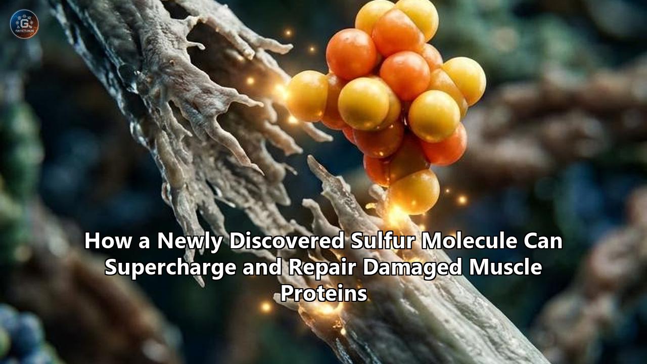
A scientific team led by Professor Ryuichi Tatsumi at Kyushu University’s Faculty of Agriculture published research in Scientific Reports detailing how a synthetic trisulfide molecule—lipoic acid trisulfide (LASSS)—can prevent age-related chemical corruption of Hepatocyte Growth Factor (HGF) and effectively double its therapeutic potency. The findings introduce a chemical biology strategy to repair damaged muscle proteins by structural modification rather than simple radical scavenging.
Skeletal muscle regeneration relies heavily on satellite cells, the resident stem cells embedded along muscle fibers. In healthy tissue, injury or mechanical strain triggers the release of HGF from the surrounding extracellular matrix. Free HGF docks onto c-met receptors on quiescent satellite cells, activating them to proliferate, differentiate, and fuse into new muscle fibers.
As tissue ages or suffers chronic oxidative strain, this regenerative cascade breaks down. The Kyushu University team discovered that the primary fault line is not a absolute deficiency of HGF, but rather a site-specific post-translational modification: nitration. Peroxynitrite and related reactive nitrogen species attach nitro groups to two critical tyrosine residues—Y198 and Y250—located squarely in HGF’s receptor-binding domain. Once nitrated, HGF loses its spatial conformation and fails to engage the c-met receptor, leaving satellite cells dormant despite abundant growth factor presence.
[Injured Muscle Matrix] ──> Releases HGF ──> Binds c-met Receptor ──> Satellite Cell Activation ──> Fiber Repair
                               │
                      (Age/Nitrative Stress)
                               ▼
                   Nitration at Y198 & Y250
                               │
                               ▼
                    [Rusted Key / Inactive HGF] ──X (Failed Docking)
                               │
                        (LASSS Treatment)
                               ▼
                     Conformational Shift
                               │
                               ▼
                    ["Super HGF" Formed] ───> 2x c-met Binding Affinity ───> Accelerated Repair
The introduction of LASSS fundamentally alters this outcome. In preclinical models, treating nitration-damaged HGF with LASSS at elevated molar ratios did not merely halt chemical modification; it induced an unexpected conformational shift. This structural altered form—termed "Super HGF" by the investigators—exhibits more than double the binding affinity for c-met receptors compared to native, unnitrated HGF, offering an unprecedented mechanism to repair damaged muscle proteins in aging and atrophied tissues.
The Molecular Mechanics: Rescuing the Rusted Key
To understand why this discovery marks a departure from existing regenerative therapies, one must examine the specific mechanics of tyrosine nitration. Tyrosine residue nitration adds a bulky, hydrophobic nitro ($-NO_2$) group to the ortho position of the phenolic ring. In HGF, residues Y198 and Y250 sit within the $N$-terminal and kringle domains, which form the primary contact interface with the c-met receptor extracellular binding domain.
When nitration occurs, the steric bulk of the nitro group physically blocks key hydrogen bonds and ionic interactions between HGF and c-met. The protein behaves like a rusted key: present in the lock, but incapable of turning the mechanism.
Native Tyrosine Residue (Y198/Y250):
    [Phenolic Ring] ─── H-Bonding Interface ───> Fits c-met Pocket

Nitrated Tyrosine Residue (Peroxynitrite Attack):
    [Phenolic Ring] ─── (-NO2 Group added) ───> Steric Hindrance / Electrostatic Repulsion ───> Docking Failure

Trisulfide-Modified Residue (LASSS Interaction):
    [Phenolic Ring] ─── Polysulfide Protection/Refolding ───> Enhanced Allosteric Fit ───> Potentiated Signal
Historically, scientists attempted to solve this issue through broad-spectrum antioxidant administration to reduce reactive nitrogen species ($RNS$) formation before nitration could occur. However, systemic scavenging of $RNS$ and reactive oxygen species ($ROS$) often disrupts necessary physiological signaling pathways required for muscle adaptation.
The Kyushu team evaluated two trisulfide compounds carrying three consecutively linked sulfur atoms ($R-S-S-S-R$): glutathione trisulfide (GSSSG) and lipoic acid trisulfide (LASSS). While both compounds prevented tyrosine nitration in vitro at low concentrations, raising the stoichiometric ratio of trisulfide to HGF to 1:8000 revealed a divergence between the two molecules.
GSSSG suppressed nitration but failed to restore functional receptor binding. In contrast, LASSS interacted directly with the HGF polypeptide chain. The three-sulfur bridge of LASSS donated persulfide groups or facilitated transient sulfur-exchange reactions with labile cysteine residues near the binding pocket. This chemical modification locked HGF into a altered, highly active spatial orientation. The resulting "Super HGF" demonstrated enhanced resistance to enzymatic cleavage and an elevated affinity for c-met, successfully awakening quiescent satellite cells even in environments dominated by inflammatory cytokines.
Evaluating Synthetic Trisulfides Against Alternative Sulfur Approaches
Sulfur compounds have long held a place in biomedical science, yet their clinical utility has suffered from poor target specificity, rapid metabolic clearance, or narrow therapeutic windows. Comparing LASSS with established sulfur-based interventions highlights what sets this novel molecule apart in its ability to repair damaged muscle proteins.
    Sulfur Therapy Spectrum
    │
    ├── Organosulfur Precursors (MSM, Cysteine)
    │   └── Low specificity; requires multi-step enzymatic processing; limited structural activity.
    │
    ├── Direct Hydrogen Sulfide Donors (GYY4137, AP39, SG1002)
    │   └── Rapid gas clearance; risk of systemic toxicity; broad off-target redox effects.
    │
    ├── Hydrophilic Trisulfides (GSSSG)
    │   └── In vitro antioxidant protection; fails in vivo due to poor membrane permeability/steric hindrance.
    │
    └── Lipophilic Smart Trisulfides (LASSS) [Kyushu Discovery]
        └── Site-specific protein refolding; high tissue penetration; creates "Super HGF" state.
1. Organic Sulfur Donors and Amino Acid Precursors
Nutritional sulfur sources—such as methylsulfonylmethane (MSM), $L$-cysteine, $L$-methionine, and $N$-acetylcysteine (NAC)—serve as general substrates for intracellular glutathione ($GSH$) synthesis. Clinical trials show that 1g to 3g daily doses of MSM can bolster total antioxidant capacity and attenuate post-exercise oxidative markers like lipid peroxides.
However, these dietary precursors rely on intact metabolic pathways (such as cystathionine $\gamma$-lyase and cystathionine $\beta$-synthase) to generate bioactive sulfur species. In aging or diseased muscle tissue, these enzymatic networks are frequently downregulated. Dietary sulfur precursors cannot directly bind to damaged extracellular proteins or alter protein folding dynamics; their action remains indirect and systemic.
2. Gaseous Hydrogen Sulfide ($H_2S$) Donors
Slow-release $H_2S$ donors—including GYY4137, mitochondria-targeted AP39, and sodium polysulfides—represent a second approach. These compounds deliver $H_2S$ gas or hydropersulfides to promote general protein persulfidation ($R-S-SH$), a reversible post-translational modification that protects active-site cysteines from irreversible oxidation.
The drawback of direct $H_2S$ delivery lies in its toxicodynamic profile. Hydrogen sulfide is a potent biological gas exchange inhibitor at high concentrations, inhibiting cytochrome c oxidase in the mitochondrial electron transport chain. Furthermore, $H_2S$ gas diffuses rapidly across cell membranes without tissue specificity, often causing systemic vasodilation before localized muscle repair can occur.
3. Hydrophilic vs. Lipophilic Trisulfides (GSSSG vs. LASSS)
The comparison between glutathione trisulfide (GSSSG) and lipoic acid trisulfide (LASSS) offers clear insights into structural requirements for therapeutic protein refolding. GSSSG is a large, highly hydrophilic peptide derivative. Although its three-sulfur chain can quench peroxynitrite radicals in an open aqueous solution, its charge profile and steric bulk prevent it from inserting into the hydrophobic clefts of HGF.
LASSS incorporates a lipoic acid backbone attached to the trisulfide motif. The hydrophobic lipophilic tail of lipoic acid enables the molecule to partition into lipid membranes and hydrophobic protein pockets. This structural property allows LASSS to dock against the $Y198/Y250$ region of HGF, delivering its reactive sulfur atoms directly to the sites of nitrative vulnerability.
| Parameter | Dietary Sulfur (MSM / NAC) | $H_2S$ Donors (AP39 / GYY4137) | Glutathione Trisulfide (GSSSG) | Lipoic Acid Trisulfide (LASSS) |
|---|---|---|---|---|
| Primary Mechanism | Substrate supply for intracellular $GSH$ synthesis | Enzymatic release of gasotransmitter $H_2S$ | Non-specific fluid-phase radical scavenging | Targeted protein refolding & anti-nitration |
| Site Specificity | None (Systemic metabolic distribution) | Moderate (Mitochondria-targeted variants) | Low (Restricted to aqueous extra/intracellular fluid) | High (Hydrophobic binding to growth factor interfaces) |
| Effect on HGF/c-met | Indirect reduction of general oxidative stress | Transient persulfidation of cysteine sensors | Prevents nitration; fails to restore receptor binding | Prevents nitration; doubles c-met binding affinity |
| In Vivo Efficacy | Modest post-exercise recovery enhancement | Variable; limited by gas diffusion rate | Ineffective in disuse atrophy mouse models | Restores satellite cell activation and fiber size |
| Toxicity Risk | Extremely low | High at elevated doses ($H_2S$ toxicity) | Low | Low to Moderate (Requires optimal stoichiometric dosing) |
Direct Growth Factor Therapies vs. Small-Molecule Protein Chaperones
The discovery of LASSS introduces a broader question in regenerative medicine: Is it better to flood the body with exogenous growth factors, or to chemically repair and supercharge the growth factors the body already produces?
APPROACH A: Direct Recombinant Protein Delivery (rHGF / IGF-1)
  [Exogenous Protein Injection] ──> High Systemic Concentration ──> Receptor Overstimulation ──> High Tumorigenic Risk & Rapid Clearance

APPROACH B: Small-Molecule Protein Chaperoning (LASSS)
  [LASSS Administration] ──> Binds Local Endogenous HGF ──> Forms "Super HGF" ──> Controlled Local Repair
Recombinant Growth Factor Administration
For decades, recombinant human HGF (rHGF) and insulin-like growth factor 1 (IGF-1) were considered primary candidates for treating severe muscle wasting conditions like sarcopenia, muscular dystrophy, and acute trauma. However, systemic administration of rHGF presents severe clinical limitations:
- Oncogenic Hazards: The c-met receptor is a known proto-oncogene. Sustained, elevated systemic concentrations of active rHGF trigger unguided cellular proliferation, significantly increasing the risk of tumor development and metastatic activation in subclinical lesions.
- Short Biological Half-Life: Free circulating rHGF is cleared by the liver within 15 to 30 minutes, requiring continuous high-dose infusions that compound off-target safety risks.
- Susceptibility to Microenvironmental Damage: Injecting pristine rHGF into an inflamed, aged, or dystrophic muscle matrix exposes the recombinant protein to the same nitrative conditions that damaged the endogenous HGF in the first place. The injected growth factor rapidly undergoes tyrosine nitration, losing efficacy shortly after administration.
Myostatin and Activin Receptor Blockers
Another major strategy in muscle biology focuses on neutralizing negative regulators of muscle mass, primarily myostatin (GDF-8). Antibodies and soluble receptors targeting the Activin type IIB receptor ($ActRIIB$) have demonstrated the ability to produce muscular hypertrophy.
Yet, myostatin inhibition faces a physiological tradeoff: it causes muscle fiber enlargement (hypertrophy) without stimulating satellite cell recruitment or resolving matrix inflammation. Patients treated with myostatin blockers often gain muscle volume, but do not show proportional gains in muscle quality, force output, or cellular repair capacity. Furthermore, off-target binding of early $ActRIIB$ inhibitors caused epistaxis and telangiectasia in clinical trials, stalling FDA approval for primary sarcopenia indications.
Cell Transplantation and Macrophage-Derived Signals
Advanced regenerative strategies attempt to introduce active cellular machinery directly into damaged muscle. Approaches include myoblast transplantation, autologous satellite cell therapies, or delivering macrophage-derived factors such as Nicotinamide Phosphoribosyltransferase (NAMPT) embedded within hydrogel scaffolds.
While hydrogel-delivered NAMPT has achieved complete muscle fiber restoration in acute severe trauma models, its translation is limited by manufacturing complexity, cold-chain logistics, invasive delivery requirements, and high cost. Cell transplant strategies face low post-injection cell viability, with over 90% of transplanted myoblasts dying within 48 hours due to exposure to a hostile, nitratively stressed host microenvironment.
Therapeutic Paradigm Comparison:

1. Recombinant Growth Factors (rHGF)
   - Potency: High
   - Tissue Specificity: Low
   - Safety Profile: Poor (High oncogenic/c-met risk)
   - Mechanism: Add synthetic signals

2. Myostatin Blockers (Anti-GDF-8)
   - Potency: Moderate
   - Tissue Specificity: Moderate
   - Safety Profile: Mixed (Vascular off-target risks)
   - Mechanism: Block brake signals

3. Cell/Hydrogel Therapies (NAMPT/Myoblasts)
   - Potency: High
   - Tissue Specificity: High
   - Safety Profile: High Complexity/Immune Risks
   - Mechanism: Replace cellular hardware

4. Trisulfide Chaperones (LASSS) [New Paradigm]
   - Potency: High (2x Native Activity)
   - Tissue Specificity: High (Matrix-bound HGF target)
   - Safety Profile: High (Modulates local native proteins)
   - Mechanism: Repair damaged muscle proteins natively
LASSS avoids these systemic and logistical barriers by operating as a small-molecule protein chaperone. It does not force non-physiological signaling cascades or require delicate living biological payloads. Instead, it works within the physiological architecture of the tissue, relying on natural mechanical strain or injury signals to release matrix-bound HGF, which it then stabilizes against chemical inactivation. By rescuing endogenous proteins, LASSS preserves the body's natural localized control over stem cell activation, offering a targeted path to repair damaged muscle proteins without elevating systemic cancer risks.
Site-Specific Persulfidation vs. Broad Antioxidant Scavenging
The discovery that LASSS induces a "Super HGF" state underscores an evolution in redox biology: the transition from non-specific radical scavenging to precision chemical modification.
For decades, the standard approach to oxidative and nitrative stress was the administration of high-dose, non-specific antioxidants like Vitamin C, Vitamin E, and high-dose $N$-acetylcysteine ($NAC$). The core logic was straightforward: neutralize reactive species ($ROS/RNS$) before they damage tissue components.
However, large-scale clinical trials repeatedly demonstrated that broad-spectrum antioxidants fail to accelerate muscle recovery or prevent sarcopenia. In many instances, high-dose antioxidant therapy actively hindered exercise-induced physiological adaptations. The biological reason is clear: reactive oxygen and nitrogen species are not merely toxic byproducts; they are essential signaling molecules required for cellular adaptation, mitochondrial biogenesis, and vascular remodeling.
Broad-Spectrum Antioxidants (Vitamin C/E/NAC):
  [ROS/RNS Production] ─── (Global Quenching) ───X Blocked Exercise Adaptation Signals
                                               ───X Fails to Reverse Established Nitration

Precision Trisulfide Chaperone (LASSS):
  [ROS/RNS Production] ─── Allowed for Physiological Signaling
                               │
                      (Nitrative Strain on HGF)
                               │
[LASSS Molecule] ──────────> Direct Binding to HGF ───> Reverses Inactivation & Supercharges Receptor Docking| Redox Mechanism | Broad-Spectrum Scavengers | Reactive Sulfur Chemistry (Trisulfides) |
|---|---|---|
| Primary Biological Target | Soluble free radicals ($O_2^{\bullet-}, \bullet OH, ONOO^-$) in bulk fluid | Specific cysteine/tyrosine residues on target proteins |
| Effect on Endogenous Signaling | Suppresses physiological $ROS/RNS$ signaling pathways | Preserves homeostatic signal cascades |
| Structural Impact on Proteins | None (Cannot alter existing post-translational modifications) | Induces conformational shifts via persulfidation/polysulfide exchanges |
| Therapeutic Window | Wide, but functionally ineffective for targeted structural repair | Narrow; dependent on specific molar stoichiometry |
The synthetic trisulfide bridge ($S_3$) in LASSS operates through nucleophilic sulfur chemistry. Rather than sweeping through fluid space to clear free radicals, the central sulfur atom in the trisulfide linkage exhibits unique electrophilic-nucleophilic dual reactivity.
When LASSS encounters a nitrated or oxidation-vulnerable protein like HGF, the trisulfide motif can undergo sulfur-exchange reactions with adjacent thiols or interact directly with the nitro-tyrosine adducts. This interaction alters the local tertiary structure of the protein, re-exposing key binding sites and neutralizing steric hindrance.
This precise mechanism offers a targeted bio-chemical strategy to repair damaged muscle proteins at the sub-molecular level, maintaining baseline physiological oxidation while restoring the function of essential signaling proteins.
Preclinical Benchmarks: Tail-Suspension Atrophy Models
To test whether the molecular recovery of HGF observed in vitro translates to functional tissue repair in live organisms, Professor Tatsumi’s team deployed a standardized model of severe muscle disuse: the rodent tail-suspension model.
Tail-Suspension Disuse Model (Unloaded Hindlimbs)
                     │
     ┌───────────────┴───────────────┐
     ▼                               ▼
[Control Group]              [LASSS Pretreatment]
     │                               │
     ├── Severe HGF Nitration        ├── Reduced HGF Nitration
     ├── Satellite Cell Quiescence   ├── Satellite Cell Activation
     ├── Fiber Atrophy (CSA Drop)    ├── Preserved Fiber Cross-Section
     └── Loss of Fast-Twitch Fibers  └── Retained Fast-Twitch Architecture
Tail suspension removes ground reaction forces from the hindlimbs of mice, inducing rapid hindlimb unloading, localized nitrative stress, microvascular regression, and acute disuse atrophy within 7 to 14 days. This model mirrors the microgravity-induced muscle loss experienced by astronauts, as well as the rapid muscle wasting seen in bedridden intensive care patients.
Key Preclinical Findings:
- Reduction of Nitrotyrosine Markers: Untreated tail-suspended mice demonstrated a dramatic rise in nitrated HGF ($Y198/Y250$) within extracellular matrix extracts. Animals pretreated with LASSS showed a marked reduction in HGF nitration, maintaining the protein in its active state despite sustained physical unloading.
- Satellite Cell Activation Rates: Immunohistochemical staining of muscle cross-sections revealed that LASSS-treated animals maintained active, proliferating satellite cells ($Pax7^+ / MyoD^+$ positive) along the basal lamina. In contrast, control animals exhibited severe stem cell quiescence and reduced regenerative capacity.
- Preservation of Fast-Twitch Muscle Fibers: Aged and disuse-atrophied muscles preferentially lose Type II (fast-twitch) muscle fibers—the primary fibers responsible for power generation, rapid balance correction, and overall force output. LASSS treatment preserved fast-twitch fiber cross-sectional area ($CSA$) and reduced fibrotic scar tissue formation.
- Comparison with GSSSG: Confirming the cell-free assay results, mice treated with glutathione trisulfide (GSSSG) showed no significant protection against disuse atrophy or stem cell decline, confirming that lipophilic trisulfide architecture is required for in vivo efficacy.
Pharmacokinetic Constraints, Stoichiometry, and Clinical Hurdles
Despite the impressive performance of LASSS in preclinical trials, translating this discovery into a human therapeutic introduces clear pharmacological and chemical challenges.
            Therapeutic Stoichiometric Window

   Low Ratio (< 1:4000)      Optimal Ratio (1:8000)      Excessive Ratio (> 1:15000)
┌─────────────────────────┬───────────────────────────┬──────────────────────────┐
│   Incomplete Protection │     "Super HGF" State     │   Risk of Off-Target     │
│   Nitration Persists    │   2x c-met Affinity       │   Redox Disruptions /    │
│   Receptor Docking Fails│   Optimal Regeneration    │   Polysulfide Cleavage   │
└─────────────────────────┴───────────────────────────┴──────────────────────────┘
1. The Stoichiometric Requirement
A key challenge identified by the Kyushu University researchers is the non-linear dose response of LASSS. At lower molar ratios of trisulfide-to-HGF (e.g., 1:4000), LASSS successfully suppressed tyrosine nitration, but failed to induce the conformational change required to form "Super HGF".
Only when the molar ratio was raised to 1:8000 did the protein undergo the allosteric shift that doubled its binding affinity for the c-met receptor. Achieving and maintaining this precise stoichiometric ratio within human skeletal muscle matrixes—where localized extracellular growth factor concentrations vary—will require sophisticated dosing algorithms and advanced drug-delivery systems.
2. Metabolic Stability and Synthetic Scalability
Because lipoic acid trisulfide (LASSS) is a synthetic molecule created in a laboratory, it is not present in natural food sources or human metabolic pathways. Once introduced systemically, the three-sulfur chain ($S_3$) is susceptible to reduction by circulating thiols, such as serum albumin and red blood cell glutathione.
If the trisulfide bridge is prematurely cleaved into mono- or disulfide forms in the bloodstream, the compound loses its ability to refold HGF. Developing targeted delivery vehicles—such as polymeric nanoparticles, lipid nanoemulsions, or localized hydrogel depots—will be essential to shield the trisulfide linkage until it reaches target muscle tissue.
3. Safety Margin and Off-Target Effects
While HGF potentiation is beneficial for injured or atrophied muscle, over-activating c-met receptors across non-target tissues (such as the liver, kidneys, or gastrointestinal tract) poses potential risks. c-met hyperactivation is implicated in oncogenesis and cellular motility in various carcinomas.
Translational studies must confirm that LASSS-modified HGF remains anchored within the local extracellular matrix of skeletal muscle, rather than circulating systemically to overstimulate c-met receptors in other organ systems.
Future Outlook: The Horizon for Sulfur Chemobiology
The identification of LASSS as a small-molecule protein refolding agent opens up new research directions in age-related degenerative disease, sports medicine, and rehabilitative care.
                     Potential Therapeutic Pipeline
                                   │
      ┌────────────────────────────┼────────────────────────────┐
      ▼                            ▼                            ▼
[Sarcopenia & ICU Atrophy]   [Neuromuscular Disease]    [Cardiomyopathy & Injury]
- Age-related muscle loss    - Duchenne Dystrophy (DMD) - Post-MI scar reduction
- Bed-rest recovery          - ALS-adjacent weakness    - Equine/Canine sports medicine
Primary Therapeutic Targets:
- Age-Related Sarcopenia and ICU-Acquired Weakness: Millions of aging adults experience severe loss of muscle mass, balance, and independence due to age-related nitrative stress. LASSS offers a potential therapeutic path to restore muscle stem cell activity, preserving fast-twitch muscle fibers without relying on invasive hormone replacement or high-risk systemic anabolic agents.
- Muscular Dystrophies: In conditions such as Duchenne Muscular Dystrophy (DMD), chronic membrane tears subject the extracellular matrix to continuous oxidative and nitrative stress, impairing native repair mechanisms. Small-molecule chaperones could be paired with gene therapies or exon-skipping treatments to protect newly synthesized muscle proteins.
- Veterinary and Regenerative Medicine: Because HGF signaling is highly conserved across mammalian species, trisulfide chaperones could find early applications in veterinary medicine—particularly in treating age-related muscle loss or soft-tissue injuries in canine and equine athletes.
- Targeted Organ Repair: Beyond skeletal muscle, HGF plays a critical role in liver regeneration, lung tissue repair, and cardiac remodeling post-myocardial infarction. Investigating whether LASSS can protect HGF from nitrative inactivation in damaged cardiac or hepatic tissues represents a promising direction for future research.
As clinical development progresses, researchers will focus on establishing optimal delivery formulations, evaluating toxicity profiles across multi-organ systems, and identifying additional protein targets vulnerable to trisulfide-mediated structural recovery.
The transition from non-specific antioxidant approaches to site-specific molecular chaperones represents a meaningful shift in chemical biology, offering new possibilities for targeted therapies designed to repair damaged muscle proteins in aging and diseased populations.
Reference:
- https://www.news-medical.net/news/20260724/Researchers-discover-molecule-to-fight-age-related-muscle-decline.aspx
- https://neurosciencenews.com/compound-restore-aging-muscles-31121/
- https://www.forbes.com/sites/maryroeloffs/2026/07/24/getting-weaker-as-you-age-may-not-be-inevitable-scientists-say/
- https://www.sciencedaily.com/releases/2026/07/260724061432.htm
- https://www.mdpi.com/1467-3045/47/9/765
- https://pubmed.ncbi.nlm.nih.gov/41731189/
- https://www.psu.edu/news/research/story/method-producing-sulfur-compounds-cells-shows-promise-tissue-repair
- https://balchem.com/news/sulfur-msm-exercise-recovery/
- https://pmc.ncbi.nlm.nih.gov/articles/PMC9668843/
- https://wellnessresources.com/blogs/news/rebuild-restore-and-renew-with-the-power-of-sulfur
- https://www.researchgate.net/publication/395595556_Reactive_Sulfur_Species_and_Protein_Persulfidation_An_Emerging_Redox_Axis_in_Human_Health_and_Disease
- https://www.dkfz.de/en/news/press-releases/detail/metabolic-enzyme-protects-proteins-with-sulfur
- https://www.eurekalert.org/news-releases/1136524
- https://www.sciencedaily.com/releases/2021/02/210210170145.htm
- https://communities.springernature.com/posts/sulfur-amino-acid-supplementation-may-hold-promise-in-muscular-dystrophy